JScholar Publishers
Different Chitosan-Based Biomaterials and their Biomedical Applications
Inês Moutinho1,*, Inês da Costa Oliveira1, Mariana Cristina Santos1, Mário Vasconcelos2, Ana Isabel Portela2
1Master degree from Faculty of Dentistry, University of Porto (Portugal) 2Professors at Faculty of Dentistry, University of Porto (Portugal)
Review Open Access
European Journal of
Medical Research and Clinical Trials
Received Date: August 13, 2019 Accepted Date: September 06, 2019 Published Date: September 09, 2019
Citation: Inês Moutinho (2019) Different Chitosan-Based Biomaterials and their Biomedical Applications. Eur J Med Res Clin
Trials 1: 1-12.
*Corresponding author: Inês Moutinho,Master degree from Faculty of Dentistry, University of Porto (Portugal); E-mail: [email protected]
Abstract
Chitin, the second most available marine biopolymer, was firstly used in the 1930s and early 1940s. [1-4] It has a linear structure that is composed by repeating β-1,4 linked N-acetylated glucosamine units [2, 3, 5, 6]. Chitosan, derived from chitin, is a natural polymer that is known for being biodegradable, non-toxic and antibacterial [1, 2, 4, 7-12]. It is cationic, has a hydrophilic surface and causes minimal foreign-body reaction [1, 6, 7, 10, 11, 13-17]. Also, chitosan shows a reasonable solubility in water and in organic acids. However, this represents the major limitation to chitosan’s preparation and use for biomedical applications.[1, 4, 6, 10, 14-16, 18] Due to these properties it can be used for drug delivery, cancer di-agnosis, immune regulation, tissue regeneration (treatment of damaged skin, nerve regeneration, cartilage and bone repair) and as antioxidant and anti-inflammatoryy [3, 4, 11, 13, 15, 17]. Also, shows an antitumor and antidiabetic capacity and hae-mostatic effects [5, 19, 20]. In what Dental medicine is concerned, chitosan is very useful too. Due to its bone regeneration capacity, it can be used as a therapy for alveolar bone repair, as well as in the implantology field. [7, 8, 21-23] Because of its antimicrobial effect, is very promising in endodontic treatment. [24] Also, it promotes a higher resistance to fungal diseases and leads to better effectiveness on decay remineralization [13, 14, 17]. In addition, although numerous treatments are used to regenerate the alveolar bone, cementum, and loss of periodontal ligaments (PDL), Chitosan-based therapies are used to treat periodontal lesions [17]. To conclude, chitosan is an innovative biomaterial which use has been increasing exponential-ly over the last years. Thus, the aim of this review is to study chitosan’s biomedical applications, especialexponential-ly in what concerns to Dental Medicine.
Keywords: chitosan; chitin; biomaterials; biomedical application; tissue regeneration; drug delivery.
©2019 The Authors. Published by the JScholar under the terms of the Crea-tive Commons Attribution License http://creaCrea-tivecommons.org/licenses/ by/3.0/, which permits unrestricted use, provided the original author and source are credited.
2
Introduction
The commercial applications of chitin arose in the 1930s and ear-ly 1940s but it was first identified in 1884. [1, 2]
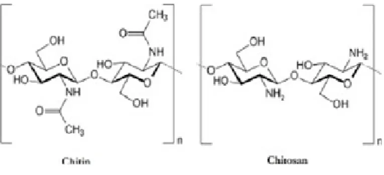
Chitin consists of a marine biopolymer which is the second most available in nature (the main one is cellulose) [2-4]. It has a linear structure that is composed by repeating β-1,4 linked N- acetylat-ed glucosamine units. [2, 3, 5, 6]
Chitin can occur as α, β or γ forms. Each one has its own struc-tural properties:
• The α form has microfibril with an antiparallel orienta-tion which are linked by strong hydrogen bonds. In nature, it is the most common form and is the best one for the industry. • The β form, unlike α, has its chains parallel orientated (linked by weak hydrogen bonds).
• The γ form, that is the less known, consists of a mixture of antiparallel and parallel chains.
Due to strong hydrogen bonds present in α- chitin, it is insolu-ble in water, most organic acids and dilutes the acid and alkaline solutions. On the other hand, since β-chitin has weak hydrogen bonds, it has better solubility in most acids and swells up in the water. [4]
This polymer can be found in cell walls of yeasts and fungi, shells of crustaceans, corals, diatoms sponges, molluscs, the cuticles of insects and worms, however, the major commercial sources of chitin are crab and shrimp shells. [1, 5, 6, 14, 25] Bearing this in mind, chitin extraction from shells unable us to minimize the waste, as well as to produce compounds with valuable biological properties and specialty applications, such as chitosan [1, 4].
Materials and Methods
In order to find reliable articles to base this review on, the re-search was done in the database Pubmed.
As a starting point, we used the terms “Chitosan collagen mem-brane”. As a result, we obtained 174 articles. Posteriorly applying filters, such as “5 years” and “free full-text”, this number reduced to 17, from which we chose 10 after reading their abstract. Once the number of articles we obtained was insufficient to elab-orate a review, we were forced to enlarge de research and, conse-quently, the theme, looking for more embracing terms.
Therefore, in the second phase, we searched for "Chitosan". It re-sulted in 21608 articles, a number that reduced to 63, after apply-ing for the filters "review", "free full-text", "5 years" and "humans". From those, 13 were found pertinent by Reading the abstract. Secondly, “chitosan membrane” was the expression searched. 2759 articles were gathered, resulting in 12 after using the fil-ters “review”, “free full-text” and “5 years”. Due to reading the abstract, the number of selected articles decreased to 5.
In another research, we searched for “Chitosan membrane AND dentistry”. We found a total of 56 articles. After using the words “free full- text” and “5 years” as filters, 8 articles remained, which became 4 after reading the abstract.
Ultimately, “Chitosan scaffold AND dentistry" was the expres-sion we looked for. 75 documents were founded, however, only 2 were selected by reading the abstract. The filters used were "free full-text" and "5 years". To conclude, some articles were common to the different researches that we made. Those were only con-sidered once.
Chitosan
Chitin is transformed into chitosan, its main derivative, by par-tial deacetylation under special conditions like a strongly alka-line environment. Chitosan is a alka-linear amino polysaccharide composed of β(1→4) glycosidic bonds linking, randomly distrib-uted, D- glucosamine, and N-acetyl-D-glucosamine
monomers. [5-8, 16, 25, 26] It is structurally similar to the gly-cosaminoglycans in the extracellular matrix. [8, 11]
Figure 1- Chemical structure of chitin and chitosan (Available
3
JScholar Publishers
Eur J Med Res Clin Trials 2019 | Vol 1: 101 Deacetylation can be performed by enzymatic preparations or
chemical process, being the last one the most used because of its low cost and as it allows massive production. (1)
Chemical Deacetylation
Chemical deacetylation can be performed by acids or alkalis. However, as glycosidic bonds are very susceptible to acid, alkalis (such as NaOH) are more often used. Depending on the NaOH concentration, reaction time, temperature and repetition of alka-line steps, final chitosan may have different characteristics, such as degree of deacetylation (DA) and molecular weight (MW). Other factors may be taken [5] into consideration: atmosphere, reaction reagent, chitin and solvent ratio, particle size and source of raw material. Despite its advantages, chemical deacetylation has some cons like energy consumption and waste of concen-trated alkaline solution. As a result, enzymatic deacetylation has occurred. [1, 25]
Enzymatic Deacetylation
Enzymatic deacetylation uses chitin deacetylases to perform a controlled, non- degradable process that allows us to obtain a novel, well-defined chitosan. Such enzymes are glycoproteins which are found in several insect species and fungi. They are extremely stable at 50 °C and show an excellent specificity for β- [1,4]-linked N-acetyl-D-glucosamine polymers. Nevertheless, chitin substrates need to be pretreated so that the accessibility of the acetyl groups to the enzyme can be improved [1].
The process and therefore the degree of deacetylation is deter-minant for the establishment of chitosan characteristics. [16] Chitosan is a very interesting polymer as it is biodegradable, non-toxic, biocompatible and has antibacterial effects, that can promote the acceleration of wound healing. It has high adsorp-tion, is capable of forming three- dimensional porous structures, is easy to handle and has a low cost. Moreover, chitosan has me-chanical stability, especially when associated with collagen. [1, 4, 6, 10-12, 14, 18] It is a cationic polymer at low pH (an excep-tion since other polysaccharides are generally neutral or anionic) which gives it a great affinity for anionic molecules. Therefore, chitosan can bond with mucous tissue (mucoadhesiveness), being a good option for tissue engineering [1, 6, 10, 13-16]. In addition, its hydrophilic surface promotes cell adhesion, prolif-eration, and differentiation and causes a minimal foreign- body reaction on implantation. [7, 11, 17]
As mentioned above, the DA and MW influence, to a great ex-tent, chitosan characteristics like crystallinity, solubility, and its mechanical performance. This polymer has low molecular
weight and a high degree of deacetylation. As a result, chitosan shows a reasonable solubility in water (that can be improved by chemical modification) and inorganic acids. This last character-istic represents the major limit to chitosan preparation and uses for biomedical applications. [1, 4, 6, 10, 14-16,18].
Due to these properties, chitosan has even more applications. It is very useful for drug delivery, cancer diagnosis, immune regu-lation, treatment of damaged skin, nerve regeneration, cartilage and bone repair, antioxidant properties and anti-inflammation. [3, 4, 11, 13, 15, 17] Also, shows an antitumor and antidiabetic capacity and hemostatic effects (as its cationic nature attracts the anionic blood cells and platelets allowing it to be used in post-partum hemorrhage, dental surgery, and other surgical appli-cations). [5, 19,20] Besides that, chitosan can interact with substances like collagen, heparin, hyaluronic acid, gelatine, alginate and minerals: [21, 25]
• Hydroxyapatite maximizes the in vivo osteogenic ca-pacity which leads to an internal bone growth with accelerated resorption of the matrix. (16, 21)
• Calcium aluminate also has an important role in bone regeneration (it releases Ca2+ that increases osteogenesis and cementogenesis). In addition, it improves chitosan’s mechanical strength, which is required for scaffolds. [21]
Despite all the information known about this biomolecule, the lack of clinical studies in humans requires more and deeper stud-ies on this matter.
Chitosan and its different morphologies Depending on environ-mental pH values and degree of acetylation, chitosan can dis-play different ways of production. At low pH (<6), amines are protonated, exhibiting a polycationic nature (contributes to cre-ating multilayer structures and/or electrostatic complexes). At high pH (>6.5), chitosan amines are deprotonated and reactive, promoting interpolymer associations. These associations lead to film, fibre, porous templates or hydrogel formation. [2, 20, 27, 28] It can also be presented as sponge, powder, bead, solution, foams, microcapsules, tubes, membranous films, nanofi-bers, scaffolds, nanoparticles. [2, 11, 16, 18, 22, 25]
Scientific literature supports that bacterias are unable to form biofilm by chitosan nanoparticles. [24] Additionally, scaffolds are very useful in bone, skin (also regenerated using fibers and hydrogels), liver, cartilage, nerve, and blood vessel wounds. Fi-bers and hydrogels allow migration of inflammatory cells to the wound site and collagen matrix deposition. Furthermore, hydro-gels have the capacity to promote angiogenesis. [3]
4
Chitosan’s applications 4.1 Tissue Regeneration
Natural occurring biomaterials (like chitosan and collagen) and Calcium phosphate ceramics (like hydroxyapatite) are common-ly used for tissue regeneration. Chitosan, due to its properties, is a great material to form different platforms where cells can adhere, grow in and differentiate. [9, 10, 18]
Nevertheless, several studies proved that better results were achieved when chitosan was combined with collagen in compar-ison with pure chitosan. [9, 16]
In addition, hydrogels can be used to produce scaffolds that have as advantages: minimal physical flexibility, invasive procedure, and easiness of incorporating cells. However, one of its disadvan-tages is that some cells have a lack of adhesion to scaffolds. Dha-nya et al. prepared a new scaffold, O-carboxymethyl chitosan (O- CMC), as an attempt to solve this limitation. It has high viscosity, good water solubility, and high hydrodynamic volume. [18] Moreover, chitosan-based platforms can also be used in a more delicate area, such as cardiac patch for defects in myocardial tis-sue. Pok et al. prepared a gelatin-chitosan hydrogel-based multi-layer cardiac patch. This compound (which the main component is chitosan) increases patch tensile strength and, due to gelatin, consolidates cell attachments. [18]
Another study has been developed to investigate in-vitro and in-vivo degradation rates of porous freeze gelated chitosan (CH) and chitosan hydroxyapatite scaffolds. It is believed that scaffold’s pore architecture is essential to an efficient cell seeding into the scaffold. Simultaneously, it may offer enough space for the regen-eration of newly formed tissue. Therefore, those two character-istics should be taken in consideration when chitosan scaffold is being produced. Biomaterial degradation rate may, also, be taken into account. It might be sustained enough so that healthy tissue regeneration can be fulfilled [28].
Chitosan hydroxyapatite scaffolds are stable and biodegradable (however its degradation rate is lower). Additionally, they pro-vide an osteoconductive environment for bone formation and have a high elastic modulus. These are the reasons why they are appropriate to bone substitution and regeneration in dental im-plants [28].
On the other hand, scaffolds are favorable for tissue regenera-tion if they show a highly porous structure to support cellular adhesion and proliferation and extracellular matrix (ECM) pro-duction. Their degradation is dependent on pore morphology and size, porosity percentage, surface area, and hydrophilicity.
Porous freeze gelated chitosan scaffolds have been tested in order to be used for periodontal regeneration [28].
Peter et al. and Mota et al. concluded that degradation rate of chi-tosan can be reduced by adding compounds (such as Bioglass™), what can contradict one of the major limitations of its use for tissue regeneration. Therefore, the conclusions of these studies prove the enormous chitosan and hydroxyapatite potential for use in composites that aim bone tissue engineering applications [28].
Even so, the lack of precise information about chitosan’s charac-teristics, its large chemical variability and studies’ methodologi-cal limitations make it difficult to reproduce and standardize its clinical application. [10]
Wound healing
Inflammation is a crucial part of healing, however, it encumbers tissue regeneration when we are dealing with chronic wounds or biomaterial implantation. [3]
Until known, in order to decrease the inflammatory response, biomaterials have been incorporated with anti-inflammatory therapeutics. Therefore, new scaffolds’ designs aim to provide instructive signals for cell attachment, function and proliferation within the native tissue and to modulate the immune response. [3]
Chitin-derived materials, such as chitosan, were discovered to be effective to chronic inflammatory diseases treatment (inflamma-tory bowel disease, arthritis, sepsis, and asthma, for example) as they are useful for drug delivery and in modulating immune re-sponses (it attracts macrophages and neutrophils to the wound, monitoring inflammatory response and reepithelization). Their mechanism, although not fully defined, consists of molecules degradation into monosaccharides, accelerating wound healing. [3, 18].
Nerve regeneration
Autografts have been used to treat peripheral nerve damage, but they have many disadvantages. Therefore, chitosan is an excellent option to treat this condition as hydrophilicity is considered to be essential for preventing fibrous scar tissue invasion and for promoting nerve regeneration. Nerve conduits formed by this biopolymer have better mechanical properties and slow biode-gradability. [29]
Eur J Med Res Clin Trials 2019 | Vol 1: 101 JScholar Publishers
5 Recently, porous hybrid membranes (such as chitosan-
-glyc-idoxypropyltrimethoxysilane - chitosan-GPTMS) were inves-tigated for peripheral nerve regeneration. They were proven to significantly improve nerve fibre regeneration and its functional recovery in rat models by demyelination. The quantity of regen-erated myelin fibres increased, as well as, myelin thickness [29].
Neuronal disorders
Chitosan can play an important role in the prevention and treat-ment of age-related diseases. Among others, it can prevent pro-tein conformational diseases and treat those which result from oxidative stress. Researchers from Fukuyama University con-ducted in vitro and in vivo studies and concluded that, by de-creasing oxidative stress (it suppresses the formation of reactive oxygen species), chitosan can have a direct antioxidant effect in the systemic circulation. [5, 19]
Chitosan oligosaccharides (COS) are also used in order to treat neuronal disorders, including Alzheimer’s disease, Parkinson’s disease, and nerve crush injury. Owing to their shorter chain lengths, COS are rapidly soluble in water which confers them new properties like being neuroprotective (such as β-amyloid and acetylcholinesterase inhibitory activities), anti- neuroinflam-matory and having anti-apoptosis effects. Moreover, Gong et al. explored the effects of this polysaccharide on nerve regeneration after peripheral nerve injuries. As a result, they concluded that it could enhance the quantity of regenerated myelinated nerve fibres, the cross-sectional area of muscle fibres, the muscle action potentials and the thickness of regenerated myelin sheaths in the nerves. [5]
Nowadays, neuroprotection aims to limit neuronal dysfunction or death after nerve injury. For that, neuroprotective agents like antioxidants and anti-inflammatory agents are used. The prob-lem is that synthetic agents are linked to several side effects. Therefore, anti-neuronal disorders agents with low toxicity have been studied. [5]
To conclude, Chitosan and its derivatives are neuroprotective through mechanisms like antioxidative stress action; suppress-ing effect on Abeta aggregation; anti-neuroinflammatory action; anti-apoptosis action; anti-excitotoxic action. [5]
Cancer
Cancer diagnosis
A cancer diagnosis is a crucial step since it gives some informa-tion about treatment effectiveness. Theragnosis is a new concept
which combines molecular imaging (through nanoparticles that are accumulated in tumor tissue) and anti-cancer drugs that are encapsulated into nanoparticles. In this line of thought, chitosan is ideal for nanoparticle-basedd theragnosis, as it can be easily transformed into nanoparticles and self- degradable after accu-rate diagnosis and drug delivery [6].
Cancer Therapy
In this field, chitosan has several applications, especially when prepared into nanoparticles. This biomaterial can be an ide-al vaccine adjuvant due to its cationic nature, biocompatibili-ty, safety and its ability to be used as an antigen carrier. It also showed a superior immune activity when compared to tradition-al immunoadjuvants, as chitosan retains the peptide antigen in the administration site for a longer period, allowing the antigen to be presented for efficient immune activity [6].
Chemotherapy
Anti-cancer drugs used in chemotherapy have several severe side effects, due to the low specificity to the tumor tissue and their sys-temic administration. To minimize this non- specificity therapy, selective embolization has been studied. It provides angiogenesis that will affect cancer cells viability (it occludes arteries formed by those cells, inducing tumor’s cells starvation). Chitosan is not a viable candidate to this approach since it is weak when pro-duced into microspheres. [18] Chemoembolization is a method that occurred later described by Park et al. It combines the effect of embolization with sustained release of the anti-cancer drug. They accomplished an efficient in vivo anti-cancer effect with a reduction of one-third of the initial tumor size. [18]
Radiotherapy
Chitosan is soluble in acidic conditions, unlike neutral or basic ones. Yuka et al. suggested that it can be chelated with several substances and then injected (as an acidic solution- 166Ho- chi-tosan complex) into human organs. It forms a solid structure that consists of a radioactive pharmaceutical device, allowing radio-activity to be localized in the administration site (when intratu-morally and intrahepatically injected) [18].
Drug delivery
The presence of amino groups with positive charge allows chi-tosan to promote chemical and physical cross-linking (chichi-tosan can adhere to negatively charged organs’ surface), which is the main advantage of its use. In addition, if chitosan is modified with hydrophobic groups, it can be used to encapsulate
hydro-6 phobic drugs, since it forms intra- and intermolecular
interac-tions [1, 18].
Therefore, chitosan can be a successful carrier for many different therapeutic agents: proteins, peptides, growth factors, DNA, vac-cines, anti-inflammatory, antibiotics, and other drugs for paren-tal and non-parenparen-tal routes of administration (like oral, ocular, topical, nasal, transdermal and pulmonary) [27]. Although the many forms of chitosan formulations, the choice to use a mi-cro or a nanoparticle depends on what is required. For exam-ple, since microparticles cause obstruction in the blood vessels, nanoparticles are the best choice for intravenous delivery. On the other hand, if nanoparticles are used to pulmonary delivery (to treat different pulmonary infections and diseases, like tuberculo-sis) they would be exhaled. So, in this case, the use of micropar-ticles would be better [1, 27].
Both chitosan’s micro and nanoparticles, when loaded with an-tibiotics and administrated by the oral route, can be used in the treatment of gastrointestinal and systemic diseases, such as ul-cerative colitis, irritable bowel syndrome, Crohn’s disease and suppression of Helicobacter pylori. Drug colon targeting is use-ful since the degradation of chitosan is done by the specific local microflora [27].
Bone regeneration
Bone regeneration is important to orthopedic, maxillofacial, and oral surgery (such as in periodontology and implantology field) in order to correct the bone defects. Therefore, the density and bone volume must be recreated, without forgetting the function. At the moment, therapies use bone tissue’s grafts from different kind of origins – heterologous, homologous or autologous (each one of them with advantages and disadvantages). As autologous grafts (which have no risk of rejection and diseases transmission) are limited, a new approach for bone regeneration is needed. [7, 8, 12, 13, 16, 21]
Furthermore, the biomaterials have been studied because of their capacity to induce a biological response, knowing that the results obtained are satisfactory. Nowadays, guided bone regeneration (GBR) is a therapy for alveolar bone repair using a membrane which has the ability to prevent the formation of fibrous connec-tive tissue [7, 8, 21, 22]. The membrane has some properties like bioactivity (promotes cellular migration, proliferation, and infil-tration), biocompatibility, bioresorption, lack of cytotoxicity and stability. This membrane can be combined with natural polymers (collagen and chitosan). The degradation rate of polymers is also a crucial factor: it should be total and not require a second
sur-gical intervention. That is why it may have a long-term influence on the success of bone regeneration [7, 12, 22].
According to Jiayu Zhang et al. [22] local drugs delivery systems (DDS) allow aspirin to be in the right place at the correct mo-ment. As a result, there will be a reduction in the number of ad-ministrations, bacterial resistance, gastrointestinal effects and we can avoid variations of large concentration. [22]
Non-steroidal anti-inflammatory drugs (NSAID) are reported to be effective in: repair of bone defects and fracture (bear in mind that dose is an important factor), reduce the percentage of tissue destruction and bone loss in periodontal disease [22].
The results show a good controlled-release pattern, the biocom-patibility and osteogenic potential of the membranes and also a significant increase of bone, in animal models. [22] On the oth-er hand, GBR membranes are vulnoth-erable to infection because of their exposure to pathogenic agents, which affects bone regener-ation. In an attempt to counteract this, it is used antibiotic oint-ments. However, it should be created a new antibacterial GBR membrane. (8) Shiqing Ma et al. [8] used membranes containing antibacterial agents such as tetracycline, chlorhexidine, mino-cycline, and amoxicillin. These ones have the ability to inhibit bacterial proliferation and induce the growth of fibroblasts and osteoblasts, as well as, stimulate angiogenesis. [8]
An asymmetric collagen/chitosan GBR membrane (CCM brane) was carried with encapsulated minocycline. This mem-brane had loose chitosan (that allows it to be osteoconductive) and dense collagen layers (act as a physical barrier), low degrada-tion and activity against Fusobacterium nucleatum and Porphy-romonas gingivalis, which are the core of periodontitis disease [8].
CCM membrane promoted new bone formation. In addition, there weren’t verified inflammatory reactions and grade of deg-radation has raised. Furthermore, it was observed a huge num-ber of mesenchymal stem cells and capillary vessels (responsible for transportation of nutrient and stem cells), which is an ideal environment of osteoinduction [8]
Jae Min Song et al. [7]. reinforced that chitosan/fibrin–hydroxy-apatite (CFB–HAP), a membrane that has also been studied, promotes haemostasis and adhesiveness and is biocompatible, anti-infective, resorbable and plastic. [7] After this study, it is possible to affirm that the bone repair capacity is similar between CFB–HAP membrane and collagen membrane. In addition, the absorbable chitosan membrane can be used as a barrier mem-brane in GBR. However, the inflammatory reaction didn’t exist
Eur J Med Res Clin Trials 2019 | Vol 1: 101 JScholar Publishers
7 in the CFB-HAP, which can comprise the success of the guided
bone regeneration [7].
Antimicrobial activity
The endodontic treatment has a significant percentage of failure, associated with inefficient decontamination of root canals and repair of changes caused by the disease [24].
In this context, antimicrobial photodynamic therapy (PDT) has been used as it promotes cross-linking between collagen and proteins and has a large spectrum of antimicrobial activity. Also, some nanoparticles like chitosan, which is able to reduce the bac-terial-biofilm, have been modified with a photosensitizer (such as rose Bengal, that produces synglet oxygen) in order to high-light PDT. As a result, bioactive polymeric chitosan nanopar-ticles functionalized with rose-bengal (CSRBnp) were studied to eliminate biofilms and stabilize the matrix (dentin-collagen) attending to its positive charge and nano size. Those character-istics, also promote a Physico-chemical interaction with some other particles [24].
The conjugation of this cationic molecule with photosensitizer allowed the penetration on the bacterial membrane. These cells were efficiently killed, as well as their biofilm structure destroyed. The antibacterial effects of chitosan are based on promoting bac-terial membrane damage, increasing its permeability and provid-ing intracellular leakage [24]. In addition, chitosan nanoparticles have been incorporated into root canal sealers in order to reduce the biofilm formation and inhibit microbial penetration in the dentin-root [13].
In another view, pulp exposure can be treated without chemical injury to pulp cells. For that, it was tested a chitosan/collagen membrane combined with calcium-aluminate micro-particles. The results were: an increase of human dental pulp cells proliferation, trailed by odontoblastic phenotypes (high expres-sion) and intensive deposition of matrix’s mineralized [30].
Implants
The tooth extraction is associated with resorption of alveolar bone in a period of three months. Residual ridge absorption is a real problem to posterior rehabilitation of the edentulous area because of the lack of bone support. Any solution present has to promote osteogenesis and also angiogenesis. [23]
The core is to preserve the level of bone in order to realize a func-tional and aesthetic restoration. A dental implant is used to re-place multiple or single missing teeth or to support a prosthesis. The lifetime of a dental implant is limited by its capacity to
up-take and hold backwater, which is related to diffusion’s coefficient of the material. [23, 28].
One of the proposals is to use Poly (L -lactic-co-glycolic) acid and nanohydroxyapatite together with chitosan to delivery ad-renomedullin (ADM). It was reported that ADM is a substance which promotes the mitogen activity and proliferation of osteo-blasts. Besides that, it seems like a promising new formula for bone regeneration, once it increases the formation of new bone tissue and is capable of stabilizing the residual alveolar ridge [13, 23].
Oral diseases
It is known that chitosan has activity against oral pathogens such as Prevotella intermedia, Porphyronomasgingivalis, and Actinobacillus actinomycetemcomitans, inhibiting the bacterial plaque formation. To some authors, this inhibition can occur as chitosan nanoparticles kill the bacteria or have a direct action in the bacteria-substrate. Additionally, it has been reported that the collagen’s turn over has increased when chitosan is present. In this line of thought, toothpaste could contain chitosan nanopar-ticles, in order to reduce bacterial biofilm formation [13, 17].
Carie (Decay)
Chitosan nanoparticles can be used to remineralize dentine and enamel caries. This occurs because chitosan’s nanoparticles, at a neutral pH, cause damage in cariogenic microorganisms, such as S.mutans’ cells. [13] There are studies using nanocom-plexes of carboxymethyl chitosan/amorphous calcium phos-phate (CMC/ ACP) to remineralize the dentine via biomimetic strategy. This remineralization is more difficult to occur due to dentine’s organic matrix when it is compared to enamel. In ad-dition, the remineralization of dentine happens by its residual crystals’ growth. Using a single-layer model, CMC/APC released ACP nanoparticles to achieve intrafibrillar mineralization forms of collagen (the minerals are deposited in the collagen fibril’s gap zones and microfibrillar space) - bottom-up strategy. The rem-ineralization of demineralized dentine happens preserving the greatest extent possible of dentinal tissue when this combination is used [31].
Candida
In the last few decades, the number of cases with microbial infec-tions increased exponentially, possibly due to the raise of immu-nosuppressive diseases or infections (eg. leukemia or AIDS), as well as secondary effects from cancer chemotherapy. Moreover, the resistance of pathogenic microorganisms to conventional
8 treatment has also increased due to primary or secondary
resis-tance [14]. Chitosan acts against the growth of hyphal, filamen-tous fungi and yeast infection (human), as well as spore germi-nation. The plasma membrane of fungi (phospholipid heads and glycoinositolphosphorylceramides), that is composed of polyun-saturated fatty acids, is the target of chitosan. [14] It is import-ant to take into consideration that the more fluid the membrane is, the more sensitive will be to the fungicides. Therefore, some fungi are resistant to chitosan because they have a low concen-tration of polyunsaturated fatty acids, such as P. chlamydospores, nematophagous and entomopathogenic fungi [14].
Chitosan can be used against Candida spp. and has the potential to, in the future, reduce the use of antifungals, such as amphoter-icin B and fluconazole [14]. Chitosan-coated nanoparticles (CS-NPs) containing fluconazole (FLZ) were tested for local treat-ment. The results show that the CS- NPs interact with mucins, extend the delivery of the drug and promote animals’ recovery from candidiasis. FLZ remains in the mucosa to act locally and it isn´t absorbed to the systemic circulation. [18, 32] Due to this, this formulation hasn´t a cytotoxic effect and can eliminate the chance of drug interaction when is compared to systemic thera-py with FLZ [32].
Application Mechanism
Chondrocyte adhesion, growth, and IL-6 secre-tion
Nabila Mighri et al. compared the chitosan-coated collagen membranes to collagen membranes. They observed more live cell density and higher metabolic activity in chitosan-coated collagen mem-branes. The pore size of membranes influences the cellular adhesion and growth, chitosan level has a direct correlation with metabolic activity. This hybrid membranes containing chitosan promote adhesion and proliferation of chondrocytes, as well as interleukin (IL-6) secretion.(9)
Repair of abdominal wall defects (Hernia Repair)
Hernia repair is a very frequent surgery procedure achieved by using conventional prostheses. How-ever, they induce the creation of a peritoneal interface that is not completely desirable. New bioma-terials like chitosan are proved to be excellent in terms of peritoneal regeneration, induce a minimal inflammatory response and promote a good collagen deposition that is crucial to mechanical behav-ior of the regenerated tissue. (33)
Neuronal disorders
Parkinson’s Disease Dibutyltin (used as a psychedelic drug for Parkinson) induces the release of lactate dehydrogenase
(LDH). Wang et al. discovered that this release could be diminished by chitosan, polymer that was able to increase cell viability too. In summary, chitosan inhibits mitochondrial membrane potential (MMP) disruption, cell apoptosis, and reactive oxygen species (ROS) formation. (5)
Huntington’s Disease There is no cure for Huntington’s disease, so therapeutics aim to improve primary symptomatology
of this condition. Xu et al. discovered that COSs may decrease neurotoxicity induced by Cu(II) by interfering with intracellular production of ROS. (5)
Periodontal disease
Although numerous treatments are used to regenerate the alve-olar bone, cementum, and loss of periodontal ligaments (PDL), new approaches are required to treat periodontal lesions [17]. Porous membranes of chitosan, combined or not with hydroxy-apatite, were prepared using freeze gelation (FG). They were to be used for GTR (guided tissue regeneration) to increase peri-odontal recovery. They are unique due to their physicochemical properties, ultrastructural morphology, degradation, mechanical properties and biocompatibility [20].
In this study, it was demonstrated that porous membranes would be used as a core layer and that the selection of solvent and quan-tity of incorporated HA benefited the properties (chemical and physical) of membranes. Additionally, it was observed the resil-ience of the membranes while they were handling in wet and dry conditions. The cellular response was positive, which indicates that cellular activity was affected by HA incorporation. [20] Some other applications of chitosan are succinctly explained in the table below.
Eur J Med Res Clin Trials 2019 | Vol 1: 101 JScholar Publishers
9
Alzheimer’s Disease
This condition may be caused by cholinergic neuron death, deposition of amyloid, acetylcholine (ACh) deficiency, metal ion dynamic equilibrium disorder, neuroinflammation, and oxidative stress. As mentioned before, chitosan and COSs have beneficial neuronal effects as they inhibit oxidative stress and neuroinflammation. Unlike this biopolymer, current synthetic materials are ineffective in decreasing oxidative stress and have many side effects. Xao et al. discovered that peracetylated chitosan oligosaccharides (PACOs) reduce lactate dehydrogenase release, reactive oxygen species production, and attenuated the loss of mitochondrial membrane potential.(5, 15)
Gene therapy Gene therapy is achieved by introducing exogenous genetic material into a target cell so that the therapeutic effect is achieved. For that purpose, viral or non-viral methods can be used to deliver nu-cleic acids into cells. Chitosan-based vectors are considered non-viral vectors that must have efficient cell uptake, escape from endolysosomal pathways, have efficient unpacking of the nucleic acid cargo and nuclear import, as well as, protect nucleic acids from nuclease degradation. (16) Chitosan-based vectors have good appeal for nucleic acid delivery applications due to its the amine groups (such as N-acetylglucosamine). When positively charged, thoseamine groups can bind to negatively charged molecules, such as nucleic acid. (16)
Cartilage Mighri et al. reported in their study that when chitosan is present in the composite chitosan-collagen scaffold, it increases the scaffolds’ biostability, which is dependent on the chitosan level. Therefore, chitosan-collagen scaffold may have potential use in the regeneration of cartilage. (9) Other study shows that chitosan attracts neutrophils and stimulate macrophages in vitro and in vivo. That can play a role in cell proliferation mediated by chitosan and in the chitosan’s implants integration in vivo. In the same study, when chitosan solution was used to inject in rat knees’ cartilage, the proliferation of chondrocyte increased.(16)
Hepatitis B Nowadays, the treatment of Hepatitis B consists in virostatic approaches, so the development of alternative treatments is urgent. Therefore, nonviral vectors such as cell-penetratingg peptides and cationic polymers (like chitosan) have grown of interest, once they facilitate cellular delivery of DNA vaccines or peptide nucleic acids. (34) In their study, Ndeboko et. al used two animal models. In the first model was investigated the capacity of peptide nucleic acids to inhibit the replication of the virus when they were combined with different cell-penetrating peptides. The authors found that they inhibited the virus replication once they reached the animals' liver. In addition, they found in the other model that the humoral and cellular responses to plasmid-encoded antigen have improved when HBV DNA vaccine was conjugated with modified chitosan. Despite the weak cell specificity of chitosan-based delivery systems (which is a problem), it can be improved to dendritic cells when coupled with mannose. This allows the stimulation of potent immune responses. Altogether, the results show that these nonviral vectors are capable of increasing the uptake and consequently the ex-pression of plasmid DNA, resulting in a specific immune response. The strategies mentioned above could represent a potential alternative treatment in the carriers of chronic hepatitis B. (34) Chitosan can also be used to produce human skin models. Mieremet et al. develop a human skin model where the dermal matrix was modulated by chitosan. In terms of morphogenesis, this model is the most similar to the native human skin, since it forms an epidermal barrier with composition and organi-zation that decreases the transepidermal water loss and improves its functionality.(35)
Skin models
10
Insulin delivery system Researchers are trying to develop a different insulin delivery system to reduce the subcutaneous
injection in type 1 diabetes mellitus’ patients. Through:
Oral Route: Using nanocarriers (like chitosan), through the liver before reaches the circulation (like the physiological insulin secretion), which reduces the peripheral hyperinsulinemia and other com-plications that occur with injected insulin. The degradation of insulin in the upper gastrointestinal tract is also protected when chitosan-based particles are used as a carrier. (26, 27)
Ocular Route: Nanoparticle-based ophthalmic formulations have high interest because of its low irritation, bioavailability, and biocompatibility. (27)
Morphine delivery system Chitosan’s mucoadhesive properties promote retention and permeation of the administered drugs in
nasal route. (27) At the moment, a chitosan-based formulation of morphine for nasal administration is in phase 2 in the UK and EU and phase 3 in USA clinical trials. Probably, it will be released on the market in the next years. (1)
Conclusion
In this review, the applications of the biopolymer chitosan were highlighted in the medical field, including dentistry. Thanks to its properties, such as biocompatibility and biodegradability, its cationic nature, it's non- toxic and antibacterial effects and the fact that causes a minimal foreign body reaction, chitosan is a useful biomaterial in drug delivery, immune regulation, tissue regeneration, cancer diagnosis, among many other. Particular-ly in dentistry, it has application in bone regeneration, alveolar bone repair and in the implantology field. It’s also very promising in endodontic treatment (since it has an antibacterial action), de-cay remineralization and treatment of fungal diseases and peri-odontal lesions.
In the near future, this innovative biomaterial, which research has been increasing exponentially in the last years, can be very helpful in the diagnosis and treatment of many prevalent diseas-es. Nevertheless, more studies are needed, in order to its mecha-nism of action and applications are fully understood and known.
References
1. Barbara Bellich IDA, Sabrina Semeraro , Amelia Gam-ini and Attilio Cesàro (2016) “The Good, the Bad and the Ugly” of Chitosans. Marine Drugs..
2. Rinaudo IYaM (2015) Chitin and Chitosan Preparation from Marine Sources. Structure, Properties, and Applications. Marine Drugs.
3. Harley RAHaBA (2016) Naturally derived biomaterials for addressing inflammation in tissue regeneration. Experimen-tal Biology and Medicine.
4. José Antonio Vázquez, Isabel Rodríguez-Amado, Javier Fraguas, María Ignacia Montemayor, Maria del Pilar González, et al. (2013) Chondroitin Sulfate, Hyaluronic Acid and Chitin/ Chitosan Production Using Marine Waste Sources: Characteris-tics, Applications, and Eco-Friendly Processes: A Review. Marine Drugs.
5. Cui Hao W, ShuyaoWang, Lijuan Zhang and Yunliang Guo (2017) An Overview of the Protective Effects of Chitosan and Acetylated Chitosan Oligosaccharides against Neuronal Dis-orders. Marine Drugs.
6. Ramesh ABaR (2017) Multifaceted Applications of Chitosan in Cancer Drug Delivery and Therapy. Marine Drugs. 2017.
7. Jae Min Song SHS, Yong Deok Kim, Jae Yeol Lee, Young Jae Baek, Sang Yong Yoon, et al. (2014) Comparative study of chitosan/fibroin–hydroxyapatite and collagen membranes for guided bone regeneration in rat calvarial defects: micro-comput-ed tomography analysis. International Journal of Oral Science. 8. Shiqing Ma AA, Zihao Liu, Meng Li, Mingyao Wu,
Eur J Med Res Clin Trials 2019 | Vol 1: 101 JScholar Publishers
11 Linghao Xiao, Yingchun Sun, et al. (2016) Asymmetric
Colla-gen/chitosan Membrane Containing Minocyclineloaded Chi-tosan Nanoparticles for Guided Bone Regeneration. Scientific Reports:10.
9. Nabila Mighri JM, Frej Mighri , Abdallah Ajji and Mahmoud Rouabhia (2015) Chitosan-Coated Collagen Mem-branes Promote Chondrocyte Adhesion, Growth, and Interleu-kin-6 Secretion. Materials.
10. José Carlos Viana Ribeiro RSV, Iracema Matos Melo, Vilana Maria Adriano Araújo, and Vilma Lima (2017) Versatility of Chitosan-Based Biomaterials and Their Use as Scaffolds for Tissue Regeneration. The Scientific World Journal: 25.
11. Bahar Uslu BB, Seçnur Denir, Suna Özbaş-Turan, Serap Arbak et al. (2015) Differences between Solution and Membrane Forms of Chitosan on the In Vitro Activity of Fibroblasts. The BMJ:10.
12. Endang W. Bachtiar1 Lra, Pradono Suhardi, Basril Abas (2016) Scaffold degradation during bone tissue reconstruction in Macaca nemestrina mandible. Interventional Medicine & Ap-plied Science 8:5.
13. Maria Justina Roxana Virlan DM, Radu Radulescu, Cristina M. Sabliov, et al. (2016) Organic Nanomaterials and Their Applications in the Treatment of Oral Diseases. Molecules. 14. Lopez-Llorca FL-MaLV (2016) Omics for Investigating Chitosan as an Antifungal and Gene Modulator. Journal of fungi. 15. Qian-Qian Ouyang SZ, Si-Dong Li and Cai Song (2017) Application of Chitosan, Chitooligosaccharide, and Their Deriv-atives in the Treatment of Alzheimer’s Disease. Marine Drugs. 16. Rosanne Raftery FJOBaS-AC (2013) Chitosan for Gene Delivery and Orthopedic Tissue Engineering Applications. mol-ecules. 2013.
17. Saad B. Qasim SN, Robin M. Delaine-Smith, Andrew Rawlinson, et al. (2016) Potential of electrospun chitosan fib-ers as a surface layer in functionally graded GTR membrane for periodontal regeneration. Elsevier: 13.
18. Seong-Chul Hong Seung-Yup Yoo HKaJL (2017) Chi-tosan- Based Multifunctional Platforms for Local Delivery of Therapeutics. Marine Drugs.
19. Kerch G (2015) The Potential of Chitosan and Its De-rivatives in Prevention and Treatment of Age-Related Diseases. Marine Drugs: 25.
20. Saad B. Qasim RMD-S, Tobias Fey , Andrew Rawlinson , Ihtesham Ur Rehman (2015) Freeze gelated porous membranes for periodontal tissue regeneration. Elsevier:12.
21. Paola Castro de Moraes ICdSM, Fernanda Gonçalves Basso3, Hebert Luis Rossetto, et al. ( 2017) Repair of Bone De-fects with Chitosan-Collagen Biomembrane and Scaffold
Con-taining Calcium Aluminate Cement. Brazilian Dental Journal :9. 22. Jiayu Zhang SM, Zihao Liu, Hongjuan Geng, Xin Lu, et al. (2017) Guided bone regeneration with asymmetric collagen- chitosan membranes containing aspirin-loaded chitosan nano-particles. Dove press: International Journal of Nanomedicine. 23. Lin Wang LZ, Chunyan Li, Shujun Dong, Lan A, and Yanmin Zhou (2013) Adrenomedullin delivery in microsphere- scaffold composite for remodeling of the alveolar bone following tooth extraction: an experimental study in the rat. BioMedical Engineering OnLine:14.
24. Annie Shrestha MRHaAK (2014) Photo activated rose bengal functionalized chitosan nanoparticles produce antibacte-rial/biofilm activity and stabilize dentin-collagen. Nanomedicine :21.
25. Maciej Nowacki DT, Katarzyna Nowacka, Marta Ras-mus, Tomasz Kloskowski, et al. (2017) Are agricultural and natu-ral sources of bio-products important for modern regenerative medicine? A review. Annals of Agricultural and Environmental Medicine:24.
26. Garima Sharma ARS, Ju-Suk Nam, George Priya C. Doss, Sang-Soo Lee and Chiranjib Chakraborty (2015) Nano-particle-based insulin delivery system: the next generation effi-cient therapy for Type 1 diabetes. Journal of nanobiotechnology. 27. Tarek A Ahmed BMA (2016) Preparation, characteriza-tion, and potential application of chitosan, chitosan derivatives, and chitosan metal nanoparticles in pharmaceutical drug deliv-ery. Drug Design, Development and Terapy- Dovepress: 25. 28. Saad B. Qasim SH, Ying Huang , Maksym Pogorielov, et al. (2016) In-vitro and in-vivo degradation studies of freeze ge-lated porous chitosan composite scaffolds for tissue engineering applications. Elsevier :8.
29. Yuki Shirosaki SH, Akiyoshi Osaka, Maria A. Lopes, et al. (2014) Challenges for Nerve Repair Using Chitosan-Siloxane Hybrid Porous Scaffolds. BioMed Research International :7. 30. Diana Gabriela SOARES HLR, Fernanda Gonçalves BASSO,Débora Salles SCHEFFEL, et al. (2016) Chitosan-colla-gen biomembrane embedded with calcium-aluminate enhances the dentinogenic potential of pulp cells. Braz Oral Res.
31. Zhen Chen SC, Haorong Wang, Yanqiu Li, Anil Kishen, Xuliang Deng, et al. ( 2015) Biomimetic Remineralization of Demineralized Dentine Using Scaffold of CMC/ACP Nanocom-plexes in an In Vitro Tooth Model of Deep Caries. PloS one. 32. Seda Rençber SYK, Fethiye Ferda Yılmaz, Bayri Eraç, Merve Nenni, Seda Özbal,et al. (2016) Development, characteri-zation, and in vivo assessment of mucoadhesive nanoparticles containing fluconazole for the local treatment of oral candidiasis.
Submit your manuscript at
http://www.jscholaronline.org/submit-manuscript.php
Submit your manuscript to a JScholar journal
and benefit from:
¶ Convenient online submission ¶ Rigorous peer review
¶ Immediate publication on acceptance ¶ Open access: articles freely available online ¶ High visibility within the field
¶ Better discount for your subsequent articles International Journal of Nanomedicine 13.
33. Gemma Pascual SS, Marta Rodríguez, Yves Bayon, Juan M. Bellón (2013) Behaviour of a New Composite Mesh for the Repair of Full-Thickness Abdominal Wall Defects in a Rabbit Model. PLOS ONE 8:16.
34. Bénédicte Ndeboko GJL, Peter. E Nielsen and Lucyna CovaBénédicte Ndeboko (2015) Therapeutic Potential of Cell-Penetrating Peptides (CPPs) and Cationic Polymers for Chronic Hepatitis B. International Journal of Molecular Sciences 12. 35. Arnout Mieremet MR, Samira Absalah, Jeroen van Smeden, Joke A. Bouwstra, Abdoelwaheb El Ghalbzouri (2017) Improved epidermal barrier formation in human skin models by chitosan modulated dermal matrices. PloS one :20.