Evaluation of the Interactions Between Polymeric Chains and
Surfaces with Different Structures Performed by an
Atomic Force Microscope
Rodrigo Lambert Oréficea, Anthony Brennanb
a
LEPCom - Laboratório de Polímeros e Engenharia de Compósitos,
Departmento de Engenharia Metalúrgica e de Materiais,
Universidade Federal de Minas Gerais, Rua Espírito Santo 35 - 2° andar,
30160-030 Belo Horizonte - MG, Brazil;
e-mail: [email protected] b
Department of Materials Science and Engineering,
University of Florida, 32611 Gainesville - FL, USA
Received: February 13, 1998; Revised: July 14, 1998
Interactions between polymers and inorganic surfaces are present in a series of phenomena involving processes such as coagulation and deffloculation of ceramic powder and adsorption of organic macromolecules on the surface of implants, among others. In this work, Atomic Force Microscopy (AFM) was modified to allow the evaluation of interactions between polymeric chains and inorganic surfaces (silica) with different structures. Polymers (sulfonated polysulfone) were grafted onto AFM cantilevers. AFM force-distance curves were obtained for this modified tip against a series of substrates produced by depositing silica films on silicon wafers. The structure of the silica layer was modified by employing heat treatments at different temperatures. The results showed that the interactions between polymer and surfaces are dependent on the structure of the surfaces. Penetration of the polymeric chains can occur through a soft gel layer (substrates treated at low temperature, 110 °C). For surfaces with dense silica layers, the results showed that not only the concentration of hydroxy groups but also their spatial distribution along the surfaces are important in defining the magnitude of interactions between polymers and surfaces. A model involving a molecular recognition process, in which interactions are maximized for inorganic surfaces with structures that can match the chemical architecture of the polymer, was then used to explain the obtained results.
Keywords:AFM, polymeric chains, inorganic surfaces, adhesion, molecular recognition
1. Introduction
1.1. Atomic force microscopy as a tool to investigate interactions across interfaces
Although the importance of interfaces in a large variety of processes is well recognized, the structure of interfaces and surfaces is much less characterized than the bulk of materials. The main reasons for this lack of information are probably the combination of difficulties of searching the top layers of the material and also the usual and notorious
and final steps of them other than investigating the mecha-nisms involved.
Atomic Force Microscopy, although being a very recent technique, has rapidly spread throughout many fields of science due to its high versatility in using different types of physical and chemical aspects of surfaces and probes to give the desired output. More than allowing the processing of images on a nanometric scale, AFM has demonstrated to be very useful in investigating the magnitude of chemical and physical-chemical forces between materials on a mo-lecular scale. Interactions between sharp AFM tips and samples can be monitored and the information obtained can be associated with physical-chemical and chemical proc-esses, such as adhesion, van der Waals forces, electrostatic forces and even primary forces.
Since 1994, AFM has been used as a way to measure interactions involving materials. Much of the work done was related to the interactions usually involved in colloidal suspensions. The DLVO (Derjaguin-Landau-Vermey-Overbeek)1 theory, which combines components due to the electrostatic repulsion and van der Waals attraction, has been fitted in AFM data, when forces between surfaces were measured. Also non-DLVO forces have been investi-gated such as depletion forces, hydrophobic2,3 and hydro-philic forces and others4. Surfaces forces between ceramic particles in different electrolytes, containing dissolved polymers, have been measured by AFM5-8.
Adhesion forces were also used to produce functionally dependent images9. In this case, instead of producing
im-ages from the deflection of the cantilever, imim-ages could be processed in a modified AFM by measuring the adhesion between the tip and surface and using the differences in values throughout the surface as the source of contrast.
More recently AFM has been employed to study defor-mation and chemical interactions between biomolecules
10-14. The adhesion forces between individual polymeric
ligand-receptor pairs were investigated by AFM10. This type of bonding is quite frequent in biological processes and is present in molecular recognition processes such as antibody-antigen and DNA complementary strands. The results showed the capability of the technique to reveal individual unbinding events11-14.
Another advantage of AFM in comparison with other analytical techniques is its ability to be used in many different types of environments, ranging from ultra-vac-uum to fluids. This fact is very important for characterizing processes occurring at surfaces, since the surrounding en-vironment at the analytical chamber can be made to mimic the real ambient conditions that define the process.
1.2. Interactions between inorganic surfaces and polymers
Interactions involving polymeric chains and inorganic surfaces are present in a large number of processes, includ-ing: coagulation and deffloculation of colloidal suspen-sions; interfacial phenomena in polymer composites; interactions between biological moieties and the surface of implants; catalytic processes induced by inorganic sur-faces; and many others. It has been proposed that in most of these processes, a molecular recognition procedure be-tween the polymeric chains and surfaces can be a critical step in determining the degree of interaction in the system. In this molecular recognition type of process, polymeric entities and species at the surfaces are continuously brought together in a attempt to minimize energies of bonding by maximizing the interaction of their chemical and structural affinities. In this picture, not only the presence of chemical and structural affinities between polymer and surfaces is important, but also how these features are distributed throughout the materials. The degree of interaction is en-hanced when the spatial distribution of the mentioned features in both inorganic surface and polymer can resem-ble each other.
It is the purpose of this work to investigate the hypothe-sis that the structure of the superficial layer on inorganic substrates can regulate the interactions involving polymers and surfaces. This type of phenomenon should be present in processes such as interfacial interactions in polymer-glass composites and coagulation-deffloculation processes involving polymers, among others. In glass reinforced composites, for example, the adhesion between the phases across interfaces is well known to play a fundamental role in the stress transfer process. For the experiments per-formed here, silica was chosen as the inorganic substrate, since the chemical structure of its surface has been exten-sively studied1,15-17. Presence of silanol groups (-SiOH) is the main chemical feature of silica surfaces and the concen-tration of these entities is basically related to the thermal history of the material15.
analysis. Sulfonated polysulfone was chosen as a probe for searching the surface of silica because it has sulfonic acid units that can readily detect structural features such as the presence of silanol groups. Interactions between this modi-fied AFM tip and sample were then measured in an AFM liquid cell (water as solvent) and used to provide informa-tion about interacinforma-tions occurring between the entities pre-sent on them. The effect of altering the structure of the silica surface on the interactions across the interface was then recorded and used to propose a model involving a molecu-lar recognition process.
2. Experimental
2.1. Preparation of modified AFM tips
2.1.1. Chemical modification of polysulfone
As indicated before, polysulfone chemically modified by a sulfonation process was used as a polymeric probe of the surfaces. Chemical modification of polymers has been explored in depth for the last fifteen years. Biomedical needs motivated the modification of some polymers from hydrophobic in nature to hydrophilic by introducing polar groups on polymer backbones. Chemical modification of polymers is also used in many other applications, such as to improve chemical resistance (fluoridization of polyeth-ylene), enhance wear resistance and others.
Polysulfone is an important material for membrane fabrication. The ability to change chemical affinities of PSF enables the production of highly selective membranes. The polysulfone repeat unit is shown in Fig. 1.
Basically, three different methods of introducing new chemical groups have been investigated to modify polysul-fones: sulfonation18-20, halomethylation21 and lithiation22.
Irradiation of the polymer with high energy sources, such as UV radiation23 have also been used to increase the hydrophilicity of polysulfone.
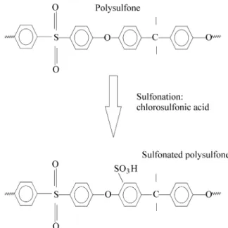
In this work, sulfonation of polysulfone was preferred as a way to introduce polar groups (sulfonic acid groups) on this polymer. The sulfonation of polysulfone was per-formed by dissolving a commercially available polysulfone (Udel®- Union Carbide, MW = 40,000 gmol) in dichlo-roethane (10% wt). Chlorosulfonic acid was added to the solution at room temperature and the resultant mixture was stirred for 30 min. The molar ratio between chlorosulfonic acid/polysulfone repeat unit was used to alter the degree of substitution.
Sulfonation of polysulfone can lead to the introduction of sulfonic acid groups (-SO3H) on the polymer. During the
synthesis of the modified polysulfone, a change in the solubility behavior of the polymer could be noted and therefore evidences of the chemical modification were observed. The sulfonated polysulfone (SPSF) could readily be dissolved in solvents that are not solvents for the original polysulfone, such as ethanol and THF with 1% water, while chlorinated solvents, such as chloroform, could not dis-solve the sulfonated polysulfone. A range of polymers with different degrees of substitution was produced by varying the initial concentration of chlorosulfonic acid. The effect of degree of sulfonation was readily observed regarding solubility. When high levels of sulfonation were used, water soluble sulfonated polymers could be synthesized.
Modified polysulfones were cast on KRS-5 crystals (IR transparent) and dried overnight at 60 °C. Transmission FTIR (Nicolet spectrometer 20SX - 128 scans with 4 cm-1 resolution) was used to characterized the sulfonation proc-ess. The FTIR spectra of sulfonated polysulfone with dif-ferent degrees of sulfonation are reproduced from 900 to 1200 cm-1 in Fig. 2. The peak at 1028 cm-1 is related to the sulfonate unit and can be correlated to the levels of substi-tution by ratioing it against a peak assigned to a group not involved in the modification reaction. Unfortunately, this peak overlaps the peak attributed to the ring vibration of a para-substituted aryl ether (1010 cm-1). In order to get the desired information from the sulfonate peak, a mathemati-cal deconvolution procedure was performed on the peak (Fig. 3, insert). The height of the 1028 cm-1 peaks was then ratioed against the 1010 cm-1 peaks. The degree of substi-tution of a specific sulfonated polysulfone was calculated using NMR data published in another manuscript24. This value was used to calibrate the FTIR data of polymers with different degrees of substitution, by associating it to the FTIR peak ratio of the same polymer. A plot of the effect of the initial ratio between reactants (polysulfone repeat unit and chlorosulfonic acid) on the degree of substitution, calculated as indicated above is shown in Fig. 3. Polymers with different levels of sulfonation could therefore be syn-thesized by changing the initial stoichiometry of the
tion. A scheme of the reaction procedure is shown in Fig. 1. The NMR study24 also demonstrated that the substitution occurs preferentially on the ortho position, in relation to the ether group, of benzene rings at the Bisphenol A unit (Fig. 1).
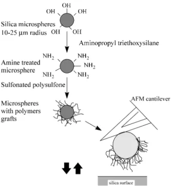
2.1.2. Grafting modified polysulfones on the surface of glass spheres
In order to simulate the interface proposed (polymer against silica), a conventional AFM tip was modified by attaching a glass sphere (Polyscience, 15 µm radius) to the cantilever. Before attaching it to the cantilever, sulfonated polysulfone, with a low degree of substitution (0.5), was grafted onto the surface of the spheres. In order to graft the sulfonated polymer on the spheres, their surfaces were firstly treated with aminopropyl triethoxysilane (APS) by dispersing the spheres in an ethanol-water-APS for one hour and curing them at 110 °C for 12 h. The APS treated spheres were then reacted with SPSF (sulfonated polysul-fone) via formation of sulfonamide bonds, by dispersing them in a SPSF-dimethylformamide solution for 2 h. The spheres were collected by filtration, dried overnight at 120 °C under vacuum and washed extensively with di-methylformamide, to remove any non-bonded polymer.
To verify the success of the grafting technique and to determine the capabilities of the new polymers to bond via primary bond formation to the silane treated glass spheres, X-ray photoelectron spectroscopy (XPS) was performed on a soda-lime silicate glass disk containing grafts of the modified polysulfone. X-ray photoelectron spectroscopy was performed in a XPS, Kratos DS800 Mg radiation, system. A 90° incident beam was used and the results were corrected using the carbon photoelectron peak at 285 eV. Prior to the analysis, the samples were dried at 90 °C and reduced pressure for 12 h.
For the XPS measurements, glass disks (1 cm in diame-ter), with composition close to the composition of the glass spheres (glass type E), were cut and polished with a SiC sand paper # 600. Disks were treated with a silane coupling agent (aminopropyl triethoxysilane) by introducing them into a solution of amino silane (1 vol.%) in ethanol-water (95 vol.% ethanol) for one hour. The treated disks were then cured at 110°C for 1 day. After this, the samples were introduced into a solution of sulfonated polysulfone in ethanol (5 wt%) for 2 h. Subsequently, disks were washed several times with ethanol and any non-bonded specie was removed by using a Sohlex extractor for 12 h.
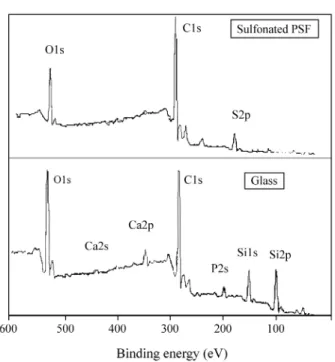
XPS data was collected in Perkin-Elmer spectrometer. The XPS spectra (from 0 to 600 eV) of a glass disk and a glass disk with grafted sulfonated polysulfone are repro-duced in Fig. 4. The peaks identified in the spectrum of the glass disk with sulfonated PSF grafts were mainly related to C (1s), S (2p) and O (1s). A semi-quantitative software routine, that computes the area under the peaks, was used to verify the amount of each element on the surface of the sample. The theoretical composition of the sulfonated PSF was calculated using the values of degree of substitution given by the NMR/FTIR studies. These calculations (Table 1) demonstrated that a large part of the surface was covered Figure 3.
of polysulfone. Top of the figure: deconvolution of FTIR spectra used to determine the degree of sulfonation.
with the sulfonated polysulfone. Very little signal coming from elements typically in the glass could be captured, such as silicon and calcium. Another important observation from these results is that the theoretical composition of sulfon-ated polysulfone is very close to the obtained results. There-fore, the success of the grafting procedure was proven. Moreover, the new polymer was grafted onto the surface of glass possibly through covalent bonds between sulfonic acid groups and amine groups on the silane layer (formation of sulfonamide bonds -SO2-NH-).
2.2. Attachment of modified glass spheres onto AFM cantilevers
The modified glass spheres (with sulfonated polysul-fone grafts) were attached onto AFM silicon nitride canti-levers by using a procedure similar to the one reported by Duker17. An epoxy (DGEBA - dyglicidyl ester biphenol A) with a diamine hardener was used to glue the sphere to the cantilever. The epoxy and sphere were transferred to the cantilever using a micromanipulator coupled with an opti-cal microscope. The epoxy was cured overnight at room temperature. Figure 5 shows a SEM micrograph of a silicon nitride AFM tip that has four cantilevers, two in each side. In Fig. 5, one modified cantilever (SPSF treated glass sphere) and a non-modified one are shown. Figure 6 shows the procedure involved on the preparation of modified AFM tips.
2.3. Preparation of Silica Substrates
The substrate chosen to accommodate the silica sur-faces in the AFM studies was a silicon wafer. Silicon wafers were firstly cleaned with acetone and dichloroethane and then submitted to a heat treatment at 420 °C for one hour in air to develop a thin silica layer at the top of the silicon wafer. Hydroxylation of the newly formed silica layer was performed by dipping the sample in boiling water for 6 h. In order to control the nanostructure of the oxide layer, a thin silica-gel layer was deposited on the surface of the grown oxide by using the sol-gel process. Silicon wafers were dipped into a tetraethoxy silane
(TEOS)/etha-nol/water solution (1% vol. TEOS, pH = 2) for one hour. The nanostructure of the silica layer was changed by heat treating the substrates at different temperatures: 110, 400,
Figure 4. XPS spectra of glass samples with grafted polysulfone.
Table 1. Comparison between theoretical composition and results obtained from XPS of glass surfaces grafted with modified polysulfone.
Elements PSF* (wt. %) Sulfonated PSF* (wt. %) Grafting SPSF** onto glass (wt. %)
C (1s) 77.1 62 61.67
O (1s) 15.3 24 23.13
S (2p) 7.6 14 13.34
N (1s) - -
-Ca (2p) - - 1.82
Si (2p) - -
-* Theoretical values.
** SPSF = sulfonated polysulfone.
Figure 5. SEM micrograph of a glass sphere attached to an AFM
600 and 800 °C. It is known that heat treatments on silica gels can change structural features of the material, such as: (1) reduce the concentration of silanol groups; (2) reduce surface area; (3) increase number of siloxane bonds, i.e., the cross-link density; (4) increase both bulk and structural density; (5) shift the distribution of silica rings to larger number of tetrahedra.
2.4. Measurements
A Nanoscope III (Digital Inst., Santa Clara, CA) was used in contact mode. In order to avoid the formation of a thin liquid film on the substrate that usually adds a capillary force component to the measured forces by AFM, a fluid cell accessory, provided also by the manufacturer, was used. Deionized water (pH = 5.0) was used during the measurements. Water is not a good solvent for the sulfon-ated polysulfone used (low degree of substitution), but swell the polymer.
Force-distance measurements were performed by ap-plying to the AFM piezoelectric a saw-tooth type of curve. In this case, instead of keeping the height or force constant, as usual when images are being processed in contact mode, the piezoelectric will move the sample against the tip in the vertical direction (z direction) in a cyclic way. In each cycle, the deflection of the cantilever is recorded as a function of the travel of the piezoelectric. When the tip is approaching the surface of the sample, an attractive force is usually present, represented by any one of the type of chemical forces (electrostatic, van der Waals, etc.). A ‘‘jump’’ of the tip towards the sample surface occurs when the elastic constant of the cantilever cannot keep up with the gradient of forces acting on the tip. When an initially in-contact tip is released from the surface, another charac-teristic jump is produced in which the tip sticks to the surface and an extra force is required to unload the tip. The magnitude of this force can be related to the adhesion between the materials on the tip and sample. The raw data provided by the equipment is usually given in a curve where the deflection of the cantilever is plotted against the dis-tance traveled by the piezoelectric. In order to convert this raw data to the force-distance curve, positions of zero force and zero displacement need to be assigned. Zero forces are defined where the cantilever is very far from the sample, and therefore no forces are present. The zero displacement is assigned to the position where the repulsive forces acting on the cantilever start to display a linear behavior, meaning that the tip is elastically compressing the sample. The parameters of the technique usually used to manipulate the way the tip and sample will come together are: z-scan start, that is basically the initial height from where it starts the approach; z-scan size, that is the total distance traveled by the piezoelectric during one extension-retraction cycle; z-scan rate, that is the rate of oscillation. By making use of those parameters, it is possible to perform measurements
far from the surface or close to the surface with different amplitudes of oscillation. Unless otherwise specified, scan rate of 1 Hz and scan size of 500 nm were used throughout this work. Many sites in each sample were used to obtain the force-displacement curves, thus ensuring reproducibil-ity of results.
In order to ensure that the procedures of attaching the glass sphere to the cantilever and measuring the AFM forces were being employed successfully, a special tip was prepared by attaching a plain glass sphere on the cantilever. The interactions between this tip and the silicon wafer sample treated at 110 °C were measured in water (pH = 5). The raw data as well as the converted data are shown in Fig. 7. The basic result is that there is no attractive forces during tip extension and no adhesive forces during tip retraction. These results are similar to the ones reported by others1,17 for a silica-silica system. At pH 5, silica surfaces are negatively charged and electrostatic repulsion will oc-cur. Moreover, hydration forces (where the water mole-cules are oriented nearby the surface) are usually employed to explain the short range repulsive forces that appear in a silica system also in pH’s closer to the isoelectric point of this material. The presence of silicate chains at the surface has also been used to explain the phenomenon1.
3. Discussion
In Fig. 6, a drawing representing the whole technique developed to measure interactions between surfaces and polymeric entities grafted on modified AFM tips (glass sphere with polymer grafts) is shown. In this scenario, the polymer chains are being brought continuously in and out of contact with the surface. This cyclic mechanism mimics
natural processes where polymeric chains hit the surface of substrates looking for positions with lower energy to ad-sorb. As show in this figure (Fig. 6), the use of a sulfonated polysulfone with a low degree of substitution favors the formation of few linkages between the polymer and the surface of the glass sphere during the tip preparation, thus allowing the presence of fairly large non-bonded chain lengths.
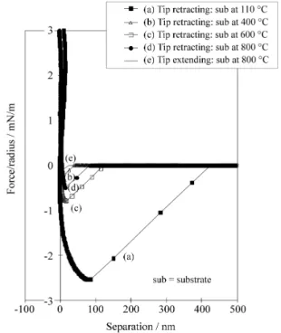
The results of force-distance curves obtained by AFM in aqueous solution (pH = 5.0) are shown in Fig. 8 for a glass sphere treated with sulfonated polysulfone (SPSF) in contact with silica substrates. For this experiment, the same tip was used in all the measurements, and therefore it was possible to use the average value of the cantilever elastic constant (provided by the manufacturer, k = 0.12 N/m) on the calculations (conversion of deflection in force). At the end of the measurements, the first sample investigated was again tested to ensure that no major degradation of the polymer grafts had occurred during previous measure-ments.
A typical force-distance curve for the interaction be-tween a sphere treated with SPSF and the silica substrate heat treated at 800 °C can be seen in Fig. 8. In this figure, the tip extending toward the surface and coming out of the surface are observed respectively in Figs. 8-e and 8-d. It is important to realize the differences between this curve and the curve in Fig. 7, where a plain sphere was used instead of the SPSF treated sphere. In the case of the polymer grafted sphere, an attractive jump can be noted as well as a large adhesive force during tip retraction, while only repul-sion was measured for the pure glass sphere. This result proves that the systems are very different, suggesting the presence of polymer on the surface of the sphere and confirming the XPS results.
Although the ionization equilibrium of sulfonated poly-sulfone is not known, it is very likely, based on the behavior of other sulfonated materials, that sulfonated polysulfone should have some of its sulfonic acid groups protonated at pH’s close to 5.0. Therefore sulfonated PSF can exhibit positive charges in some locations. These positive charges located far from the surface can then interact electrostati-cally with the approaching negatively charged silica sur-face, leading to the attractive jump seen in Fig. 8-e. After being compressed onto the silica surface during tip exten-sion, many more groups on the polymer will interact strongly with the silica surface not only by electrostatic forces, but mainly by hydrogen forces between silanol groups and sulfonic acid groups.
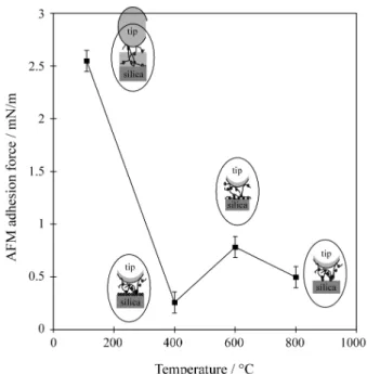
The retracting path of the tip, shown for all the sub-strates in Fig. 8, reveals that the adhesion of the polymer to the silica sample is a function of the silica structure. A maximum AFM adhesion was obtained for the silica sam-ple treated at 110 °C. At this temperature (110 °C), the silica top layer is fully hydrated and has an open and less packed
structure with lower cross-link density. This situation fa-cilitates penetration of free chain ends through the open network, maximizing chemical interactions (hydrogen bonds between silanol groups and sulfonic acid groups) and entanglements. Figure 9 plots the effect of the temperature
Figure 7. Force-distance curve for a glass sphere attached to an AFM
cantilever against a silica surface in water. Raw data shown on the top.
Figure 8. AFM force-distance curves for a sulfonated polysulfone tip and
of heat treatment of the substrates on the adhesion meas-ured by AFM.
As it can be observed in Figs. 8 and 9, a minimum value of adhesion was measured for the substrate treated at 400 °C. At 400 °C, the surface of silica retains a large concentration of silanol groups15, but the network is much more dense due to the progressing of condensation reac-tions during the thermal treatment.
On the other hand, higher levels of adhesion were measured for interactions between the tip and substrates treated at 600 and 800 °C. From 400 °C to 600 °C, silanol groups are removed from the surface and, thus, isolation of silanol groups occurs. From 600 °C to 800 °C, the isolated silanol groups are progressively removed. Therefore, it is proposed that the presence of isolated silanol groups allows higher levels of interactions between a polymer (intrinsi-cally capable of interacting with silanol groups via hydro-gen bonds) and silica than a highly hydrated surface. In a highly hydrated silica surface, the overlapping of chemical potential fields of nearby silanol groups restricts the ad-sorption of the polymer by reducing the number of confor-mational modes that the polymer can have on the surface. Only conformational modes that can limit the interaction between hydrophobic groups on the polymer and silanol groups should be able to contribute to adhesion.
The fundamental result obtained from the AFM study was that the nanostructure of the silica surface can defi-nitely regulate interactions between species present on the interface of polymer-inorganic material systems and
there-fore can define interfacial properties, such as adhesion and others.
Based upon the AFM results, it can be proposed that two different group of mechanisms of interactions between polymer chains and silica surface were detected. The first one occurs when polymer chain interact with a less packed, low density and soft silica-gel layer. In this case, penetra-tion of the chains through this gel layer is allowed. Large degrees of adhesion are obtained due basically to both mechanical interlock of chains inside the gel network as well as to chemical interactions between silanol groups and sulfonic acid groups on the polymer. This situation is illustrated in the drawings shown in Fig. 9. This type of mechanism is used to explain adhesion between polymer chains and silane coated reinforcing agents in polymer composites24,25,26.
When the gel layer is heat treated at higher temperatures (above 400 °C), densification of the network leads to an increase in the bulk density, reduction in the surface area and an increase in the elastic modulus of the layer. As a consequence, penetration of the polymeric chains through the network is restricted and interactions will basically resume to chemical bonding between silanol groups on the surface and sulfonic acid groups on the polymer (second group of mechanisms detected), via formation of hydrogen bonds. In this case, adhesion between polymeric chains and the surface is improved when the number of silanol-sul-fonic acid linkages per unit area is maximized. Although the opportunities for a silanol-sulfonic acid bond to occur should be maximum for a surface filled with surface silanol groups (surface at 400 °C), this situation can also maximize the probability of a less energetically possible interaction between hydrophobic units of polysulfone (benzene rings, for example) and silanol groups. Conformation modes of the polymeric chains that promote this type of interaction are forbidden, leading to a reduction in the total number of conformation modes and consequently a reduction in en-tropy and an increase in free energy for bonding. The overall result of this phenomenon is a reduction in adhesion between the surface and polymer.
When the silica surface is submitted to temperatures above 400 °C, silanol groups on the surface are progres-sively eliminated and become isolated. This process will allow the formation of a chemically patterned surface, where chemical functionalities, such as silanol groups, are equally spaced on average from each other. The isolation of silanol groups will allow conformation modes of the polymer chains, forbidden on substrates treated at 400 °C, to exist by restricting interactions between hydrophobic units on the polymer and hydrophilic silanol groups. This phenomenon will eventually lead to a situation where the spatial arrangement of silanol groups on the surface of the silica substrate can match the distribution pattern of the sulfonic acid groups on the polymer structure. In this case, Figure 9. Effect of the temperature of heat treatment of silica surfaces on
the inorganic surface acts as a template for the interaction of polymeric chains, leading to a maximized adhesion. Figure 9 also illustrates this phenomenon.
From the discussion above, it is possible to propose that a molecular recognition phenomenon can regulate interac-tions between polymers and patterned surfaces. Based upon the obtained results, it can be proposed that a series of processes can be improved by either adapting the chemical architecture of the polymers to a specific surface (such as in the case of coagulation of ceramic powder) or designing structures of surfaces to interact in an optimized way with polymeric chains. In the biomaterial field of science, for example, the structure of the surface of implants has to be designed to either favor protein and cell adhesion or reduce it. In the case of intraocular lens (for the treatment of cataracts), it was observed that lens with highly hydropho-bic or hydrophilic surfaces can reduce cell adhesion (that is aimed), while surfaces with an intermediary character favors protein and cell adhesion27. Based upon the results of this work, it is clear that the combination of hydrophobic and hydrophilic groups on the surface of the implant can possibly match the sequence of distribution of hydrophobic and hydrophilic groups on polymeric biomolecules, lead-ing to high levels of adhesion.
4. Conclusion
In this work, interactions between polymeric entities and the surface of inorganic materials were monitored by using an Atomic Force Microscope (AFM).
The interface between polymers and inorganic surfaces was mimic by modifying an AFM tip and using this tip to investigate the surface of a series of silica substrates with different nanostructures. The modification of AFM tips was accomplished by grafting sulfonated polysulfone onto it. Sulfonated polysulfone was prepared by sulfonating a commercial polysulfone. The newly introduced sulfonic acid groups on polysulfone can examine the surface of the silica substrate by interacting with silanol groups.
The results of this work revealed that AFM can be an important tool to investigate phenomena occurring at the interfaces of materials and systems. In this work, mecha-nisms involved in the interfacial phenomena, such as chain penetration, adsorption, chemical bond formation, entan-glements could be studied using the developed AFM pro-cedure.
The results of the AFM force-displacement curves showed that adhesion of the polymeric chains to the surface is a function of the structure of the surface. It was proposed that penetration of polymer chains through surfaces with low cross-link density and high porosity was responsible for the high levels of interactions measured. For dense surfaces, the interactions between polymer and surfaces were shown to be dependent on both the concentration and spatial distribution of silanol groups on the surface. A
molecular recognition type of process, in which interac-tions between the phases are enhanced for structures of surfaces that matches the chemical architecture of the poly-mer, was proposed to be responsible for the obtained re-sults.
Acknowledgment
The authors gratefully acknowledge the financial assis-tance of CNPq (Conselho Nacional de Desenvolvimento Científico e Tecnológico) of the Brazilian Government and the NIH (National Institutes of Health) of the Government of the United States, grant # DEO 9307-07.
References
1. Israelachvili, J.N. Intermolecular and Surface Forces, Academic Press, San Diego, 1991.
2. Mantel, M.; Rabinovich, J.P.; Wightman, J.P.; Yonn, R.-H. A study of hydrophobic interactions between stainless steel and silanated glass surface using atomic force microscopy. J. Colloid Interface Sci., v. 170, p. 203-14, 1995.
3. Rabinovich, Y.I.; Yoon, R.-Y. Use of atomic force microscopy for the measurements of hydrophobic forces between silanated silica plate and glass sphere. Langmuir, v. 10, p. 1903-9, 1994.
4. Ackler, H.D.; French, R.H.; Chiang, Y.M. Compari-sons of Hamaker constants for ceramic systems with intervening vacuum or water from force laws and physical-properties. J. Colloid Interface Sci., v. 179, p. 460-469, 1996.
5. Toikka, G.; Hayes, R.A.; Ralston, J. Surface forces between spherical ZnS particles in aqueous-electro-lyte. Langmuir, v. 12, p. 3783-3788, 1996.
6. Biggs, S. Steric and bridging forces between Surfaces bearing adsorbed polymer. An atomic force micros-copy study. Langmuir, v. 11, p.156-162, 1995. 7. Biggs, S. Nonequilibrium interaction forces between
adsorbed polymer layers. J. Chem. Soc.-Faraday Trans., v. 92, p. 2783-2789, 1996.
8. O’Shea, S.J.; Welland, M.E.; Rayment, T. An atomic force microscope study of grafted polymers on mica. Langmuir, v. 9, p. 1826-35, 1993.
9. Berger, C.E.H.; Vanderwerf, K.O.; Kooyman,R.P.H.; Degrooth, B.G.; Greve, J. Functional-group imaging by adhesion afm applied to lipid monolayers. Lang-muir, v. 11, p. 4188-4192, 1995.
10. Florin, E-L.; Moy, V.T.; Gaub, H.E. Adhesion forces between individual ligand-receptors pairs. Science, v. 264, p. 415-7, 1994.
12. Grubmüller, H.; Heymann, B.; Tavan, P. Ligand bind-ing: molecular mechanics calculation of the strepta-vidin-biotin rupture force. Science, v. 271, p. 997-9, 1996.
13. Lee, G.U.; Chrisey, L.A.; Colton, R.J. Direct measure-ment of the forces between complemeasure-mentary strands of DNA. Science, v. 266, p. 771-3, 1994.
14. Rief, M.; Oesterhelt, F.; Heymann, B.; Gaub, H.E. Single molecule force spectroscopy on polysaccha-rides by atomic force microscopy. Science, v. 275, p. 1295-7, 1997.
15. Zhuravlev, L.T. Concentration of hydroxyl groups on the surface of amorphous silicas. Langmuir, v. 3, p. 316-8, 1987.
16. Vigil, G.; Xu, Z.; Steinberg, S.; Israelachvile, J. Inter-actions of silica surfaces. J. Colloid Interface Sci., v. 165, p. 367-385, 1994.
17. Ducker, W.A.; Senden, T.J.; Pashley, R.M. Measure-ment of forces in liquids using a force microscope. Langmuir, v. 8 (1992), 1831-6.
18. Brousse, C.L.; Chapurlat, R.; Quentin, J.P. New mem-branes for Reverse Osmosis 1. Characteristics of the base polymer: sulphonated polysulphones. Desalina-tion, v. 18, p. 137, 1976.
19. Pozniak, G.; Krajewska, B.; Trochimczuk, W. Urease immobilized on modified polysulphone membrane: Preparation and properties. Biomaterials, v. 16, p. 129, 1995.
20. Chen, M.; Chiao, T.; Tseng, T. Preparation of sulfon-ated polysulfone/polysulfone and aminsulfon-ated
polysul-fone/polysulfone blend membranes. J. Appl. Polym. Sci., v. 6, p. 1205-1209, 1996.
21. Higuchi, A.; Koga, H.; Nakagawa, T. Surface-modi-fied polysulfone hollow fibers. IV. Chloromethylated fibers and their derivatives. J. Appl. Pol. Sci., v. 46, p. 449, 1992.
22. Guiver, M.D.; Robertson, G.P. Chemical Modifica-tion of Polysulfones: An facile Method of Preparing Azide Derivatives from Lithiated Polysulfone Inter-mediates. Macromolecules, v. 28, p. 294, 1995. 23. Nyström, M.; Jaervinen, P. Modification of
polysul-fone ultrafiltration membranes with UV irradiation and hydrophilicity increasing agents. J. Membr. Sci. v 60, p. 275-296, 1991.
24. Oréfice, R.L.; Brennan, A.B. An Atomic Force Micro-copy study of the interactions involving polymers and silane networks. Submitted for publication in Revista Polímeros: Ciência e Tecnologia, 1998.
25. Suzuki, N.; Ishida, H. A review on the structure and characterization techniques of silane/matrix inter-phases. Macromol. Sym., v. 108, p. 19-53, 1996. 26. Schutte, C.L. Environmental durability of glass-fiber
composites. Mat. Sci. Eng. R-reports, v. 13, p. 265-323, 1994.