Cardiology Unit, Santa Casa de Misericórdia de Araraquara Email: [email protected]
Received for publication on July 29, 2005. Accepted for publication on September 19, 2005.
ORIGINAL RESEARCH
THE INFLUENCES OF POSITIVE END EXPIRATORY
PRESSURE (PEEP) ASSOCIATED WITH
PHYSIOTHERAPY INTERVENTION IN PHASE I
CARDIAC REHABILITATION
Audrey Borghi-Silva, Renata Gonçalves Mendes, Fernando de Souza Melo Costa, Valéria Amorim Pires Di Lorenzo, Claudio Ricardo de Oliveira, and Sérgio Luzzi
Borghi-Silva A, Mendes RG, Costa F de SM, Di Lorenzo VAP, Oliveira CR de, Luzzi S. The influences of positive end expiratory pressure (PEEP) associated with physiotherapy intervention in phase I cardiac rehabilitation. Clinics. 2005;60(6):465-72.
PURPOSE: To evaluate the effects of positive end expiratory pressure and physiotherapy intervention during Phase I of cardiac rehabilitation on the behavior of pulmonary function and inspiratory muscle strength in postoperative cardiac surgery.
METHODS: A prospective randomized study, in which 24 patients were divided in 2 groups: a group that performed respiratory exercises with positive airway expiratory pressure associated with physiotherapy intervention (GEP, n = 8) and a group that received only the physiotherapy intervention (GPI, n = 16). Pulmonary function was evaluated by spirometry on the preoperative and on the fifth postoperative days; inspiratory muscle strength was measured by maximal inspiratory pressure on the same days. RESULTS: Spirometric variables were significantly reduced from the preoperative to the fifth postoperative day for the GPI, while the GEP had a significant reduction only for vital capacity (P < .05). When the treatments were compared, smaller values were observed in the GPI for peak flow on the fifth postoperative day. Significant reductions of maximal inspiratory pressure from preoperative to the first postoperative day were found in both groups. However, the reduction in maximal inspiratory pressure from the preoperative to the fifth postoperative day was significant only in the GPI (P < .05).
CONCLUSIONS: These data suggest that cardiac surgery produces a reduction in inspiratory muscle strength, pulmonary volume, and flow. The association of positive expiratory pressure with physiotherapy intervention was more efficient in minimizing these changes, in comparison to the physiotherapy intervention alone. However, in both groups, the pulmonary volumes were not completely reestablished by the fifth postoperative day, and it was necessary to continue the treatment after hospital convalescence.
KEYWORDS: Cardiac surgery. Pulmonary function. Respiratory muscle strength. Physiotherapy. PEEP.
Cardiac surgery reverts symptoms for individuals with specific cardiopathologies and measurably increases their chances of survival and quality of life.1–3 However,
pulmo-nary complications are quite frequent and represent an im-portant cause of morbidity and mortality for patients under-going cardiac surgery with cardiopulmonary bypass.2,4-6
These patients can develop various degrees of a sys-temic inflammatory response syndrome due to factors such as surgical trauma, contact of blood with nonendothelial surfaces of the bypass circuit, and alterations known as reperfusion post-cardiopulmonary bypass lesions, mainly affecting the cardiac and pulmonary regions.2,4–6
increases in the respiratory effort during the postoperative (PO) period.7,8
In spite of modernization of procedures, cardiac surgery can damage pulmonary function, with decreases of respi-ratory muscle strength and spirometric measurements oc-curring postoperatively, in addition to the occurrence of at-electasis in more than 90% of the patients.9
Reduction in oxygenation,10 pulmonary function,11,12,13,14
and respiratory muscle strength,4,5,9,12 as well as
radiologi-cal changes such as atelectasis8,12,15 have been cited as
com-mon alterations in postoperative cardiac surgery. The re-duction of respiratory muscle strength, resulting from di-rect or indidi-rect lesion of respiratory muscles during sur-gery and the secondary diaphragmatic dysfunction due to phrenic nerve lesion, has also been related to reduced pul-monary function tests, worsened gas exchange, and increase in the rate of pulmonary complications.4,5,9,12 Considering
this, some authors8,15-19 have investigated the application of
different physiotherapeutic treatment techniques in an at-tempt to minimize the alterations in the respiratory and car-diovascular system and thereby reduce the incidence of complications.
Physiotherapy intervention in phase I of cardiac reha-bilitation (PPI) is routinely performed with patients who have undergone cardiac surgery.11,13,18,19 The application of
deep breathing exercises, cough stimulation, thumping and vibration of the rib cage, and continuous positive airway pressure may prevent further deterioration in pulmonary function and reduce the incidence of pulmonary compli-cations.18 However, Jenkins et al.19 observed that deep
breathing exercises, thumping and vibration of the rib cages, and cough stimulation did not result in significant increases in spirometric measurements when compared to the control group.
With the identification of communication between the respiratory bronchioles in human lungs, some authors have concluded that collateral ventilation is important in normal pulmonary function17 and thereby confirm that the
appli-cation of positive end-expiratory airway pressure (PEEP) can promote a more homogenous distribution of pulmonary ventilation through interbronchial collateral channels and prevent expiratory collapse.17 Thus, PPI associated with the
application of PEEP through a circuit of expiratory posi-tive airway pressure (EPAP) using a face mask coupled to a PEEP valve could be effective in minimizing complica-tions that occur postoperatively after cardiac surgery.
Campbell et al.20 found that PEEP assists with the
re-moval of secretions from the main bronchi, which can be expectorated, in those hypersecretive patients who undergo upper abdominal surgery. In a study by Larsen et al.,15 the
tendency for reduced complications was observed in a
group that was administered PPI associated with PEEP, when compared to a group treated only with PPI. However, in another study, the prophylactic application of PEEP did not present benefits when compared to PPI in patients who had undergone thoracic surgery.16
In view of the conflicting results of these studies, the objective of this study was to investigate the efficacy of the association of PEEP with a protocol of physiotherapy intervention in Phase I of cardiac rehabilitation, through the evaluation of pulmonary function and inspiratory mus-cle strength in patients who had undergone elective cardiac surgery.
MATERIALS AND METHODS
This study was approved by the Ethics Committee for Human Research of the institution. The patients were in-formed about the procedures to be carried out, and all signed an institutionally reviewed informed consent form agreeing to participate in the study in accordance with the Brazilian National Health Council Resolution 196/96.
Thirty patients were recruited for participation, but only 24 patients concluded the study. The patients included in this study presented coronary insufficiency diagnosed by coro-nary angiography. These patients underwent elective cardiac surgery with cardiopulmonary bypass, and the surgical in-cision utilized was sternotomy. All patients received medi-cal prescriptions for the physiotherapy procedures. Patients who presented hemodynamic instability, associated neuro-logical sequelae, or difficulty in comprehension or adherence to the procedures performed in this study were excluded.
Patients were randomly distributed into 2 groups in a 1:2 proportion, as follows: 1. a group in which EPAP as-sociated with PPI was performed after cardiac surgery (GEP, n = 8) and 2. a group receiving physiotherapy inter-vention only (GPI, n = 16) The anthropometrical, clinical, and surgical characteristics of the groups are presented in the Table 1.
Experimental Procedure
In the preoperative period, all the patients underwent a standardized evaluation that consisted of personal data, anthropometrics, medical diagnosis, vital signs, and per-sonal antecedents. The body mass index (BMI) was calcu-lated as:
BMI = body weight (kg)/[height(cm)]2
cardiop-ulmonary bypass surgery time were recorded. Heart rate (HR) and peripheral saturation of oxygen (SpO2) were monitored and recorded during the procedures with a port-able pulse oximeter (Nonim 8500A, Plymouth, Mn., USA). After an initial evaluation, all patients were informed of the proposed protocol, surgical procedure, tracheal in-tubation, course of treatment, and the importance of physi-otherapy for recovery during hospitalization. This was fol-lowed by pulmonary function and respiratory muscle strength evaluations.
Pulmonary function test: Spirometry was performed us-ing the Vitalograph® Hand-Held 2120 spirometer (Ennis,
Ireland). During the pulmonary function tests, patients re-mained in the sitting position, with the nostrils occluded by a noseclip, while the maneuvers of vital capacity (VC) and forced vital capacity (FVC) were performed. The tech-nical procedures, acceptable criteria, and reproducibility followed American Thoracic Society guidelines.21
Measure-ments for VC, FVC, PF, and FEF25-75% were obtained, and these values were analyzed as percentages of predicted values. Reference values from Knudson et al.22 were used.
The results obtained were expressed in BTPS (liters at body temperature and pressure saturated with water vapor).
Inspiratory Muscle Strength (IMS): To measure IMS, a manovacuometer Ger-Ar (SP-Brazil) was used, with a scale varying from 0 to 150 cm H2O, according to the method-ology proposed by Black & Hyatt.23 The maximal
respira-tory pressures were assessed by maximal inspirarespira-tory pres-sure (MIP) at residual volume. Using a noseclip, patients were asked to produce maximal efforts against an ob-structed mouthpiece with a small leak to prevent patients from closing their glottis during the maneuver. Patients sus-tained maximal effort for 1 second, and the best of 3 con-secutive attempts was used.
The 2 groups were reevaluated regarding pulmonary function on the fifth postoperative day (5th PO) and
regard-ing the inspiratory muscle strength at the 1st PO and 5th PO.
The evaluations described above were performed by the
same professional, with the patient in the sitting position.
Proposed Treatments
Physiotherapy intervention in phase I of cardiac rehabilitation. (PPI)
The patients underwent 2 physiotherapeutic interven-tions dailyeach lasting approximately 40 minutes, from the immediate postoperative day (IPO) until hospital discharge. The physiotherapeutic sessions carried out were elaborated according to the following protocol:
IPO: Weaning from mechanical ventilation assistance, thumping and vibrating patients’ rib cages (airway clear-ance maneuvers), endotracheal tube aspiration and extuba-tion, which occurred at a maximum of 12 hours after sur-gery.
1st PO: Airway clearance maneuvers in the prone
posi-tion; cough assist with the head of bed inclined at 45° (ap-proximately 10 min), respiratory diaphragmatic exercises (3 series of 20 repetitions), inspiration in three stages (2 series of 20 repetitions) of room air, assisted active exer-cises of the extremities (ankles and wrists, 3 series of 10 repetitions);
2nd PO: Airway clearance maneuvers in prone and
semilateral positions and cough assist in a sitting position (approximately 10 min); respiratory diaphragmatic exer-cises (3 series of 20 repetitions) and inspiration in three stages (2 series of 20 repetitions) in a sitting position. In addition, the following assisted active exercises of upper and lower limbs associated with respiration were per-formed: 1) flexion-extension of the elbow and elevation of the arms, respecting the articular amplitude range and pain tolerance (2 series of 10 repetitions for each exercise); 2) flexion-extension of the knee, respecting articular ampli-tude and pain tolerance (2 series of 10 repetitions for each exercise);
3rd PO: Airway clearance maneuvers in a semilateral
position (approximately 10 minutes), cough assist in a sit-ting position, and the respiratory exercises described for the 2nd PO. The following active free exercises of upper and
Table 1 - Anthropometrics, clinical, and surgical characteristics of the population studied (mean ± SD)
GEP (n = 8) GPI (n = 16) P value
Age (years) 59.9 ± 9.8 55.9 ± 11.9 .24
Weight (kg) 70.2 ± 12.3 64.5 ± 10.1 .14
Height (m) 1.7 ± 0.1 1.6 ± 0.1 .09
Body mass index (BMI) (kg/m2) 24.7 ± 3.0 24.1 ± 3.0 .30
Surgery time (min) 170.7 ± 32.1 188.7 ± 43.9 .19
Cardiopulmonary bypass time (min) 70.1 ± 17.7 89.2 ± 25.9 .06
Duration of ischemia (min) 64.1 ± 28.9 56.2 ± 20.7 .44
lower limbs of items 1 and 2 associated with respiration were performed; maintaining an orthostatic position and walking in place for a 5-minute period.
4th PO: Airway clearance maneuvers when necessary
and cough assist (approximately 10 min); respiratory ex-ercises, exercises of the upper and lower limbs (as in the protocol for the 2nd PO). During this phase, all the patients
were in the medical ward, and walking was performed in the corridor for 10 minutes.
5th PO: The protocol for the 4th PO. Walking in the
hos-pital corridor for 10 minutes, and walking up and down 1 flight of stairs.
Expiratory Positive Pressure in Airways (EPAP)
The application of PEEP was performed through an EPAP circuit using a facial mask coupled to a unidirectional valve containing, at its extremity, a PEEP valve of 10 cm H2O15,24 for all patients in the GEP. This group performed
60 repetitions of respiratory exercises divided into 3 series of 20 respirations in 2 daily sessions until discharge from the hospital. The patient inhaled room air through the mask, without additional oxygen, and exhaled against the referred resistance. The patients in this group also went through the PPI protocol after the EPAP exercises, in accordance with the standard hospital physiotherapy treatment routine.
Data Analysis
Based on the means and standard deviations of data for the spirometric variables, a power size calculation was per-formed with Graphpad StatMate version 1.01, 1998.This revealed that a power of 80% and a significance levelof 5% would be obtained. To verify the data distribution, data was plotted on a gaussian curve and did not distribute ac-cording to a normal distribution. Therefore, for matched-pair comparisons, the nonparametric Wilcoxon test and the Friedman test for matched variables (MIP) were used; the Dunn test was used for differentiation between conditions. For comparison between groups, the Mann-Whitney test was used. The level of significance was set at P≤ .05.
RESULTS
From a total of 30 eligible patients, only 24 patients constituted the final research study population of 15 men (62.5%) and 9 women (37.5%), aged 57 ± 11 years. Of the 6 patients excluded from the study, 2 presented hemodynamic instability and were not released by the medical team for spirometric and respiratory strength meas-urement, 1 presented neurological sequelae, 2 presented difficulties in performing the spirometric test and exhib-ited a comprehension deficit, and 1 refused to continue the treatment. Table 1 shows the age, weight, height, IMC, du-ration of surgery, hospitalization, and perfusion of the pa-tients included in this study. No significant differences were found in the anthropometric parameters, clinical, or surgi-cal aspects between the groups analyzed.
Concerning angina, in the GEP, 25% were functional class III and 75% were functional class IV; for the GPI, 31.2% were functional class III and 68.8% were class IV, according to Campeau.25 Concerning drains, 87.5% of the
GEP patients and 81.2% of the GPI patients used the subxiphoid drain, in addition to the mediastinal drain ap-plied to all patients in the postoperative recovery. Of the total grafts, 85% were performed with the left internal tho-racic artery plus saphenous vein, and 15% with the radial arteries plus saphenous vein or only the saphenous vein.
The spirometric results obtained in preoperative and 5th
PO are presented in Table 2. No differences were found between preoperative spirometric variable values for the groups studied. However, it can be observed that for all spirometric values, previous values for the GPI were not reestablished by the 5th postoperative day, while for the
GEP, only VC did not return to its preoperative values (P
< .05). Intergroup analysis revealed a significant difference only in PF, with greater values for the GEP when compared to the GPI postoperatively.
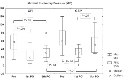
Inspiratory muscle strength, evaluated through MIP val-ues, was significantly reduced on the 1st PO for both groups
studied, with MIP increasing from the 1st PO to 5th PO only
Table 2 - Spirometric variables in the preoperative and postoperative treatment (5th PO) with statistical results for
intra-and inter-groups (mean ± SD)
GEP GPI
Preoperative 5th PO P value Preoperative 5th PO P value
VC (%) 84.7 ± 21.2 57.6 ± 16.8 .0078* 71.5 ± 21.6 53.6 ± 19.4 .0006*
FEV1(%) 73.6 ± 23.8 57.4 ± 14.1 .1094 70.5 ± 19.3 46.3 ± 28.2 .0001*
FEF25-75(%) 57.1 ± 37.1 34.0 ± 21.0 .1563 54.3 ± 17.8 38.3 ± 20.8 .0015*
FVC (%) 83.1 ± 24.4 67.4 ± 23.8 .1094 83.0 ± 38.0 49.3 ± 16.0 .0004*
PF (%) 69.1 ± 37.9 62.8 ± 13.2 .4063 64.0 ± 26.0 40.5 ± 21.9 .0020*.0189†
in the GEP. However, in the GPI, significant reductions are observed when comparing preoperative to 5th PO values.
In relation to intergroup analysis, greater values of MIP were found on the 1st PO and 5th PO for the GEP compared
to the GPI (P < .05). Figure 1 illustrates the behavior of this variable for inspiratory muscular strength.
DISCUSSION
Patients undergoing cardiac surgery with cardiopulmo-nary bypass were studied to determine the effects of a physiotherapy intervention in phase I of cardiovascular re-habilitation, associated or not with the application of PEEP on pulmonary and inspiratory muscular strength.
Alterations in pulmonary function can be associated with various factors such as the type of surgical incision,26
the anesthetic modality employed,27 diaphragmatic
dysfunc-tion,27 postoperative pain,26 and the positioning of the
pleu-ral drain.11 In the present study, all the patients were
oper-ated through a sternotomy with the thoracic drain posi-tioned in the subxiphoid (pleural and/or mediastinal) re-gion, which minimized the possible differences that might result from the procedure.
Additionally, some authors have demonstrated that a large number of patients who undergo cardiac surgery with cardiopulmonary bypass present alterations in pulmonary functions in postoperative evaluations.10,12 Therefore, the
performance of procedures to improve their recovery be-comes necessary in an effort to minimize the deleterious effects on pulmonary function and of immobility.
Alterations in pulmonary function after cardiac surgery were observed in this study, in agreement with other find-ings, which supports that a reduction in functional residual capacity (FRC)27, VC,13,14,23,28,29 and expiratory flows11, 13,14
occurs following cardiac surgery. According to published
studies, the FVC presents a general reduction for a mini-mum period of 10 to 14 days.28,30 In the present study, the
FVC was analyzed until the 5th day PO, and no difference
was found between the preoperative level and that for 5th
PO in the GEP, indicating this variable had returned to pre-vious values. However, in the GPI, in which only PPI was performed to the 5th PO, the FVC was not reestablished.
These results corroborate those reported by Guizilini et al.11
in patients who underwent cardiac surgery without cardi-opulmonary bypass.
Westerdahl et al.13 evaluated pulmonary function up to
4 months after cardiac surgery in patients who did PPI and found that VC and FEV1 were still significantly reduced when compared to preoperative values. In the present study, the only preoperative measurements reestablished by the 5th postoperative day occurred for the GEP that is, when
EPAP was associated with PPI.
The importance of a postoperative physiotherapeutic intervention protocol for cardiac surgery has been justified by some authors in that it can lower the incidence of pul-monary complications brought on by reductions in spiro-metric measurements.13,18 Additionally, the application of
PEEP has been shown to be effective in increasing the re-turn to pulmonary volumes and the resolution of atelecta-sis.17
Differing from findings in our study, Larsen et al. ,15
found no difference between the group treated with PPI plus EPAP and the group treated with PPI alone. However, a tendency for the reduction of complications was observed in the group that received PEEP. Ricksten et al.29 concluded
that the administration of EPAP or continuous positive air-way pressure was superior to PPI regarding gas exchange, the preservation of pulmonary volumes, and the prevention of atelectasis, in accordance with the findings of the present study, although those findings were from postoperative ab-dominal surgery patients.
In another study, the application of PEEP did not con-fer additional prophylactic benefits regarding atelectasis and the reduction of hypoxemia, when compared to physi-otherapy intervention.16 The FVC was not improved
postoperatively in patients receiving EPAP compared to those receiving incentive spirometry or physiotherapeutic interventions.30 In constrast, our results show superiority for
the variables analyzed after the application of EPAP asso-ciated with PPI in comparison to isolated physiotherapeu-tic intervention. As in this study, other authors20,29 have also
concluded that the application of PEEP should be used as an adjuvant in the routine physiotherapeutic intervention for surgical patients.
Reduced pulmonary function, worsening of gas ex-change, and higher rates of pulmonary complications have
been associated with the reduction of IMS.4, 5,9,12 In this
study, the values of MIP showed significant reductions from preoperative to 1st PO measurements for the groups
stud-ied, and the reestablishment of this variable was found only for the GEP. These results suggest an additional effect by EPAP regarding an earlier reversion of IMS when compared to isolated physiotherapeutic intervention, since signifi-cantly higher values were found for MIP on the 1st and 5th
PO for the GEP than for the GPI.
The improvement of MIP in the postoperative period was confirmed by Elias et al.,9 even without training
di-rected towards inspiration muscles, in agreement with our results in that no specific muscle training had been done. This increase can be related to a possible improvement in the mechanics of thoracic-abdominal movement and con-sequently an increase in the amplitude of respiratory move-ments,9 which were not measured in this study.
This study presents some limitations such as the absence of a group with only EPAP application without PPI, which would meet the objective of verifying whether patients who underwent this treatment would present higher pulmonary function values and inspiratory muscle strength, when
com-pared to patients who underwent physiotherapeutic inter-vention. However, in our study, all patients followed a rou-tine established by the hospital’s physiotherapeutic section. Another important aspect was the limited number of EPAP kits available, which kept the sample size small. While some additional benefits were observed with the use of PEEP in this study, it is necessary to consider the cost/ benefit ratio of using this equipment in addition to the pro-posed physiotherapeutic treatment.
In conclusion, patients who underwent elective cardiac surgery with cardiopulmonary bypass exhibited reductions in postoperative pulmonary function and muscle strength . Physiotherapeutic intervention associated with the applica-tion of positive end-expiratory pressure improved the re-covery of these patients in comparison to physiotherapeu-tic intervention alone. However the pulmonary volumes were not completely reestablished until the 5th PO,
suggest-ing the need to continue treatment after the period of hos-pital convalescence. Due to the small sample size in this study, the performance of new studies to better establish the results obtained in this study is suggested.
RESUMO
Borghi-Silva A, Mendes RG, Costa F de SM, Lorenzo VAP Di, Oliveira CR de, Luzzi S. A influência da pressão positiva expiratória final (PEEP) associada à intervenção fisioterapêutica na fase I da reabilitação cardiovascular. Clinics. 2005;60(6):465-72.
OBJETIVO: Avaliar os efeitos da pressão positiva expiratória final e da intervenção fisioterápica na fase I da reabilitação cardiovascular sobre o comportamento da função pulmonar e da força muscular inspiratória e sobre o pós-operatório de cirurgia cardíaca.
MÉTODO: Estudo prospectivo, randomizado, com 24 pacientes, separados em 2 grupos: GEP (n=8), que realizaram exercícios respiratórios com pressão positiva expiratória nas
vias aéreas associados à intervenção fisioterápica; e GFI (n=16), que realizaram somente a intervenção fisioterápica. A função pulmonar foi avaliada pela espirometria no pré e 5º dia pós-operatório; a força muscular inspiratória pela pressão inspiratória máxima no pré, 1º e 5º dias pós-operatório.
REFERENCES
1. Timerman A, Cesar LAM. Manual de Cardiologia. São Paulo: Editora Atheneu; 2000.
2. Brasil LA, Mariano JB, Santos FM, Silveira AL, Melo N, Oliveira NG, et al. Revascularização do miocárdio sem circulação extracorpórea: experiência e resultados iniciais. Rev Bras Cir Cardiovasc. 2000;15(1):6-15.
3. Sundt TM, Bailey MS, Moon MR, Mendeloff EN, Huddleston CB, Pasque MK et al. Quality of life after aortic valve replacement at the age of > 80 years. Circulation. 2000;102(19 Suppl 3):III 70-4. 4. Beluda FA, Bernasconi R. Relação entre força muscular respiratória e
circulação extracorpórea com complicações pulmonares no pós-operatório de cirurgia cardíaca. RSCESP. 2004;14(5):1-9.
5. Schuller D, Morrow LE. Pulmonary complications after coronary revascularization. Curr Opin Cardiol. 2000;15:309-15.
6. Butler J, Rocker GM, Westaby S. Inflammatory response to cardiopulmonary bypass. Ann Thorac Surg. 1993;55:552-9. 7. Magnusson L, Zemgulis V, Wicky S, Tyden H, Thelin S, Hedenstierna
G. Atelectasis is a major cause of hypoxemia and shunt after cardiopulmonary bypass: an experimental study. Anesthesiology. 1997;87:1153-63.
8. Oikkonen M, Karjalainen K, Kahara V, Kuosa R, Schavikin L. Comparison of incentive spirometry and intermittent positive pressure breathing after coronary artery bypass graft. Chest. 1991;99:60-5. 9. Elias DG, Costa D, Oishi J. Efeitos do treinamento muscular inspiratório
no pré e pós-operatório de cirurgia cardíaca. Rev Bras Ter Intens. 2000;12(1):9-18.
10. Barbosa RAG, Carmona MJC. Avaliação da função pulmonar em pacientes submetidos à cirurgia cardíaca com circulação extracorpórea. Rev Bras Anestesiol. 2002;52:689-99.
11. Guizilini S, Gomes WJ, Faresin SM, Carvalho ACC, Jaramillo JI, Alves FA, et al. Efeitos do local de inserção do dreno pleural na função pulmonar no pós-operatório de cirurgia de revascularização do miocárdio. Rev Bras Cir Cardiovasc. 2004;19(1): 47-54.
12. Johnson D, Hurst T, Thomson D, Mycyk T, Burbridge B, To T, et al. Respiratory function after cardiac surgery. J Cardiothorac Vasc Anesth. 1996;10:571-7.
13. Westerdahl E, Lindmark B, Bryngelsson I, Tenling A. Pulmonary function 4 months after coronary artery bypass graft surgery. Respir Med. 2003;97:317-22.
14. Kristjansdottir A, Ragnarsdottir M, Hannesson P, et al. Respiratory movements are altered three months and one year following cardiac surgery. Scand Cardiovasc J. 2004;38:98-103.
15. Richter Larsen K, Ingwersen U, Thode S, Jakobsen S. Mask physiotherapy in patients after heart surgery: a controlled study. Intensive Care Med. 1995;21:469-74.
16. Frolund L, Madsen F. Self-administered prophylactic postoperative positive expiratory pressure in thoracic surgery. Acta Anaesthesiol Scand. 1986;30:381-5.
17. Andersen JB, Jespersen W. Demonstration of intersegmental respiratory bronchioles in normal human lungs. Eur J Respir Dis. 1980;61:337-41. 18. Stock MC, Downs JB, Cooper RB, et al. Comparison of continuous positive airway pressure, incentive spirometry, and conservative therapy after cardiac operations. Crit Care Med. 1984;12:969-72.
19. Jenkins SC, Soutar SA, Loukota JM, Johnson LC, Moxham J. Physiotherapy after Coronary artery surgery: are breathing exercises necessary? Thorax. 1989;44:634-9.
significativas da situação pré para o 5º pós-operatório somente no GFI (p<0,05).
CONCLUSÕES: Estes dados sugerem que a cirurgia cardíaca produz reduções da força muscular inspiratória, dos volumes e fluxos pulmonares e que a pressão positiva associada à intervenção fisioterápica foi mais eficiente em minimizar essas alterações do que quando a fisioterapia foi realizada de forma isolada. Entretanto, os volumes
pulmo-nares não foram completamente restabelecidos até o 5° dia pós-operatório em ambos os grupos, sendo necessária a continuidade dos tratamentos após a convalescença hospitalar.
PALAVRAS-CHAVE: Cirurgia Cardíaca. Função
20. Campbell T, Fergunson N, McKinlay RGC. The use of a simple self-administered method of positive expiratory pressure (PEP) in chest physiotherapy after abdominal surgery. Physiotherapy. 1986;72:198-200.
21. American Thoracic Society. Pulmonary Rehabilitation. Am J Respir Crit Care Med. 1999;159:1666-82.
22. Knudson RJ, Lebowitz MD, Holberg CJ, Burrows B. Changes in the normal maximal expiratory flow-volume curve with growth and aging. Am Rev Respir Dis. 1983;127:725-34.
23. Black LF, Hyatt RE. Maximal respiratory pressures: normal values and relationship to age and sex. Am Rev Respir Dis. 1969;99:696-702. 24. Christensen EF, Schultz P, Jensen OV, Egebo K, Engberg M, Gron I, et
al. Postoperative pulmonary complications and lung function in high-risk patients: a comparison of three physiotherapy regimens after upper abdominal surgery in general anesthesia. Acta Anaesthesiol Scand. 1991;35:97-104.
25. Campeau L. The Canadian Cardiovascular Society grading of angina pectoris revisited 30 years later. Can J Cardiol. 2002;18:371-9. 26. Senra DF, Iasbech JA, Oliveira SA. Pós-operatório em cirurgia cardíaca
de adultos. RSCESP. 1998;8:446-54.
27. Irwin S, Tecklin J. Fisioterapia Cardiopulmonar. 1994. 2nd ed. São Paulo: Manole; 315-324.
28. Craig, DB. Postoperative recovery of pulmonary function. Anaesth Analg. 1981;60:46-51.
29. Ricksten SE, Bengtsson A, Soderberg C, Thorden M, Kvist H. Effects of periodic positive airway pressure by mask on postoperative pulmonary function. Chest. 1986;89:774-81.