Study of the cephalometric features
of Brazilian long face adolescents
Omar Gabriel da Silva Filho*, Gleisieli C. Petelinkar Baessa Cardoso**, Mauricio Cardoso***, Leopoldino Capelozza Filho****
Objective: To determine skeletal and dental cephalometric values for Brazilian long-faced adolescents. Methods: The sample comprised lateral cephalograms of 30 long-faced pa-tients, 17 females and 13 males, and 30 Pattern I adolescent papa-tients, 15 males and 15 females, with permanent dentition, during adolescence. The features that characterized the long face pattern were defined clinically by subjective facial analysis. The following cepha-lometric landmarks were assessed: 1) Sagittal behavior of the apical bases (SNA, SNB, ANB, NAPog, Co-A, Co-Gn), 2) Vertical behavior of the apical bases (SN.PP, SN.MP, go-nial angle, TAFH, LAFH, MAFH, PFH, TAFHperp, LAFHperp), 3) Dentoalveolar behavior (1-PP, 6-PP, 1-MP, 6-MP, 1.PP, IMPA), and 4) Facial height ratios (LAFHPerp/TAFHPerp, LAFH/TAFH, MAFH/LAFH). Results and Conclusions: The vertical error of the long face pattern was concentrated in the lower third. The maxilla exhibited greater dento-alveolar height and the mandible, given its more vertical morphology, displayed greater clockwise rotation. These morphological and spatial features entail sagittal and vertical skeletal changes as well as vertical dentoalveolar changes. The facial convexity angles were increased in the sagittal direction. Vertically, the total and lower anterior facial heights were increased. The dentoalveolar component was found to be longer.
Abstract
Keywords: Face. Adolescent. Cranial circumference.
* MSc, Orthodontist, Hospital for Rehabilitation of Craniofacial Anomalies, University of São Paulo.
** Dentistry Graduate - Resident, Department of Corrective Orthodontics, Hospital for Rehabilitation of Craniofacial Anomalies, University of São Paulo (HRAC/USP), Bauru/SP.
*** PhD in Dentistry, Júlio de Mesquita Filho São Paulo State University (UNESP), Araçatuba/SP. Professor, Specialization and Master Pro-gram in Orthodontics, Sacred Heart University (USC), Bauru/SP.
**** PhD in Oral Rehabilitation, area of Periodontics, School of Dentistry of Bauru, São Paulo University (FOB/USP), Bauru/SP. Coordinator, Specialization Program in Orthodontics, Society for the Social Promotion of Cleft Lip and Palate Patients (PROFIS), Bauru/SP.
INTRODUCTION
From Michelangelo’s “Belvedere Torso” to the “Venus at the mirror,” by Velazquez, the sensitivity of the human eye has captured the beauty behind the sublime conception of human anatomy. The pursuit of the beauty concealed in
A
B
C
This morphogenetic connotation of facial morphology entails two fundamental impli-cations. When the face is normal, it retains its morphology throughout its growth. When it de-viates from normality, it is not possible to per-form any orthodontic or orthopedic treatment capable of exerting any significant clinical im-pact, particularly in the frontal view. Thus, the first concern of orthodontists when performing a facial analysis in the frontal view is to determine whether there are any errors in facial morphol-ogy. Among the errors diagnosed in the frontal view of the face is the so-called long face, an error in the vertical direction.
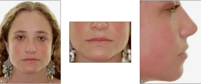
A “long face” is characterized by an exces-sively vertical face, also referred to as Long Face Pattern,4,5 “long face syndrome”,11 hypodivergent facial type13 or, erroneously, “mouth breathing syndrome”. Disparity between facial thirds (Fig 2) can be identified clinically. The lower third is increased, resulting in incompetent lip seal, over-exposed maxillary incisors at rest, gingival expo-sure on smiling and double chin in an attempt at lip seal competence.2,10,29
As is the case with other frontal view errors, long faces cannot be corrected by orthodontics
FIGURE 1 - Characteristics of the Pattern I face as defined by facial analysis. A) A pleasant frontal morphology of the face results from symmetry and propor-tionality between facial thirds. B) Lip competence results from compatibility between skeletal and soft tissue lengths. C) Lateral analysis shows a balanced sagittal behavior between the apical bases.
Nevertheless, orthodontists are concerned with the morphological—not transcendental—diag-nosis involving the shape and proportions of the face. Hence the connotation of “morpho-logical diagnosis”. In this light, there is nothing metaphysical about discovering beauty in the morphology of the face. This task consists in an analysis of the face, a morphological evaluation, both qualitative and subjective.
A B
A B
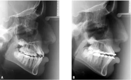
and/or orthopedics alone. Patients and clinicians share an identical perception of this issue (Fig 3). Orthodontists are therefore aware of the vital role played by orthognathic surgery in reducing the vertical excess that characterizes this facial pattern. Two morphological criteria lead to the indication of orthognathic surgery for long face reduction, i.e., compromised facial esthetics and
inability to treat the existing malocclusion. The former issue is subjective and depends mainly on what the patient expects from the treatment and from the facial change. The latter refers to the severity and direction of the interarch error found in the malocclusion. For example, an an-terior open bite in a long face pattern is a strong indication for surgery.
FIGURE 2 - Features of the Long Face Pattern. A) In lateral view, the downward and backward rotation of the mandible may favor the diagnosis of mandibular deficiency. B) In frontal view the diagnosis is unmistakable: a disproportion between the facial thirds, with a disproportionate increase of the lower third, compromises lip competence and exposes the upper incisors at rest.
In the history of orthodontics, the diagnosis of excessively vertical facial growth was initially based on cephalometric measurements. However, the criterion used for defining the long face that prevailed in the literature until the late 1970s8 was mistaken as it was based on the occlusal con-dition. From a cephalometric standpoint, a long face is characterized by increased total anterior facial height due to increased lower anterior fa-cial height.12,15,22 The mandible is considered the main culprit in the long face condition,11 exhib-iting a short ramus, obtuse gonial angle,11,24 in-creased mandibular plane angle, both relative to the cranial base2,11-14,20,29 and the palatal plane.16 The literature suggests no difference in the sag-ittal and vertical dimensions of the maxilla in patients with excessive vertical growth. The distance from the palate to the cranial base, the length of the maxilla and the palatal plane angle relative to the cranial base do not differ from nor-mal.24 Changes in the midface are concentrated in the dentoalveolar process, with an increase in distance from the molars, and from the incisors to the palatal plane.
Attuned to the esthetic demands of the facial analysis era and to the main complaint voiced by patients,11 orthodontists confirm vertical maxillary errors by reference to upper incisor exposure at rest and exposure of the gingival tissue upon smiling. The clinical reference for diagnosing the maxilla in long face cases is so important that the vertical re-duction surgery includes impaction of the maxilla, whose key advantage is high stability.21
The incidence of individuals with excessively vertical face growth patterns is controversial, even when diagnosis is based on facial analysis. This is due to difficulties in standardizing the magnitude of the excess to determine a long face. For example, the impact of increased LAFH in young Caucasian Americans is 18%5,6 while in young Brazilians it is 35,00%.8 When the diag-nosis refers to the long face itself, probably with an indication for surgical correction, incidence
drops to around 1.5% of the population.6 The cephalometric characteristics are well defined for adult patients who are no longer in the growth phase, including Brazilians.4 The literature also confirms that facial morphology is established at an early stage,17,18,22 and the long face is no excep-tion.Characteristics such as total anterior facial height, mandibular plane angle, gonial angle, an-gle formed by the palatal and mandibular planes are increased from pre-adolescence,11 while the proportion of the facial thirds remains16 or even worsens during adolescent growth.11 The num-ber of studies on the long face in adolescents is proportionate to the importance of the subject. Within this scenario, our investigation aimed to put into perspective the cephalometric charac-teristics of the long face pattern in adolescents.
MATERIAL AND METHODS Material
For this retrospective study, pretreatment lateral cephalograms of White patients of both genders, with permanent dentition and exces-sively vertical faces, enrolled in the orthodon-tics specialization program at Profis-Bauru were selected. Vertical excess was diagnosed by the presence of incompetent lip seal and exposure of upper incisors with the upper lip at rest, as seen in facial photographs. Among the selected
patients, 9 were female and 21 were male.The
selected patients had a mean age of 13 years, ranging between 10 years and 8 months and 15 years and 8 months. These patients comprised the Long Face Pattern group.
A B
CC The technical requirement for cephalogram selection was adequate bone and tooth image quality.
Methods
The radiographs were scanned and the images were analyzed using the program Radiocef 2.0, ac-cording to the manufacturer’s instructions.26 The landmarks were defined (Fig 4) on the scanned im-ages of the lateral radiographs by a single examiner
following the same method adopted by Cardoso et al.4 The results were stored and then submitted to statistical evaluation. The angular and linear mea-surements were grouped in the following order: 1) Sagittal behavior of the apical bases (Fig 5) (SNA, SNB, ANB, NAPog, Co-A, Co-Gn), 2) Vertical be-havior of the apical bases (Fig 6) (SN.PP, SN.MP, gonial angle, TAFH, LAFH, MAFH, PFH, TAFH-perp, LAFHperp), and 3) Dentoalveolar behavior (Fig 7) (1-PP, 6-PP, 1-MP, 6-MP, 1.PP, IMPA).
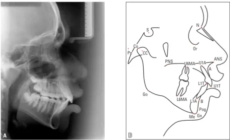
FIGURE 4 - A) Lateral cephalogram, and B) cephalometric tracing illustrating the points used as cephalometric landmarks in a long-faced patient.
FIGURE 5 - Cephalometric landmarks represen-tative of the sagittal behavior of apical bases: SNA, SNB, ANB, NAPog, Co-A, Co-Gn.
FIGURE 6 - Cephalometric landmarks repre-sentative of the vertical behavior of the apical bases: SN.PP, SN.MP, gonial angle, TAFH, LAFH, MAFH, PFH, TAFHperp, LAFHperp.
FIGURE 7 - Cephalometric landmarks represen-tative of the dentoalveolar behavior: 1-PP, 6-PP, 1-MP, 6-MP, 1.PP, IMPA.
Gonial angle PFH AnPP TAFHperp. AnMP LAFHperp. TAFH
Frankfurt horiz. plane LAFH Nperpendicular MAFH 1-MP 6-MP 1.PP 1-PP IMPA 6-PP
Mandibular plane (Go-Me) Palatal plane (ANS-PNS)
N S N
Co Po Go PNS Or ANS Gn Me S Co A B Pog Gn S Po CC Co PNS Go U6MA
Landmark 1
st Measurement 2nd Measurement
t p Error
mean SD mean SD
Co-A 90.43 3.63 90.66 3.53 1.544 0.139 0.47
Co-Gn 114.53 5.22 114.68 5.29 1.632 0.119 0.30
CoGo 114.53 5.22 114.68 5.29 1.693 0.107 0.30
Dif. Mx Md 24.10 3.98 24.02 4.01 0.641 0.529 0.37
LAFH 67.50 5.04 67.65 5.20 2.107 0.049 0.25
TAFH 118.14 6.63 117.97 6.82 1.221 0.237 0.44
MAFH 52.87 3.49 52.58 3.58 2.202 0.040 0.46
PFH 55.46 5.99 55.45 5.99 0.126 0.901 0.24
TAFH perp 117.66 6.44 117.50 6.66 1.094 0.287 0.45
LAFH perp 65.35 4.13 65.50 4.32 1.856 0.079 0.28
1-PP 4.34 2.24 4.52 2.13 1.634 0.119 0.35
6-PP 21.59 2.08 21.66 2.21 1.103 0.284 0.19
1-MP 15.81 2.47 16.05 2.73 2.721 0.014 0.33
6-MP 29.77 2.94 29.79 2.95 0.461 0.650 0.14
SNA 82.49 2.96 82.86 2.67 2.376 0.028 0.55
SNB 77.98 3.36 78.37 3.17 2.671 0.015 0.53
ANB 4.51 2.44 4.50 2.43 0.189 0.852 0.23
Gonial Angle 124.09 5.86 124.25 5.91 3.070 0.006 0.20
SN.MP 92.88 3.20 93.00 3.19 2.260 0.036 0.19
SN.PP 8.32 4.16 8.09 4.16 1.832 0.083 0.41
1.PP 64.43 5.42 64.18 5.30 0.913 0.373 0.86
IMPA 95.33 7.89 95.08 7.72 1.138 0.269 0.72
Lower third angle 101.77 3.68 101.97 3.81 2.009 0.059 0.34
NAPog 8.72 5.16 8.61 5.47 0.668 0.512 0.48
LAFHperp. / TAFHperp 0.56 0.02 0.56 0.02 0.567 0.577 0.00
LAFH/ TAFH 0.57 0.02 0.57 0.02 0.809 0.428 0.00
MAFH/LAFH 0.79 0.06 0.78 0.06 2.668 0.015 0.01
PFH/ TAFH 0.47 0.05 0.47 0.05 0.567 0.577 0.00
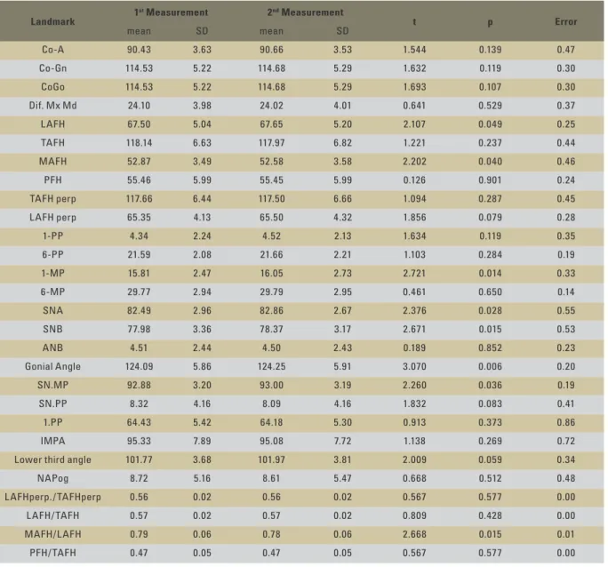
TABLE 1 - Application of the t-test in two measurements made with the intent to establish the method error.
STATISTICAL ANALYSIS
The means and standard deviations of all variables were calculated. In order to detect dif-ferences between the groups, the t-test for inde-pendent data was used. We compared the Long Face Pattern and Pattern I groups in terms of gender. Comparisons were made using a 5% (p <0.05) level of significance.
RESULTS
For didactic purposes, the statistical treat-ment of the cephalometric measuretreat-ments was organized in tables (Tables 2, 3, 4 and 5) con-taining the mean, standard deviation and the result of Student’s t-test considering facial morphology and gender.
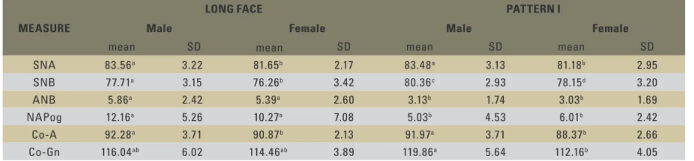
Table 2 assesses the sagittal behavior of the apical bases. The maxilla behaved similarly in both the Long Face and Pattern I groups, regard-less of gender. In boys the maxilla was larger both in linear Co-A and angular SNA measure-ments. The position of the mandible relative to the cranial base (SNB) was influenced by two variables, facial morphology and gender, ex-hibiting greater retrusion in the Long Face and in girls. Mandibular length (Co-Gn) was influ-enced only by the gender variable, and less so in girls. Facial convexity of long-faced subjects was less pronounced. Therefore, in the sagittal direction, mandibular behavior was changed in the Long Face Pattern.
Table 3 evaluates the vertical behavior of the apical bases. Angular measurements (go-nial angle, mandibular plane angle and pala-tal plane angle) were influenced only by the gender variable. The angular behavior of the mandible was affected by facial morphology. Mandibular angles (gonial angle and mandibu-lar plane angle) were increased in long-faced subjects while the palatal plane was identical in both facial patterns. Therefore, in terms of angular measurements, the mandible behaved differently in the Long Face Pattern. As for the linear measurements, the midface (MAFH) is not influenced by either gender or morpholo-gy. Total facial heights and lower facial heights tended to be higher in long-faced subjects and lower in Pattern I girls.
Table 4 evaluates dentoalveolar behavior, i.e., the heights of the upper and lower dental arches, and inclination of the incisors in their respective apical bases (1.PP and IMPA). The only
quanti-ties influenced by gender were the first molar heights (6-PP and 6-MP), which were higher in males. Dental arch heights were increased among female long-faced subjects. Maxillary incisors also behaved identically in Long Face and Pattern I subjects, whereas mandibular incisors were more proclined in long face subjects.
Table 5 evaluates facial heights and the ratios between them. The facial proportions were in-fluenced only by facial morphology, and tended to show a greater involvement of the lower face in Long Face subjects. The heights of the apical bases were lower in Pattern I girls.
DISCUSSION
The cephalometric characteristics of the Long Face pattern in adolescents is a largely unexplored subject in the orthodontic lit-erature, particularly through a cephalomet-ric study based on the clinical morphology of the face. Even in adults, very few investiga-tions have hitherto performed facial analyses to quantify the vertical error3,11,19 or assessed patients with an indication for vertical excess reduction surgery.2,10,28
This study investigated the morphological features of long-faced patients, as defined by facial analyses. It was inspired by previous re-search, which defined the cephalometric means of long-faced adult patients.4,6 These data lay the groundwork for the discussion of these find-ings in adolescents.
For didactic reasons the results were distrib-uted in Tables 2-5, following the order of cepha-lometric landmarks displayed under Material and Methods: 1) Table 2: Sagittal behavior of the cal bases, 2) Table 3: Vertical behavior of the api-cal bases, 3) Table 4: Dentoalveolar behavior, and 4) Table 5: Ratios between facial heights.
TABLE 2 - Mean values, standard deviations and application of the t-test for sagittal values.
MEAsurE
LONG FACE PATTErN I
Male Female Male Female
mean SD mean SD mean SD mean SD
SNA 83.56a 3.22 81.65b 2.17 83.48a 3.13 81.18b 2.95
SNB 77.71a 3.15 76.26b 3.42 80.36c 2.93 78.15d 3.20
ANB 5.86a 2.42 5.39a 2.60 3.13b 1.74 3.03b 1.69
NAPog 12.16a 5.26 10.27a 7.08 5.03b 4.53 6.01b 2.42
Co-A 92.28a 3.71 90.87b 2.13 91.97a 3.71 88.37b 2.66
Co-Gn 116.04ab 6.02 114.46ab 3.89 119.86a 5.64 112.16b 4.05
MEAsurE
LONG FACE PATTErN I
Male Female Male Female
mean SD mean SD mean SD mean SD
Mand. Plane Angle 93.91a 2.81 94.23a 3.67 92.32b 4.21 91.79b 3.25
Pal. Plane Angle 7.85a 3.11 9.24a 2.30 7.94a 4.88 8.80a 4.23
TAFH 122.29b 6.07 120.80b 4.62 120.57b 4.92 112.85a 3.63
LAFH 71.74b 4.97 71.11b 4.93 68.13b 4.03 62.91a 2.22
MAFH 54.24a 3.20 53.12b 3.38 54.20a 3.39 51.44b 2.97
PFH 56.57ab 3.95 55.64ab 4.84 60.64b 5.59 53.63a 4.34
TAFH perp. 121.67b 6.02 119.63b 4.32 120.35b 5.01 112.50a 3.60
LAFH perp. 68.77a 4.98 67.09b 2.90 66.78c 4.08 61.43d 2.30
TABLE 3 - Mean values, standard deviations and application of the t-test for vertical values.
TABLE 4 - Mean values, standard deviations and application of the t-test for dentoalveolar values.
MEAsurE
LONG FACE PATTErN I
Male Female Male Female
mean SD mean SD mean SD mean SD
6-PP 22.81b 2.50 23.00ab 1.38 23.07b 1.53 20.78a 1.64
1-MP 16.81bc 2.02 18.25c 3.18 15.85ab 0.99 14.74a 1.87
6-MP 31.09b 1.93 31.00b 3.12 30.23b 1.57 27.63a 1.99
1.PP 7.85a 3.11 9.24a 2.30 7.94a 4.88 8.80a 4.23
IMPA 96.87a 4.43 95.26a 7.75 89.85b 6.30 92.09b 5.66
TABLE 5 - Mean values, standard deviations and application of the t-test for facial height values.
MEAsurE
LONG FACE PATTErN I
Male Female Male Female
mean SD mean SD mean SD mean SD
LAFH/ TAFH 0.59a 0.02 0.59a 0.03 0.57b 0.02 0.56b 0.02
MAFH/LAFH 0.75a 0.06 0.75a 0.07 0.80b 0.07 0.82b 0.06
Co-A 92.28a 3.71 90.87b 2.13 91.97a 3.71 88.37b 2.66
Co-Gn 116.04ab 6.02 114.46ab 3.89 119.86a 5.64 112.16b 4.05
LAFH 71.74a 4.97 71.11b 4.93 68.13b 4.03 62.91a 2.22
Groups with the same letters presents no statistically significant difference between them.
Groups with the same letters presents no statistically significant difference between them.
Groups with the same letters presents no statistically significant difference between them.
The anteroposterior position of the maxilla in long-faced subjects can play an important role, especially when planning includes orthog-nathic surgery. A correctly positioned maxilla, for example, can eliminate the sagittal maxillary procedure during surgery. The present cephalo-metric data point to the fact that vertical facial excess does not interfere with this maxillary re-lationship. The maxilla remains well positioned in relation to the cranial base (SNA) and its length (Co-A) is similar to that of Pattern I. The sagittal behavior of the maxilla in adolescents is consistent with the literature,12,13 but disagrees with the findings of Cardoso et al,4 who report-ed maxillary retrusion.
The quantification of the maxilla using cephalometric point “A” as a reference (SNA and Co-A) does not disclose the zygomatic de-ficiency that tends to be present in these pa-tients when their faces are evaluated clinically. It is likely that the position of the maxilla in the Long Face Pattern tends towards a sagittal deficiency, with considerable individual varia-tion, which may not be amenable to identifi-cation by cephalometry.
If it is true that the long face pattern does not exhibit any cephalometric sagittal changes in the maxilla, such is not the case in the man-dible. Vertical excess interferes with the sagit-tal relationship of the mandible by keeping it retruded (SNB), although without interfering with its length (Co-Gn). The literature associ-ates the appearance of retrognathia in the man-dible with a retrusion, or posterior displace-ment, of the chin12-15,24,29 caused by a clockwise rotation of the mandibular plane, as shown in Table 2.
This mandibular retrusion affects the facial convexity angles (NAPog and ANB), making the face more convex. Facial convexity angular values are increased in the Long Face Pattern due to the positioning of point “B”.
The pattern of facial growth in the
verti-cal direction was examined through the spatial behavior of the maxilla (SN.Palatal Plane) and mandible (SN.Mandibular Plane), mandibular morphology (gonial angle) and facial heights (TAFH, LAFH, MAFH, PFH, TAFHperp., LAFHperp.).
The palatal plane angle relative to the cranial base showed no difference, suggesting that the maxilla retains its angulation relative to the cra-nial base in long-faced patients. We can there-fore conclude that the maxilla does not undergo any inclination changes in this facial type. This behavior is similar to that of adult patients with the same facial pattern.4
Mandibular morphology was typical of ver-tical growth, exhibiting a more open gonial an-gle, consistent with the literature.4,11,22,24 This morphology suggests clockwise rotation during facial growth, confirmed by a greater inclina-tion of the mandibular plane, which is unani-mously supported in the literature.2,4,11-15,19,29 This angulation of the mandibular base justi-fies the use of “hyperdivergent” when referring to patients with a Long Face Pattern.17,18
Anterior, midface, lower and total facial heights were measured. Facial height was mea-sured directly at the landmarks and was also measured perpendicularly to the Frankfurt hori-zontal plane in order to identify any possible flaw in the numerical evaluation of this dimen-sion due to the clockwise rotation of the man-dible. Clearly, the perpendicular distances were smaller, confirming the influence of mandibular rotation on facial height readings.
The total anterior facial height was increased in long-faced patients in the two readings (TAFH and TAFHperp). This increase in facial height is corroborated by the literature.3,12,14,15,24
problem and, as such, it is often found in the literature.2,10,12-15,19,22,24,29
The maxilla showed no vertical increase in long-faced individuals, at least when measured from the anterior nasal spine.2,17 In other words, the nasal floor, or palatal plane, was not more distant from the cranial base in long face ado-lescents. Maxillary height might have been in-creased if alveolar ridge height had been taken into account. This is suggested in the clinical evaluation by incisor exposure at rest and by the excessive gingival exposure on smiling. This be-havior justifies the assertion that vertical excess is located below the palatal plane.
Posterior facial height was lower only in boys, coinciding with the behavior of long-faced adult men.4 The literature is conflicting with re-spect to the behavior of the mandibular ramus height. Studies have demonstrated a reduction in the posterior face,4,14,15,22 but have also shown similarity11,17,24 and even increase.12,13
In the sagittal direction, the lower incisors of long-faced patients are proclined, a finding con-sistent with the literature.1 This position of the lower incisors can be considered a dental com-pensation to mandibular retrusion, i.e., to the clockwise rotation of the mandible. The occlusal analysis showed that 71% of long-faced adult pa-tients exhibit a Class II, Division 1 relationship.6 This sagittal compensation was present only in mandibular incisors, whereas the upper incisors showed no difference in sagittal behavior be-tween Long Face Pattern and Pattern I subjects.
Vertically, the maxillary incisors showed greater height in the alveolar ridge and this is probably responsible for the excessive exposure of the upper incisors at rest and the gummy smile. This greater height has also been confirmed in lower incisors. The greatest distance found be-tween lower incisors and the symphysis base can be easily identified both clinically and radio-logically by the greater length of the symphysis, which leads to the need for genioplasty in most
long face reduction surgeries. Vertical dentoal-veolar excess in the incisor region reflects den-tal compensation, i.e., an attempt to camouflage the vertical skeletal discrepancy. This finding has been confirmed in the literature.2,13,14,15,24,29
In the posterior region, the upper molars tended to show a height that was greater than the palatal plane, while the incisors and molars exhibited a greater vertical distance from the mandibular plane.
In long-faced individuals, the vertical excess tends to be located in the lower third (LAFH). In this sample of long-faced adolescents, vertical excess was present in females. Because patients are growing, it is likely that vertical excess will persist after adolescent growth. But just as im-portant as the absolute values of facial heights are the ratios between facial heights. The ratio between the lower anterior and total heights (LAFH/TAFH) appears increased in long-faced individuals, corroborating the findings of Car-doso et al4 for adults.
The ratio between the midface and lower face was smaller for long-faced patients, prob-ably due to lower third excess. These data agree with the data on adult patients.4,6
CONCLUSIONS
1. Bell WH, Creekmore TD, Alexander RG. Surgical correction of the long face syndrome. Am J Orthod. 1977 Jan;71(1):40-67. 2. Cangialosi TJ. Skeletal morphologic features of anterior open
bite. Am J Orthod. 1984 Jan;85(1):28-36.
3. Capelozza L Filho. Diagnóstico em Ortodontia. Maringá: Dental Press; 2004.
4. Cardoso MA, Bertoz FA, Capelozza L Filho, Reis SAB. Características cefalométricas do Padrão Face Longa. Rev Dental Press Ortodon Ortop Facial. 2005 mar-abr;10(2):29-42.
5. Cardoso MA, Bertoz FA, Reis SAB, Capelozza L Filho. Estudo das características oclusais em portadores de padrão face longa com indicação de tratamento ortodôntico-cirúrgico. Rev Dental Press Ortod Ortop Maxilar. 2002 nov-dez;7(6):63-70.
6. Cardoso MA. Epidemiologia do padrão face longa em escolares do ensino fundamental do município de Bauru - SP. [tese]. São Paulo (SP). Faculdade de Odontologia de Araçatuba, Universidade Estadual Paulista “Júlio de Mesquita Filho”; 2007. 7. Dung DJ, Smith RJ. Cephalometric and clinical diagnosis of
open bite tendency. Am J Orthod Dentofacial Orthop. 1988 Dec;94(6):484-90.
8. Epker BN. Superior surgical repositioning of the maxilla: long term results. J Maxillofac Surg. 1981 Nov;9(4):237-46.
REFERENCES
9. Fields HW, Profit WR, Nixon WL, Phillips C, Stanek E. Facial
pattern differences in long-faced children and adults. Am J Orthod. 1984 Mar;85(3):217-23.
10. Fish LC, Wolford LM, Epker BN. Surgical-orthodontic correction of vertical maxillary excess. Am J Orthod. 1978 Mar;73(3):241-57.
11. Fitzpatrick BN. The long face and V.M.E. Aust Orthod J. 1984 Mar;8(3):82-9.
12. Haralabakis NB, Yiagtzis SC, Toutountzakis NM.
Cephalometric characteristics of open bite in adults: a three dimensional cephalometric evaluation. Int J Adult Orthodon Orthognath Surg. 1994;9(3):223-31.
13. Houston WJ. The analysis of errors in orthodontic measurements. Am J Orthod. 1983 May;83(5):382-90. 14. Isaacson JR, Isaacson RJ, Speidel TM, Worms FW. Extreme
variations in vertical facial growth and associated variation in skeletal and dental relations. Angle Orthod. 1971 July; 41(3):219-29.
Contact address Omar Gabriel da Silva Filho
Rua Rio Branco, 20-81 - Altos da Cidade CEP: 17.014-037 - Bauru / SP, Brazil E-mail: [email protected] Submitted: May 2009
Revised and accepted: April 2010 16. Nahoum HI. Anterior open-bite: A cephalometric analysis
and suggested treatment procedures. Am J Orthod. 1975 May;67(5):523-21.
17. Nanda SK. Patterns of vertical growth in the face. Am J Orthod Dentofacial Orthop. 1988 Feb;93(2):103-16.
18. Nanda SK. Growth patterns in subjects with long and short faces. Am J Orthod Dentofacial Orthop. 1990 Sep;98(3):247-58 19. Opdebeeck H, Bell WH, Eisenfeld J, Mishelevic D. Comparative
study between the SFS and LFS rotation as a possible morphogenic mechanism. Am J Orthod. 1978 Nov;74(5):509-21. 20. Prittinen JR. Orthodontic management of long face syndrome.
Gen Dent. 1997 Nov-Dec;45(6):568-72.
21. Sassouni V, Nanda S. Analysis of dentofacial vertical proportions. Am J Orthod. 1964;50:801-23.
22. Schendel SA, Carlotti AE Jr. Variations of total vertical maxillary excess. J Oral Maxillofac Surg. 1985 Aug;43(8):590-6. 23. Subtelny JD, Sakuda M. Open-bite: diagnosis and treatment.
Am J Orthod. 1964; 50:337-58.
24. van der Beek MC, Hoeksma JB, Prahl-Andersen B. Vertical facial growth: a longitudinal study from 7 to 14 years of age. Eur J Orthod. 1991 Jun;13(3):202-8.
25. van der Linden PGM. O desenvolvimento das faces longas e curtas e as limitações do tratamento. Rev Dental Press Ortod Ortop Facial. 1999 nov-dez;4(6):6-11.
26. Vasconcelos MHF. Avaliação de um programa de traçado cefalométrico. [tese]. São Paulo (SP). Faculdade de Odontologia de Bauru, Universidade de São Paulo; 2000. 27. Vig KW, Turvey TA. Surgical correction of vertical maxillary
excess during adolescence. Int J Adult Orthodon Orthognath Surg. 1989;4(2):119-28.
28. Wah PL, Cooke MS, Hägg U. Comparative cephalometric errors for orthodontic and surgical patients. Int J Adult Orthodon Orthognath Surg. 1995;10(2):119-26.