175
CLINICS 2006;61(2):175-8
Departments of Surgery and Pathology, Servidor Público State Hospital -São Paulo/SP, Brazil.
Department of Pathology, São Paulo University Medical School - São Paulo/ SP, Brazil.
Email: [email protected]
LETTER TO THE EDITOR
PANCREATIC CARCINOID: A RARE CAUSE OF
DIARRHEOGENIC SYNDROME
Jaques Waisberg, Leandro Luongo de Matos, Honória Virginea Brom dos Santos, Angela Batista dos Santos, Gema Carollo Reis, and Vera Luiza Capelozzi
Pancreatic endocrine tumors (PET) are extremely rare, accounting for less than 1% of all pancreatic neoplasms and may present with a variety of clinical syndromes de-pending on the presence and type of hormone production.1
Pancreatic endocrine tumors include insulinomas, gastrinomas, VIPomas (tumors that secrete vasoactive in-testinal peptide), glucagonomas, somatostatinomas, and nonfunctioning pancreatic endocrine tumors. Insulinoma is the most common PET, and gastrinoma is the second most common; consequently, in most cases, PET produces hypoglycemic or hypergastrinemic syndromes.1 The
re-maining PETs are very rare indeed.1 Thus, the
presenta-tion of these tumors with a clinical feature of diarrheogenic syndrome is an exceedingly rare clinical entity. The clini-cal features of VIPoma are a severe but intermittent diarrhea, often of a watery nature. A true carcinoid tumor of the pancreas accounts for less than 1% of gastrointestinal carcinoids2–9 and generally has a poor prognosis.8–11 The
most frequent symptoms found in patients with pancreatic carcinoids are abdominal pain and diarrhea.5,8–11
Although the term carcinoid is well established in medi-cal terminology, it is no longer adequate to cover the en-tire morphological and biological spectrum of neoplasms of the disseminated neuroendocrine cell system. Therefore, the WHO classification published in 2000 uses instead the general terms, neuroendocrine tumor and neuroendocrine carcinoma. Although the pancreas is an uncommon loca-tion for a carcinoid tumor, the complex cell populaloca-tion of the pancreas may give rise to a large group of endocrine tumors with pluripotential secretory capabilities.2
Pancre-atic carcinoids originate from the enterochromaffin cells (Kultschitsky cells) that are usually present in the exocrine glands of the pancreatic tissue and in the pancreatic islet cells, which retain the capacity to secrete serotonergic
de-rivatives, 6,7,12 carcinoids also originate from multipotent
stem cells of the duct epithelium that may differentiate into any one of a variety of adult endocrine-secreting cells, mainly into monoamine and/or various peptide hormone-producing cells.4,13 It is apparent that the endocrine
com-ponent cells of the pancreas consist not only of the cells of the islets of Langerhans, but also of endocrine cells in its ducts and acini.14
When compared with carcinoids in other sites, the pan-creatic carcinoid has specific features: (i) the lowest inci-dence; (ii) argyrophil stain is more likely to be positive than argentaffin; (iii) tendency to be the largest; and (iiii) high-est incidence of metastases and carcinoid syndrome, result-ing in the most unfavorable prognosis.8
The objective of this report was to describe an addi-tional case of carcinoid tumor of the pancreas with a clini-cal feature of diarrheogenic syndrome, adding to those pre-viously reported, and to review the world literature in an attempt to define the clinical presentation, morphologic findings, treatment, and prognosis of this unusualtumor.
CASE REPORT
ce-176
CLINICS 2006;61(2):175-8 Pancreatic carcinoid: a rare cause of diarrheogenic syndrome
Waisberg J et al.
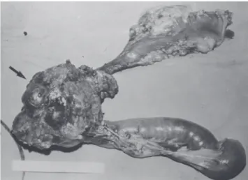
phalic portion of the pancreas, with an irregular surface, measuring 12 x 8 cm in its largest diameter, that was squeezed and stretched to the 1st and 2nd portions of the duodenum (Fig. 2). The liver exhibited a normal and con-sistent aspect, and the remaining areas of the abdominal cavity did not present alterations. The pancreatic lesion was extirpated by partial pancreatoduodenectomy (Whipple’s operation). Anatomopathological exam of the surgical specimen revealed a pancreatic carcinoid tumor with angiolymphatic invasion that infiltrated the Vater’s ampulla and the duodenal wall (Fig. 3). The lymph nodes, in a to-tal of 15 structures, and the margins of the lesion were free from neoplastic involvement. Immunohistochemical stud-ies were performed on paraffin sections using commercially available primary antibodies directed against a set of neu-roendocrine antigens as well as different polypeptides. The
neoplasia exhibited reactivity to neuron-specific enolase (NSE), EMA, CEA, factor VIII, serotonin (Fig. 4), gluca-gon, and somatostatin, and was not reactive to PS100, vimentin, cytokeratins AE1/AE3, or insulin. In the post-operative period, the patient developed venous thrombosis of the lower limbs, without other complications, and was discharged on postoperative day 15. She remains well, with-out symptoms, 5 years after the operation.
Although the histogenesis of carcinoid tumors of the pancreas has yet to be fully clarified, embryologic studies have demonstrated that argentaffin cells are present throughout the foregut and migrate to the bronchioles, pan-creatic ducts, and Langerhans islets.7,8 Wilson et al14
dem-onstrated immmunoreactivity to enterochromaffin cells in Figure 3 - Micrograph of a histological section of the pancreatic tumor showing monomorphic tumor cells growing in an organoid pattern characterized by nests, sheets, and ribbons. The tumor cells are small, round to oval, with nuclei showing “salt and pepper” chromatin and with prominent granular eosinophilic cytoplasm. The histological pattern is consistent with a carcinoid tumor (hematoxilin & eosin staining; original magnification, X200)
Figure 1 - Computed tomography of the abdomen with a solid mass (large arrow) in the area at the level of the head of the pancreas, showing the proximity between the mass and the inferior vena cava (small arrow)
Figure 2 - Resected product of partial gastroduodenopancreatectomy (Whipple’s procedure) with a well-circumscribed nodular mass in the head of the pancreas (arrow)
177
CLINICS 2006;61(2):175-8 Pancreatic carcinoid: a rare cause of diarrheogenic syndrome
Waisberg J et al.
islet cell tumors, and in non-neoplastic ducts and ductule, acini and Langerhans islets. It has been suggested that the islet cell and argentaffin cell are derived from the same neu-roectodermal precursor or from the same multipotential pre-cursor cells of the ductal epithelium, with the capacity to proliferate and differentiate themselves from peptide or monoamine hormone-producing cells.7,8
Carcinoid tumors are immunohistochemically positive for the pan-endocrine markers, neuron-specific enolase and chromogranin A, and also for antiserotonin antibodies, which are considered specific markers for such tumors.3,4,8,9
When immunohistochemical techniques are positive for serotonin in the tumor cells, the tumor can be classified as a carcinoid tumor.3,7 In the present case, the
immunohisto-chemical study of the neoplastic tissue exhibited reactiv-ity to serotonin, but also to glucagon and somatostatin, among others. This fact suggests that the tumor probably originated from pancreatic islet cells. As summarized by Mao et al,8 several carcinoids of the pancreas have stained
positively for other islet cell hormones.
Pancreatic neuroendocrine tumors are extremely rare in children, but occur in all age groups and in males and fe-males with equal frequency,1 and they can occur in all parts
of the pancreas.1 Pancreatic carcinoids, because of their
lo-cation, tend to produce relatively vague, nonspecific symp-toms resulting in larger, more advanced tumors at presen-tation.5,8,9
Less than 10% of carcinoids present diarrhea caused by systemic serotonin release. The explanation lies in the ef-ficient hepatic metabolism of vasoactive amines, and that is also the reason why carcinoid syndrome rarely occurs in the absence of liver metastasis. Exceptions are circum-stances in which venous blood from a large tumor was drained directly into systemic circulation11 as may have
occurred in the present case.
The diarrhea associated with pancreatic carcinoids is generally a consequence of pancreatic exocrine insuffi-ciency and or intestinal action of serotonin produced by the neoplasia.3,8,15One might speculate that perhaps different
mediators produce diarrheas of different intensities.15
Stud-ies of the possible role of vasoactive intestinal polypeptide (VIP), prostaglandin E, and serotonin in the cause of the watery diarrhea syndrome have attracted more attention. Machado et al16 described a patient with a Verner-Morrison
diarrheogenic syndrome with a high plasma concentration of VIP due to a neuroendocrine tumor located in the body and tail of the pancreas and multiple liver metastases. The diarrheogenic syndrome was controlled by surgical resec-tion of the pancreatic neoplasm and postoperative hepatic artery embolization.
In our patient, the diarrhea was intermittent and of lesser
severity than that found in VIPomas. Unfortunately, serum values for the potential mediators of humoral diarrhea are not available. Nonetheless, the results from the immuno-histochemical studies were positive for serotonin, suggest-ing that it might be involved in caussuggest-ing the diarrhea syn-drome.
Jaundice is an unusual finding in pancreatic carcinoids, as explained by the tumor location in the pancreatic body and tail in 70% of patients and the slow and late infiltra-tive growth.5,8,9 Although the lesion of the present case was
situated at the head of the pancreas and presented large di-mensions, there was no clinical or laboratory jaundice, probably due to the noninfiltrative nature of the pancreatic mass.
Foregut carcinoids, similarly to pancreatic carcinoids, tend to produce an atypical carcinoid syndrome11 with
in-creases in 5-hydroxytryptophan plasma levels, but usually not serotonin plasma levels because they lack the appro-priate decarboxylase.5 Nevertheless, urinary
5-hydroxy-in-dole-acetic acid (5-HIAA) levels are markedly increased in functioning pancreatic carcinoids, suggesting a 5-hydroxytryptophan decarboxylation in the intestine and other tissues.8
From a radiological point of view, the pancreatic car-cinoid tumor may be indistinguishable from pancreatic is-let cell tumors, as they have some common features.16
Echography of pancreatic carcinoid reveals the presence of round or oval tumors that are generally well-defined and hypoechoic in contrast to the normal neighboring pancre-atic tissue. A hyperechoic capsule may be delimited by the gland itself or may protrude from its surface. CT exami-nation may detect well-circumscribed, homogeneous, hypodense areas that may contain calcification. Following administration of intravenous contrast material, there is a marked enhancement of the tumor, as opposed to the pan-creatic ductal carcinoma.17 Angiography of the celiac axis
and superior mesenteric artery may confirm the diagnosis of an endocrine tumor by its marked vascularization and may help to distinguish invasion from displacement of ad-jacent large vessels.5 Fine-needle aspiration biopsy with
cytological examination and immunohistochemical tech-niques may evidence serotonin or other hormones in tumor cells and thereby enable a diagnosis of a carcinoid or a neu-roendocrine pancreatic tumor.5
Although bulky tumors are frequently resectable as a curative measure, the high incidence of distant metastases precludes long-term survival for most patients, leading to an unfavorable overall prognosis.5,8-10 Pancreatic carcinoids
178
CLINICS 2006;61(2):175-8 Pancreatic carcinoid: a rare cause of diarrheogenic syndrome
Waisberg J et al.
is no metastatic disease, surgical excision offers the best chance for recovery or long-term survival.5,8,9
The high incidence of metastases described in pancre-atic carcinoids probably results from the late onset of clini-cal symptoms and the consequent late diagnosis, thereby allowing sufficient time for metastasis of the tumor.5,8,9
Thus, tumor stage tends to be significantly more advanced in pancreatic carcinoids, with a very strong negative im-pact on outcome; hence it may be said that pancreatic carcinoids are not necessarily more aggressive than other carcinoids.9 Additionally, tumor diameter does not
corre-spond with the presence or absence of metastases.5
In cases with hepatic metastatic lesions, partial hepatic resection or intra-arterial embolization of the metastatic nodules has been attempted.18 For symptomatic treatment
of inoperable tumors, subcutaneous injection of somatostatin-like substances or the administration of sero-tonin or histamine antagonists have been used in addition to supportive chemotherapy with streptozotocin and 5-fluor-ouracil.18
Recognition of carcinoid tumors of the pancreas is im-portant, since this type of tumor has better a prognosis than the adenocarcinomas if detected early; additionally, effec-tive treatment is available to combat the distressing symp-toms due to the carcinoid syndrome. Accordingly, the re-ports of cases of primary pancreatic carcinoid should sup-port the differential diagnosis of diarrheogenic syndrome and provide new insight into the biology of carcinoid tumors and the mechanisms of their secretagogue action in the release of amines and peptides.
REFERENCES
1. Klöppel G, Perren A, Heitz PU. The gastroentreropatic neuroendocrine cell system and its tumors. The WHO classification. An N Y Acad Sci. 2004;1014:13-27.
2. Prasad S, Patankar T, Joshi A, Deshmukh H. Pancreatic carcinoid: an unusual tumour in an uncommon location. J Postgrad Med. 1998;44:97-8.
3. Prinz RA, Bemer RW, Ghejfee G. A serotonin and HCG producing islet cell carcinoma associated with focal nodular hyperplasia of the liver. J Surg Oncol. 1983;24:30-2.
4. Nagai E, Yamaguchi K, Hashimoto H, Sakurai T. Carcinoid tumor of the pancreas with obstructive pancreatitis. Am J Gastroenterol. 21992:87:361-4.
5. Maurer CA, Baer HU, Dyong TH, Mueller-Garamvoelgyi E, Friess H, Rutchti C, et al. Carcinoid of the pancreas: clinical characteristics and morphological features. Eur J Cancer. 1996;32A:1109-16.
6. Gettenberg G, Zimbalist E, Marini C. Chronic pancreatitis and pseudocyst formation secondary to carcinoid tumor of the pancreas. Gastroenterology. 1988;94:1222-4.
7. Hiller N, Berlowitz D, Fisher D, Blinder G, Hadas-Halpern I. Primary carcinoid tumor of the pancreas. Abdom Imaging. 1998;23:188-190. 8. Mao C, El Attar A, Domenico DR, Kim K, Howard JM. Carcinoid tumors
of the pancreas. Status report based on two cases and review of the world’s literature. Int J Pancreatol. 1998;23:153-64.
9. Kirshbom PM, Kherani AR, Onaitis MW, Hata A, Kehoe TE, Feldman JM, et al. Foregut carcinoids: a clinical and biochemical analysis. Surgery. 1999;126:1105-10.
10. Migliori M, Tomassetti P, Lalli S, Casadei R, Santini D, Corinaldesi R, et al. Carcinoid of the pancreas. Pancreatology. 2002;2:163-6. 11. Marisavljevic D, Petrovic N, Milinic N, Cernerikic V, Krstic M, Bilanovic
D, et al. An unusual presentation of “silent” disseminated pancreatic neuroendocrine tumor. World J Gastroenterol. 2004:10:2919-21. 12. Greene JF, Doyle WF. Pancreatic islet cell carcinoid: a highly malignant
form of carcinoid tumor. J Surg Oncol. 1974;6:183-90.
13. Wilander E, El-Salhy M, Willén R, Grimelius L. Immunocytochemistry and electron microscopy of an argentaffin endocrine tumour of the pancreas. Virchows Arch [Pathol Anat]. 1981;392:263-9.
14. Wilson RW, Gal AA, Cohen C, DeRose PB, Millikan WJ. Serotonin immunoreactivity in pancreatic endocrine neoplasms (carcinoid tumors). Mod Pathol. 1991;4:727-32.
15. Lee CH, Ching KN, Lui WY, P’eng F-K, Franklin WA, Kaplan EL. Carcinoid tumor of the pancreas causing the diarrheogenic syndrome: report of a case combined with multiple endocrine neoplasia, type I. Surgery. 1986;99:123-9.