Short Communication
Diversity in anti-N-methyl-D-aspartate receptor encephalitis:
Case-based evidence
pcn_2308153..156João Pinho,
MD, João Rocha,
MD, Margarida Rodrigues,
MD, João Pereira,
MD,
Ricardo Maré,
MD, Carla Ferreira,
MD, Esmeralda Lourenço,
MDand Pedro Beleza,
MD*
Neurology Department, Hospital de Braga, Braga, Portugal
Antibodies against N-methyl-D-aspartate receptor (NMDAR) are identified in the form of immune-mediated encephalitis in which typical manifesta-tions include neuropsychiatric symptoms, seizures, abnormal movements, dysautonomia and hypoven-tilation. The authors report two cases of anti-NMDAR encephalitis with different presentations and patterns of progression. The first patient presented with status epilepticus and later developed psychosis, pyramidal signs and diffuse encephalopathy. The second patient
presented with acute psychosis followed a week later by seizures, dystonia, rigidity, oromandibular dyski-nesias and dysautonomia. Possible mechanisms responsible for the clinical manifestations of this disease are discussed in light of recently described additional clinical and laboratory findings.
Key words: encephalitis, immune-mediated, N-methyl-D-aspartate receptor.
E
NCEPHALITIS WITH ANTIBODIES againstN-methyl-D-aspartate receptors (anti-NMDAR) is an immune-mediated encephalitis in which antibod-ies against neuronal surface antigens are found.1In 2005, patients with paraneoplastic encephalitis pre-senting with psychiatric manifestations, short-term memory loss, hypoventilation and autoantibodies against unknown neuropil antigens were described.2,3 Subsequently, antibodies against NMDAR-NR1/NR2 heteromers were identified, and NR1 was considered the crucial epitope.4,5 Patients were young adult women with typical manifestations: initial psychiat-ric symptoms followed by seizures, dyskinesias, chorea, autonomic instability and central hypo-ventilation. Psychiatric manifestations are variable, including anxiety, bizarre behavior, mania, halluci-nations, delusions, and pose diagnostic difficulties at onset. Lymphocytic pleocytosis, high protein or posi-tive oligoclonal bands in cerebrospinal fluid (CSF)
are frequent.5Pattern of progression and spectrum of manifestations led to the hypothesis of immunologi-cal response spreading against other central nervous system antigens.6,7We report two patients with anti-NMDAR encephalitis and discuss underlying patho-physiologic mechanisms.
CASE REPORTS
Case 1
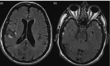
A 63-year-old Caucasian male with a medical history of febrile seizures was admitted for left facial motor status epilepticus. Despite treatment with phenytoin and valproate, he developed convulsive status epilep-ticus, requiring intensive care unit admission. CSF revealed high protein (0.78 g/L), positive unmatched oligoclonal bands, but was otherwise normal, includ-ing herpes simplex virus polymerase chain reaction (HSV-PCR). IV acyclovir was administered for 14 days. Brain magnetic resonance imaging (day 1) showed multiple cortical lesions (Fig. 1). On day 4, he had decreased consciousness, severe dysarthria and dysphagia, left hemiparesis and electroencepha-lography (EEG) showed a right temporal status
*Correspondence: Pedro Beleza, MD, Neurology Department, Hospital
de Braga, Sete Fontes, São Victor, 4701-243 Braga, Portugal. Email: [email protected]
Received 23 December 2010; revised 12 October 2011; accepted 4 November 2011.
Psychiatry and Clinical Neurosciences2012;66: 153–156 doi:10.1111/j.1440-1819.2011.02308.x
153
©2012 The Authors
epilepticus. Subsequently he developed persecutory delusions, and visual and auditory hallucinations which improved with olanzapine. During psychosis, EEG revealed moderate encephalopathy and no epileptiform activity. Extensive blood testing was normal. Between days 20 and 58 he became progres-sively stuporous with bilateral extensor plantars. High-dose IV methylprednisolone, followed by a 5-day course of IV immunoglobulins (IgIV), led to significant clinical improvement. Investigation for occult tumor was negative. Recurrence of psychosis occurred 1 month later, and examination revealed dysarthria, multifocal myoclonus and ataxic gait. Phenytoin intoxication was found, it was replaced by levetiracetam and after a 5-day course of IgIV, he recovered rapidly and has been asymptomatic for 3 years. Anti-NMDAR NR2B analysis was per-formed using a Western blot technique:8 antibodies in serum were IgG-positive and IgM-negative; in CSF both IgG and IgM were negative (samples collected several weeks after presentation and IV methylprednisolone).
Case 2
A 21-year-old previously healthy Caucasian woman was admitted for acute onset of delusions, ideas of grandeur, and visual and auditory hallucinations. Recent hallucinogenic drug ingestion was suspected. She had received a booster vaccination against tetanus 3 days earlier and since then had flu-like symptoms. She was admitted to the Psychiatry
Department, treated with risperidone, diazepam and a week later had a generalized seizure. At first neuro-logical evaluation she was drowsy, with no verbal response, had generalized right-side predominant rigidity, with catatonia cerea, dystonia of the upper limbs, oromandibular dyskinesias and episodes of sinus tachycardia and peripheral desaturation. Exten-sive blood testing and urine toxicological screen were normal. CSF showed lymphocytic pleocytosis (10 cells/uL, 95% lymphocytes) and was otherwise normal including HSV-PCR and oligoclonal bands. Brain magnetic resonance imaging was normal (including T2, diffusion-weighted imaging and contrast-T1). EEG revealed left frontotemporal status epilepticus. Status epilepticus persisted despite phenytoin, levetiracetam and clonazepam. Brain positron emission tomography (PET) scan revealed left frontotemporal and left basal ganglia hyperme-tabolism (Fig. 2). Treatment with IgIV during 5 days and, later, a 5-day course of IV methylprednisolone led to progressive clinical improvement. At discharge she had attention and arithmetic deficits, global dys-phasia and normal motor exam. She scored 17 in a Mini Mental State Examination. Investigation for occult tumor was negative, slow cognitive and lan-guage improvement was documented as the patient gradually recovered functional independence, with more than 18 months of follow up. Immunofluores-cent cell-based assay was used to determine
anti-NMDAR NR1/NR2:6 serum and CSF collected
3 weeks after admission were positive for anti-NMDAR NR1/NR2.
Figure 1. Brain magnetic resonance imaging, fluid attenuated inversion recovery – hyperintense lesions in right fronto-opercular, (a) right insular and (b) bilateral temporal cortex. Absent diffusion-weighted imaging abnormality or contrast enhancement.
154 J. Pinhoet al. Psychiatry and Clinical Neurosciences2012;66: 153–156
©2012 The Authors
DISCUSSION
Early clinical manifestations in both patients (convul-sive status epilepticus and psychiatric manifestations) were likely secondary to cortical dysfunction. Sug-gested underlying mechanisms include direct neuronal damage of anti-NMDAR antibodies, cell deposition of membrane attack complex, antibody-mediated NMDAR internalization and decreased synaptic NMDAR-mediated postsynaptic currents.9 Additional manifestations developed in a time-dependent pattern: acute psychosis and other deficits in the first patient were possibly secondary to wide-spread cortical dysfunction, while the second patient developed basal ganglia and brainstem signs. Findings must be interpreted with caution. Antibodies against NMDAR-NR2B determined by Takahashi and col-laborators’ technique were found in a heterogeneous population of patients.10–12 NMDAR are transmem-brane proteins consisting of a combination of NR1, NR2 (A-D) and NR3 (A-B) subunits.13 Since NR2B subunits have neocortical and hippocampal expres-sion, this may explain the early cortical dysfunction of anti-NMDAR encephalitis. Widespread expression
of NR1 subunits (neurons, oligodendrocytes, astro-cytes14,15) could explain the spectrum of manifesta-tions, but fails to explain the temporal pattern previously described. Myelin basic protein autoanti-bodies were identified in a patient with anti-NMDAR encephalitis who later developed transverse myelitis, optic neuritis and white matter lesions.7Evidence of unmatched CSF oligoclonal bands in a later stage of anti-NMDAR encephalitis suggests the possibility of spreading of immunological response to other epitopes, leading to extra-cortical manifestations.6An important limitation of the current report consists of the low specificity of anti-NMDAR-NR2B technique developed by Takahashi and collaborators, and paired analysis of serum and CSF from the same patients using both techniques could further clarify this point.
ACKNOWLEDGMENTS
We would like to thank Professor Yukitoshi Taka-hashi at the Shizuoka Institute of Epilepsy and Neu-rological Disorders, Professor Angela Vincent and Dr Isabel Leite at the University of Oxford. The authors have no conflicts of interests.
REFERENCES
1. Graus F, Saiz A, Dalmau J. Antibodies and neuronal autoimmune disorders of the CNS.J. Neurol.2010;257: 509–517.
2. Vitaliani R, Mason W, Ances B, Zwerdling T, Jiang Z, Dalmau J. Paraneoplastic encephalitis, psychiatric symp-toms, and hypoventilation in ovarian teratoma. Ann. Neurol.2005;58: 594–604.
3. Ances BM, Vitaliani R, Dalmau J et al. Treatment-responsive limbic encephalitis identified by neuropil antibodies: MRI and PET correlates. Brain 2005; 128: 1764–1777.
4. Dalmau J, Tüzün E, Lynch DR et al. Paraneoplastic N-methyl-D-aspartate receptor encephalitis associated with ovarian teratoma.Ann. Neurol.2007;61: 25–36. 5. Dalmau J, Gleichman AJ, Lynch DR et al.
Anti-NMDA-receptor encephalitis: case series and analysis of the effects of antibodies.Lancet Neurol.2008;7: 1091–1098. 6. Irani SR, Bera K, Waters P et al. N-methyl-D-aspartate
antibody encephalitis: temporal progression of clinical and paraclinical observations in a predominantly non-paraneoplastic disorder of both sexes. Brain2010;133: 1655–1667.
7. Kruer MC, Koch TK, Bourdette DNet al. NMDA receptor encephalitis mimicking seronegative neuromyelitis optica.
Neurology2010;74: 1473–1475.
Figure 2. Brain positron emission tomography scan – left frontotemporal and left basal ganglia hypermetabolism.
Psychiatry and Clinical Neurosciences2012;66: 153–156 Anti-NMDA receptor encephalitis 155
©2012 The Authors
8. Takahashi Y, Sakaguchi N, Kondo Net al. Epitope analysis of auto-antibodies against GluR e2 in patients with
chronic progressive epilepsia partialis continua of child-hood.Epilepsia2002;43(Suppl. 9): 74.
9. Hughes EG, Peng X, Gleichman AJet al. Cellular and syn-aptic mechanisms of anti-NMDA receptor encephalitis.
J. Neurosci.2010;30: 5866–5875.
10. Takahashi Y. Epitope of autoantibodies to N-methyl-N-aspartate receptor heteromers in paraneoplastic limbic encephalitis.Ann. Neurol.2008;64: 110–111.
11. Takahashi Y, Mori H, Fujiwara Tet al. Autoantibodies and cell-mediated autoimmunity to NMDA-type GluRe2 in
patients with Rasmussen’s encephalitis and chronic pro-gressive epilepsia partialis continua. Epilepsia 2005; 46
(Suppl. 5): 152–158.
12. Takahashi Y, Mori H, Kondo N et al. Autoanti-bodies to NMDA receptor in patients with chronic forms of epilepsia partialis continua. Neurology2003;61: 891– 896.
13. Cull-Candy S, Brickley S, Farrant M. NMDA receptor subunits: diversity, development and disease.Curr. Opin. Neurobiol.2001;11: 327–335.
14. Salter MG, Fern R. NMDA receptors are expressed in devel-oping oligodendrocytes processes and mediate injury.
Nature2005;438: 1167–1171.
15. Lalo U, Pankratov Y, Kirchhoff F, North RA, Verkhrastky A. NMDA receptors mediate neuron-to-glia signaling in mouse cortical astrocytes. J. Neurosci. 2006; 26: 2673– 2683.
156 J. Pinhoet al. Psychiatry and Clinical Neurosciences2012;66: 153–156
©2012 The Authors