Role of NMDA receptors in the
trigeminal pathway, and the
modulatory effect of magnesium in a
model of rat temporomandibular joint
arthritis
Cavalcante ALC, Siqueira RMP, Araujo JCB, Gondim DV, Ribeiro RA, Quetz JS, Havt A, Lima AAM, Vale ML. Role of NMDA receptors in the trigeminal pathway, and the modulatory effect of magnesium in a model of rat temporomandibular joint arthritis.
Eur J Oral Sci 2013; 121: 573–583.©2013 Eur J Oral Sci
Temporomandibular joint (TMJ) arthritis is a common cause of orofacial pain. In the present study, the modulatory effects of N-methyl-D-aspartate receptors
(NMDA-Rs) and magnesium were investigated in TMJ arthritis hypernociception. Male Wistar rats received an intra-articular injection of carrageenan (Cg) in the TMJ, and mechanical hypernociception was measured. The NMDA-R antagonist, MK-801, and magnesium chloride (MgCl2) were administered before arthritis
induction. Magnesium deficiency was promoted by feeding rats a synthetic magne-sium-free diet for 9 d before injection of Cg. The Cg induced mechanical hypernoci-ception that lasted for 120 h. MK-801 inhibited this hypernociceptive state. MgCl2
pretreatment prevented Cg-induced hypernociception and altered the nociceptive threshold in the absence of Cg. Magnesium deficiency increased hypernociception and induced spontaneous hypernociceptive behavior. TMJ arthritis increased the expression of mRNA for allNMDA-Rsubunits and immunostaining of phosphorylated NR1 (phospho-NR1). MgCl2inhibited expression of NR2B mRNA and
phospho-NR1 immunostaining and increased expression of NR3 mRNA. Magnesium deficiency increased expression of both NR1 and NR3 mRNAs and phospho-NR1 immunostaining in the trigeminal subnucleus caudalis. We found that magnesium modulates nociceptive behavior and inducesNMDA-Rsubunit rearrangement in the subnucleus caudalis. The present results may lead to a better understanding of cen-tral processing in the nociceptive trigeminal pathway and the development of new approaches to treat orofacial pain with a TMJ origin.
Andre L. C. Cavalcante1, Rafaelly M. P. Siqueira2, Joana C. B. Araujo1, Delane V. Gondim3, Ronaldo A. Ribeiro2, Josiane S. Quetz4, Alexandre Havt2,4, Aldo A. M. Lima2,4, Mariana L. Vale1,2,3 1
Medical Sciences Post-Graduation Program, Department of Clinical Medicine, Federal University of Ceara, Fortaleza;
2
Pharmacology Post-graduation Program, Department of Physiology and Pharmacology, Federal University of Ceara, Fortaleza; 3Department of Morphology, Federal University of Ceara, Fortaleza;4Institute of Biomedicine for Brazilian Semi-Arid & Clinical Research Unit, Federal University of Ceara, Fortaleza, Brazil
Delane V. Gondim, Department of Morphology, Faculty of Medicine, Federal University of Ceara, R. Delmiro de Farias, s/n - Rodolfo Teofilo, Fortaleza,
CE 60430-170, Brazil
E-mail: [email protected]
Key words: magnesium;N-methyl-D-aspartate receptor (NMDA-R); NR3; orofacial pain; temporomandibular joint
Accepted for publication September 2013
Temporomandibular disorders (TMDs) are often
related to chronic orofacial pain and are probably the most common non-tooth-related orofacial pain condi-tion associated with a significant worsening of the patient’s quality of life (1).
Temporomandibular disorders may be associated with the phenomenon of central and peripheral sensiti-zation. Constant nociceptive input stimulus from the injured sites may result in excessive depolarization of the central nociceptive neurons, which can promote excitotoxicity and loss of inhibitory mechanisms and eventually provoke prolonged pain (2).
Some data show that excitatory amino acids (e.g. glutamate) and neuropeptides, through receptor
mecha-nisms and involving mainly N-methyl-D-aspartate
recep-tors (NMDA-Rs), have been implicated in these neuroplastic changes, enlarging receptor fields in the
central nervous system for nociceptive input. N
-methyl-D-aspartate receptors also play an essential role in
the ‘wind-up’ phenomenon of central sensitization med-iated by the trigeminal brainstem sensory nuclear com-plex (3–8).
N-methyl-D-aspartate receptors play an essential role in
these nociceptive processes, and NMDA-R antagonists or modulators may be useful in the treatment of persistent
pain in the temporomandibular joint (TMJ) area (7, 9–16).
Magnesium is a natural modulator of NMDA-Rs, functioning as a physiological channel blocker that
DOI: 10.1111/eos.12093
Printed in Singapore. All rights reserved European Journal of
reduces ion currents (17). Magnesium is also a calcium antagonist in some systems, reducing neuronal excitability
and neurotransmitter release (17–19). The
depolariza-tion process removes the magnesium-induced blockade of NMDA-Rs in a voltage-dependent manner, result-ing in hyperexcitability and consequently a sustained state of depolarization and increased synaptic strength (17, 19, 20).
N-methyl-D-aspartate receptors are composed of the
NR1, NR2, and NR3 subunits. The combination of NR1, an essential channel-forming subunit, with the other subunits can change the kinetics of receptor depolarization and sensitivity to magnesium-induced blockade and calcium entry (16, 17). Some gene-expres-sion studies showed the differential expresgene-expres-sion of these subunits according to different stimulation that reaches the central nervous system through the trigeminal
path-way (21–23), but no evidence has been provided on the
regulatory mechanism of magnesium inNMDA-R
sub-unit expression in trigeminal subnuclei.
Considering the sparse evidence on the importance of NMDA-Rs in acute and chronic orofacial pain, and the possibility that magnesium can modulate NMDA-R responses in orofacial pain, we aimed to highlight the role of these receptors in the hypernociceptive process in the TMJ region. Additionally, the present study investigated the role of magnesium in the nociceptive response and NMDA-R gene expression in rats with TMJ arthritis.
Material and methods
Animals
We used male Wistar rats that weighed 180–200 g and were housed in a temperature-controlled room (22°C). The experimental protocol was approved by the Animal Ethics Committee of the Faculty of Medicine, Federal University of Ceara (protocol no. 30/10) and was per-formed in accordance with the Guide for the Care and Use of Laboratory Animals. Efforts were made to use the smallest number of rats possible and to minimize the stress to which they were subjected.
Drugs and reagents
Carrageenan (Cg), MK-801, and tribromoethanol were purchased from Sigma-Aldrich (St Louis, MO, USA). Magnesium chloride (MgCl2) (Vetec, Rio de Janeiro,
Bra-zil) was diluted in mineral water. PCR primers were pur-chased from Invitrogen (San Diego, CA, USA). The immunohistochemistry ABC kit was obtained from Santa Cruz Biotechnology (Santa Cruz, CA, USA).
Experimental model
Joint inflammation was induced by an intra-articular injec-tion of Cg (5% or 500lg per articulation; 10ll volume). A 5% Cg solution (10ll) in sterile saline was injected into the supradiscal space of the left TMJ (ipsilateral) using a microsyringe (Hamilton model 702RN; Hamilton, Reno,
NV, USA) coupled to a 30-gauge gingival needle (BD, Franklin Lakes, NJ, USA). As a control procedure, another group of rats was injected intra-articularly with saline unilaterally in the left joint.
Before injection with Cg or saline, the rat was anesthe-tized with tribromoethanol, and the TMJ skin region was carefully shaved. The posteroinferior border of the zygo-matic arch was palpated, and the needle was inserted inferior to this point and advanced in a medial and ante-rior direction until the needle made contact with the con-dyle. This contact was verified by moving the mandible, and the puncture of the needle into the joint space was confirmed by the loss of resistance. Gentle aspiration excluded intravascular placement, after which the specified volume of Cg or saline was injected.
Mechanical hypernociception in the TMJ was evaluated by measuring the threshold of force intensity, in grams, needed to be applied to the TMJ region until a reflex response of the rat occurred (e.g. head-withdrawal or vocali-zation). The measurements were performed by an examiner unaware of the treatments, making use of a digital device (Insight; Ribeir~ao Preto, SP, Brazil). Each rat was housed individually in a plastic cage, 30 min before beginning the tests, and was subjected to a conditioning session of head-withdrawal threshold measurements in the test room for three consecutive days under controlled temperature and low illumination and noise. On the 3rd d, the basal force threshold value was recorded before intra-articular injection of either Cg or vehicle. The measurement of force thresholds was performed (in triplicate) from the ipsilateral TMJ, 4, 6, 12, 24, 48, 72, and 168 h after injection of Cg (24). For each experiment, the nociceptive threshold was assessed before the induction of arthritic hypernociception to determine baseline values before the treatment induction when hypern-ociception developed (i.e. control values) and then once per day after injection of Cg.
Trigeminal spinal nuclei extraction: subnucleus caudalis region
Six, 24, 72, and 120 h after injection of Cg into the TMJ, the rats were anesthetized with an intraperitoneal injection of 2.5% tribromoethanol (250 mg kg 1) and transcardially perfused with 4% paraformaldehyde in 0.1 M phosphate buffer (pH 7.4).
The brain and brainstem were quickly removed, and the trigeminal nuclei area was dissected, postfixed in 4% for-malin for 60–90 min, then sliced into longitudinal 10-lm slices for the immunohistochemistry assay.
Other groups of rats were anesthetized with an intra-peritoneal injection of tribromoethanol (0.03 mg kg 1) and decapitated. The brain and brainstem were quickly removed, and the trigeminal nuclei area was dissected, immediately frozen in liquid nitrogen, and kept frozen at 80°C until further processing for real-time PCR. The sec-tions used in this study were cut 10.5 to 14.04 mm from the bregma (25).
Induction of magnesium deficiency in rats
cornstarch (397 g kg 1), casein (200 g kg 1), sucrose (100 g kg 1), soybean oil (70 g kg 1), fiber (50 g kg 1), L -cystine (3 g kg 1), choline bitartrate (2.5 g kg 1), AIN-93VX vitamin mix (10 g kg 1), and magnesium-free mineral mix (35 g kg 1; AIN93 magnesium free; Rhoster, S~ao Paulo, Brazil). After the 9th d, the rats received an intra-articular injection of Cg in the left TMJ and continued to feed on the special diet for five more days.
The assessment of head-withdrawal thresholds (mechan-ical hypernociception) was performed before beginning the special diet and was repeated after 9 d of feeding on this diet. The control group was fed a normal diet and was also subjected to the same conditions as the rats that received the special diet.
To verify magnesium deficiency, serum magnesium con-centrations were determined before and after 9 d of special diet feeding, as described below.
Magnesium supplementation
Magnesium supplementation was implemented with oral administration of MgCl2 dissolved in mineral water. We
tested dosages of 10, 30, and 90 mg kg 1per day,
admin-istered orally in a volume of 0.5 ml twice per day for 3, 5, and 7 d before the injection of Cg. The treatment was con-tinued after injection with Cg until the 7th d.
Determination of serum magnesium
The rats were anesthetized with 2.5% tribromoethanol (250 mg kg 1 of body weight, intraperitoneally). Blood was collected from the orbital venous plexus into heparin-ized tubes. Serum samples were obtained after low-speed centrifugation (1125 g for 15 min). A standardized
diag-nostic kit for magnesium determination (Labtest Di-agnostica, Minas Gerais, Brazil) was used. The spectrophotometric determination of serum magnesium was performed at 540 nm, as determined by the kit’s man-ufacturer.
Drug treatment
Pharmacological blockade of NMDA-Rs was performed with a single intraperitoneal administration of the NMDA receptor antagonist, MK-801 (0.1, 0.25, and 0.5 mg kg 1),
30 min before injection with Cg. The nociceptive threshold was measured before, and 4, 6, 10, 24, and 168 h after, injection of Cg.
Total RNA isolation
The adult rat brainstem, dissected in the region of the trigeminal subnucleus caudalis, was obtained as described above. The average quantity of tissue isolated from each rat sample was approximately 100 mg. The tissues were weighed and stored at 80°C until used for the analysis. Total RNA was isolated from each sample using the RNeasy Mini kit (Qiagen, Valencia, CA, USA) according to the manufacturer’s instructions. Briefly, sample grind-ing was performed with QIAzol lysis reagent (Qiagen) using a cell disruptor composed of a rotary tool (Dremel 300-N/10; Racine, WI, USA.) and autoclavable steel rotating blades (in-house design). After membrane disrup-tion and release of nucleic acid into the medium, the solution was centrifuged at 20,000g for 5 min, and
200ll of 1-bromo-3-chloropropane (BCP; Fluka, Mil-waukee, WI, USA) was added to the supernatant. After homogenization and centrifugation at 15,000g for
15 min at 4°C, the 2-ml tube contents were separated into three phases, and the impure RNA was found in the upper limpid phase. The following steps consisted of assembling the impure RNA in a spin column to remove impurities using 70% ethanol and RW1 and RPE buffers (Qiagen). The elution of highly pure nucleic acids occurs in RNase-free water. The RNA obtained was quantified using NanoDrop (Thermo Fisher Scientific, Waltham, MA, USA).
Reverse transcription reaction
To evaluate the gene expression of NR1 (NMDA1), NR2B (NMDA2B), and NR3 (NMDA3), the isolated RNA was transformed to cDNA by a reverse transcription reaction using a SuperScript III Reverse Transcriptase Sys-tem (Invitrogen). Briefly, RNA samples (1lg) were incu-bated with 4ll of 59iScript Reaction Mix and 1ll of iScript Reverse Transcriptase (Bio-Rad, Hercules, CA, USA) in Milli-Q water to a final volume of 20ll. This reaction was cycled at 25°C for 5 min, 42°C for 30 min, and 85°C for 5 min. The synthesized cDNA was kept in a freezer at 20°C until amplification by real-time quantita-tive PCR (qPCR).
Real-time qPCR
The expression of NMDA-1, NMDA-2, and NMDA-3 genes was assayed using an iQ5 Real-Time PCR Detection System (Bio-Rad), based on the detection of SYBR Green
Table 1
Oligonucleotide sequence of primers used in the qPCR assays
Gene Target Primer sequence (5′–3′)
Product size (bp)
Accession no. (GenBank, NCBI)
YHAWZ Rattus norvegicustyrosine 3-monooxygenase/ tryptophan 5-monooxygenase activation protein
F- GCTACTTGGCTGAGGTTGCT R- TGCTGTGACTGGTCCACAAT
61 NM_013011.3
NMDAR-1 Rattus norvegicusNMDAR1 glutamate receptor subunit
F- GCAGAACGTTTCCCTGTCCA R- CCCCTGCCATGTTCTCAAAA
140 U11418.1
NMDAR-2 Rattus norvegicusNMDAR glutamate receptor subtype 2C
F- ATGGCGGGGGTCTTCTACAT R- GTCCAGCTGGGATGAGTTGG
120 M91563.1
NMDAR-3 Rattus norvegicusNMDAR3 glutamate receptor subunit
F- TTCATGTGGCCACTCCACTG R- CCAAAGGGGCTCTTCCATTC
101 AF073379.1
incorporation. Phospholipase A2 (tyrosine 3-monooxygen-ase/tryptophan 5-monooxygenase activation protein zeta polypeptide) was used as the reference gene (YHWAZ). DNA primers for all of the genes (Table 1) were designed on the basis of mRNA sequences obtained from the National Center for Biotechnology Information (http:// www.ncbi.nlm.nih.gov; accessed December 26, 2011). Real-time PCR assays were performed in a final volume of 25ll of a medium that contained 12.5ll of iQ SYBR Green Supermix (a standard buffer for amplification con-taining reagents, such as DNA polymerase, deoxynucleo-tide triphosphates (dNTPs), and saline buffer, at optimal concentrations for RT-PCR assays), 200 nM primers, and 2ll of cDNA of the samples. Negative samples were also tested, in which the cDNA was replaced with autoclaved Milli-Q water. The PCR conditions were as follows: an initial denaturation step of 7 min at 95°C, followed by 40 cycles for gene amplification. Each cycle consisted of an initial denaturation phase of 30 s at 95°C, followed by an annealing phase of 30 s at 61°C and an extension phase of 45 s at 72°C. The samples were then subjected to an exten-sion step of 3 min at 72°C. To ensure the specificity of the amplifications applied (i.e. to determine whether the prod-ucts formed were specific for the tested genes), we con-structed a melting curve after each reaction, in which the reaction temperature was subsequently increased by 0.5°C every 20 s, beginning at the annealing temperature of a given primer and ending at 95°C. Throughout the qPCR amplification process, changes in fluorescence were mea-sured, and the data obtained from iQ5 Optical System software (version 2.0; Bio-Rad) were based on the values of the threshold cycle, in which the observed fluorescence is 10-fold higher than the basal fluorescence for each PCR assay. Gene expression was obtained by applying the mathematical method of PFAFFL(26).
Immunohistochemistry
Immunohistochemistry for phosphorylated NR1 was per-formed using the streptavidin–biotin–peroxidase method in formalin-fixed, paraffin-embedded tissue sections (5lm thick) mounted on poly-L-lysine-coated microscope slides. The sections were deparaffinized and rehydrated using xylene and a graded series of alcohol. After antigen retrie-val with citrate buffer (pH 6.0) at 95°C (15 min), endoge-nous peroxidase was blocked twice (10 min) with 3% (v/v) hydrogen peroxide and washed in PBS. The sections were incubated overnight at 4°C with primary rabbit anti-(phos-pho-NR1) (Millipore, S~ao Paulo, Brazil), diluted 1:200 in PBS containing 5% BSA (PBS/BSA). The slides were then incubated with biotinylated goat anti-rabbit (Santa Cruz Biotechnology) diluted 1:400 in PBS/BSA. After washing, the slides were incubated with avidin–biotin–horseradish peroxidase conjugate (Strep ABC complex; Santa Cruz Biotechnology) for 30 min according to the manufacturer’s protocol. Immunostaining was visualized with the chromo-gen 3,3′-diaminobenzidine (DAB; ABC staining system; Santa Cruz Biotechnology). Negative control sections were processed simultaneously, as described above, but with the first antibody replaced with PBS/BSA. None of the nega-tive controls showed immunoreactivity to phospho-NR1. The slides were counterstained with Mayer’s hematoxylin, dehydrated in a graded series of alcohol, cleared in xylene, and coverslipped. Each group consisted of six rats. Posi-tive labeling for phospho-NR1 was determined by brown staining.
The slides were photographed with a Leica microscope coupled with a DFC232 camera (Leica, Wetzlar, Ger-many). After focusing the microscope, the left Sp5C area was identified and photographed at 2009magnification.
The area stained in the pictures was quantified by differ-entiating the areas (pixels) with higher color saturation associated with immunostaining (brown). For this, we used theIMAGE J-NIHsoftware (http://rsb.info.nih.gov). The procedure was based on color saturation associated with positive staining for a particular marker. The limits required for defining selected pixels were previously defined by color threshold. The percentage of the area stained in each photograph was calculated by dividing the selected pixel areas by the total area of the photograph. At least four photographs per group were evaluated.
Statistical analysis
The results are expressed as the mean standard error of the mean of the measurements from at least six rats per group. ANOVA followed by Bonferroni’s test was used to compare means. The Student’st-test was used to compare two means. Values of P<0.05 were considered statisti-cally significant.
Results
Time-course of the hypernociceptive effect
The intra-articular Cg injection resulted in time-depen-dent and long-term hypernociception in response to
mechanical stimulation, measured by a marked
decrease in the mechanical head-withdrawal threshold. The mechanical hypernociception induced by Cg began at the 4-h time-point, peaked between 6 and 12 h
(P <0.001), with the peak lasting until 96 h (P<0.05),
and had returned to baseline by 168 h. The rats injected with intra-articular saline in the left TMJ pre-sented no significant changes in mechanical withdrawal thresholds at any of the time-points evaluated (Fig. 1).
Effect of NMDA-R antagonist
We investigated the role NMDA-Rs in the nociceptive effect of Cg-induced TMJ arthritis. The effect of the nonselective NMDA-R antagonist, MK-801, was
inves-tigated at doses of 0.1, 0.25, and 0.5 mg kg 1,
intraper-itoneally. Our results showed that 0.25 and
0.5 mg kg 1 of MK-801 dose-dependently increased
mechanical withdrawal thresholds compared with the control group (Fig. 2). Four hours after injection with Cg, no significant effect of MK-801 was observed com-pared with the control group. Withdrawal thresholds were increased by treatment with MK-801 only,
start-ing at 6 h (i.e. the peak of hypernociception;
P< 0.001) and remained at levels significantly different
from controls up to 144 h (P< 0.05). The dose of
0.5 mg kg 1 had a maximum inhibitory effect among
treated groups, at 48 h (P<0.001), that lasted until
120 h (P<0.05) after injection with Cg.
Effect of oral MgCl2supplementation
An experiment was first designed to determine the dose
at which MgCl2 would exert an antihypernociceptive
effect. We tested doses of 10, 30, and 90 mg kg 1
(given twice per day; 3-d pretreatment). The results
showed that the 90-mg kg 1 dose inhibited
hypernoci-ception 4, 6, 24, and 120–168 h after injection with Cg,
reaching the maximal effect at 6 h (P <0.001).
Interest-ingly, we found that after 3 d of treatment with
90 mg kg 1 of MgCl
2, withdrawal thresholds were
sig-nificantly increased (P <0.01), even before injection
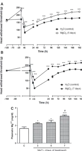
with Cg, compared with the non-treated group (Fig. 3). We then evaluated the time-course of the effect of
90 mg kg 1of MgCl2over 5 and 7 d of pretreatment. Our
results showed a progressive increase in withdrawal thresh-olds with 5 and 7 d of treatment. Withdrawal threshthresh-olds were significantly higher at all time-points evaluated after
administration of Cg (P<0.001; Fig. 4A,B).
To verify whether plasma magnesium concentrations
increased in rats supplemented with MgCl2, we
deter-mined the serum magnesium concentrations 3, 5, and
7 d after starting MgCl2 supplementation, before
injec-tion with Cg. Plasma magnesium levels were
signifi-cantly increased in all groups treated with MgCl2
(P<0.05) compared with baseline serum magnesium
levels (Fig. 4C).
Effect of magnesium deficiency
Nine days after starting the magnesium-free diet, the rats showed a significant decrease in head-withdrawal
thresholds (P< 0.001) compared with the normal diet
group (Fig. 5B). Head-withdrawal thresholds were
assessed before injection of Cg. Plasma magnesium lev-els were also assessed. After 9 d of the special diet, the rats displayed a significant decrease in plasma magne-sium concentrations compared with rats receiving the
normal diet (P <0.001; Fig. 5A).
Four hours after injection of Cg, a significant
differ-ence (P<0.001) was observed between the group treated
with a normal diet and the group treated with the magne-sium-free diet; this difference lasted for 6 h (maximum
effect,P<0.001) and had disappeared by the 24-h time
point. Interestingly, 96 h (P <0.05) after injection of Cg,
a difference between these groups reappeared that
remained until the 120-h time point (P <0.001).
Treatment with MK-801 prevented the decrease in head-withdrawal thresholds induced by magnesium
deficiency in Cg- and saline-injected rats (P< 0.001).
The inhibition induced by MK-801 reached 100%
(P< 0.001) of the hypernociceptive effect of Cg and
magnesium deficiency.
Fig. 2. Evaluation of the antinociceptive effect of the N -methyl-D-aspartate receptor (NMDA-R) antagonist, MK-801,
in carrageenan-induced temporomandibular joint (TMJ) arthritis. MK-801 (0.1, 0.25, or 0.5 mg kg 1) or vehicle (con-trol) was injected intraperitoneally 30 min before the intra-articular injection of 5% carrageenan (10ll) into the left TMJ. The nociceptive threshold (head withdrawal) in response to mechanical stimulation was measured before and after (4–
168 h) the injection. The data are expressed as the mean standard error of the mean of the force threshold, in grams, of six rats per group. Asterisks indicate significant differences from vehicle (ANOVA followed by Bonferroni’s test; ***P<0.001,**P<0.01,*P<0.05).
Fig. 3. Antinociceptive effect of magnesium chloride (MgCl2)
in carrageenan-induced temporomandibular joint (TMJ) arthritis. Vehicle (H2O) or MgCl2(10, 30, or 90 mg kg 1) was
NMDA-R gene expression in rat trigeminal subnucleus caudalis
Expression of the genes encoding three NMDA-R
subunits was investigated in tissue samples obtained from the trigeminal subnucleus caudalis. NMDAR1 (NR1), NMDAR2B (NR2B), and NMDAR3 (NR3) were expressed (Figs 6 and 7), and their expression profiles
changed according to MgCl2treatment, magnesium
defi-ciency, and Cg-induced TMJ inflammation (arthritis). Carrageenan-induced arthritis in the TMJ
signifi-cantly increased expression of the NR1 subunit
(P <0.05) compared with naive rats. Treatment with
MgCl2 did not modify expression of the NR1 subunit
compared with the non-treated group but still induced a significant difference compared with the naive group
(P <0.05; Fig. 6A).
No difference in expression of the NR2B subunit was found between naive rats and rats in the Cg-induced arthritis group. A decrease in expression of the NR2B
subunit was found in arthritic rats treated with MgCl2
compared with rats in the naive group (P <0.05;
Fig. 6B) and in the non-treated group (P <0.05; data
not shown).
Expression of the NR3 subunit was weak but signifi-cantly increased in the Cg-induced arthritis group
compared with naive rats (P <0.05; Fig. 6C), and
treatment with MgCl2 resulted in a two-fold increase in
expression of the NR3 subunit (P< 0.05; Fig. 6C). A
two-fold increase in expression of the NR3 subunit was also observed when the treated rats were compared
with non-treated arthritic rats (P <0.05; data not
shown).
Magnesium deficiency significantly increased
expres-sion of the NR1 subunit (P <0.05) compared with
naive rats (Fig. 7A). When TMJ arthritis was induced by Cg in magnesium-deficient rats, expression of the NR1 subunit decreased compared with that in the naive
group (P <0.05; Fig. 7A) and in the
magnesium-defi-cient group not injected with Cg (P <0.05; data not
shown). In summary, treatment of magnesium-deficient rats with Cg decreased expression of the NR1 subunit.
Magnesium deficiency did not modify NR2B expres-sion in either arthritic or non-arthritic rats (Fig. 7B). Expression of the NR3 subunit was significantly
upregulated in magnesium-deficient rats (P<0.05),
with no differences found between Cg-treated and non-treated rats (Fig. 7C).
Immunohistochemistry for NR1 subunit phosphorylation
The immunohistochemical analysis showed an increase in phosphorylation of the NR1 subunit (phospho-NR1)
in the subnucleus caudalis region in rats with
Cg-induced arthritis (P< 0.001) compared with naive
rats (Fig. 8B,C,G). Treatment with MgCl2 decreased
(P <0.05) this immunostaining (Fig. 8D,G) compared
with Cg-treated rats. Magnesium deficiency also
increased (P <0.001) phospho-NR1 immunostaining in
both the saline- and Cg-treated groups (Fig. 8E-G).
Discussion
A previous study showed that a single Cg injection caused a persistent hypernociceptive response. Our results showed that Cg-induced orofacial hypernocicep-tion lasted until day 6. After this point, the withdrawal threshold was almost equal to baseline (24).
To understand, in more detail, the central processing of arthritic TMJ hypernociception via the trigeminal pathway, we examined the role of NMDA-Rs in the subnucleus caudalis region. MK-801, a specific antago-A
B
C
Fig. 4. Time-course of the antinociceptive effect of magnesium chloride (MgCl2) in carrageenan-induced temporomandibular
joint (TMJ) arthritis. Vehicle (V; H2O) or MgCl2
(90 mg kg 1) was administered orally, twice daily (at 12-h intervals) for (A) 5 d and (B) 7 d, before intra-articular injec-tion of 5% carrageenan (10ll) into the left TMJ and also until day 168. The nociceptive threshold (head withdrawal) in response to mechanical stimulation was measured before and after (4–168 h) the injection. The data are expressed as the meanstandard error of the mean of the force threshold, in grams, of six rats per group. Asterisks indicate significant dif-ferences compared with the vehicle group (ANOVAfollowed by Bonferroni’s test; ***P<0.001, **P<0.01, *P<0.05). (C) Animals were treated with vehicle (V) or magnesium chloride (MgCl2; 90 mg kg 1; twice daily (at 12-h intervals)). The
results are expressed as the meanstandard error of the mean of plasma magnesium (Mg2+; mg dl 1of blood) levels. Asterisks indicate significant differences compared with the vehicle group (t-test; 95% CI; ***P<0.001, **P<0.01, *P<0.05;#P<0.05, day 5
nist, completely reduced ongoing hypernociceptive
behavior related to arthritic TMJ and oral MgCl2
sup-plementation. Furthermore, the MK-801-induced
blockade of hypernociception had a long-lasting effect of up to 120 h after a single injection.
The same doses used in our study rapidly and robustly reduced mechanical and thermal hypernocicep-tion in neuropathic models in previous studies (9, 12).
Magnesium is a physiological NMDA-R modulator that plays a significant role in the blockade of gluta-mate-induced channel opening (19, 21, 22). We investi-gated the effect of magnesium supplementation and magnesium deficiency in hypernociceptive processing in the trigeminal pathway. Previous studies demonstrated that magnesium deficiency sensitized nociceptive path-ways in the spinal cord, with the involvement of both NMDA and non-NMDA receptors (9, 27). Magnesium supplementation decreased the effect of excitatory amino acids in the central nervous system (28).
Our data showed that repeated administration of
MgCl2 partially reversed mechanical hypernociception
in an animal model of TMJ arthritis. Other authors who used different experimental models found similar
results using MgSO4(27, 29).
In the present study, on day 7, plasma magnesium
levels were approximately 1.1 mM in the MgCl2-treated
group, below toxic levels. Considering that no rigid
guidelines were used to choose the optimal dose of magnesium, we nonetheless attempted to avoid toxic effects by not exceeding plasma magnesium levels of 3 mM (13). We also considered whether supple-mentation with oral magnesium can cause magnesium to reach the central nervous system. Previous studies
reported significant magnesium concentrations in
cerebrospinal fluid and in the spinal cord after admin-istration of intraperitoneal magnesium sulfate and a magnesium-enriched diet, demonstrating that
magne-sium is indeed able to cross the blood–brain barrier
and reach central NMDA-Rs (27, 30). Therefore, we assumed that magnesium could play a central role in blocking NMDA-Rs and consequently in the hyperno-ciceptive process.
In the present study, we induced hypomagnesemia
(i.e. magnesium levels<1.8 mg dl 1 or< 0.74 mM) (9,
27) by administering a special diet. We also measured plasma magnesium levels and found a significant differ-ence from baseline. The response to mechanical stimu-lation was investigated both before and after the induction of magnesium deficiency. Magnesium defi-ciency induced a hyperalgesic state. Thresholds in the magnesium-deficient group remained lower than in
A
B
C
Fig. 6. Relative quantitative mRNA expression of the N -methyl-D-aspartate receptors (NMDA-Rs) NR1 (NMDA1), NR2B (NMDA2), and NR3 (NMDA3) in samples of the tri-geminal subnucleus caudalis in arthritic rats treated with mag-nesium chloride (MgCl2). Water (V; vehicle) or MgCl2
(90 mg kg 1) was administered orally for 7 d (twice daily, at
12-h intervals) before intra-articular injection of 5% carra-geenan (10ll) into the left temporomandibular joint (TMJ). The expression of the NR1, NR2B, and NR3 genes was eval-uated in samples of the trigeminal subnucleus caudalis region. The data are expressed as the meanstandard error of the mean of six rats. *P<0.05, compared with control (naive; Student’st-test).
A
B
Saline + MgFD
MK801 + Cg + MgFD
Cg + MgFD
Cg + ND
the normal diet group during the entire experiment. The hypernociceptive effect of magnesium deficiency was reversed by MK-801, demonstrating that NMDA-Rs play a role in this process. These results are consistent with those of previous studies (9, 10, 12, 27).
We attempted to determine whether magnesium defi-ciency or supplementation influences expression of NMDA-R subunits. We found that expression of the NR1 subunit increased in response to treatment with Cg, which is consistent with other work (31). We also found an increase in the level of phosphorylated NR1 in the Cg-induced arthritis group. Several studies have suggested that NR1 phosphorylation is correlated with the presence of neuropathic and inflammatory pain (22, 32, 33). Moreover, conditional deletion of NR1 mRNA reduced NMDA-mediated hyperalgesia induced by peripheral injury (34), and NR1 subunit knockout mice exhibited a reduction in hypernociception and allodynia (35).
The induction of magnesium deficiency also resulted in upregulated expression of NR1 and increased
phos-pho-NR1 immunostaining, possibly explaining the
decrease in nociceptive thresholds in rats that did not receive Cg treatment.
In addition to the increase in hypernociceptive behavior, downregulation of expression of the NR1 subunit occurred in the magnesium-deficiency group treated with Cg. This may reflect a necessary adaptive response to avoid excessive neuronal excitability,
gluta-mate-induced NMDA-R overstimulation, and an
excitotoxic process that leads to cell death, all of which
can be observed in many neurological conditions (36–
39). However, we did not observe a decrease in phospho-NR1; rather, we observed an increase of phospho-NR1 in the magnesium-depleted Cg group in comparison with the Cg group. These findings suggest that the decreased mRNA expression did not affect protein levels at this time point or the phosphorylation process.
Treatment with MgCl2 did not modify expression of
NR1 mRNA but did result in a decrease of NR1
phos-phorylation. A recent study demonstrated that MgSO4
supplementation reversed the increase in phospho-NR1 in diabetic mice but did not modify total NR1 expres-sion (40).
We assessed expression of NR2 because this subunit
has been directly implicated in pain perception (41–44)
in response to tissue injury and TMJ injury (10). This subunit appears to be less sensitive to magnesium blockade during the potentiation of protein kinase C that occurs during inflammatory hyperalgesia (41).
In the present study, expression of the NR2B subunit
was downregulated by treatment with MgCl2. We
sug-gest that magnesium exerts an antinociceptive effect and downregulates expression of this subunit. Magne-sium deficiency did not have any apparent effects on expression of NR2B, but induced hypernociception. This may be attributable to the absence of magnesium-induced blockade of NMDA-Rs. Similarly to previous studies, we also observed a tendency toward upregulat-ed expression of NR2B in Cg-inducupregulat-ed arthritis (15, 19, 23).
We observed an upregulation of NR3 subunit
expres-sion with magnesium deficiency and MgCl2
supplemen-tation.To our knowledge, this is the first work that has
demonstrated these findings. No study of which we are aware has related NR3 subunit expression with pain and magnesium deficiency or supplementation. The lit-erature shows that NMDA-Rs formed with this subunit are insensitive to magnesium blockade, and that neuro-nal and glial cells express this type of receptor in
response to injury (45–47). Additionally, when
co-expressed with the NR1 and NR2 subunits, NR3 modulates NMDA-R activity by decreasing receptor conductance, calcium permeability, and magnesium sensitivity (48, 49).
With regard to the modulatory effect of magnesium on glutamate release, we speculate that magnesium defi-ciency induced by dietary defidefi-ciency upregulates the NR3 subunit through a physiological compensatory
mechanism. A compensatory mechanism increases
expression of NR3 mRNA, which has an inhibitory effect on NMDA-evoked glutamate currents because NR3 is neuroprotective (47). This subunit is insensitive to glutamate and also exhibits a reduced probability of A
B
C
Fig. 7. Relative quantitative mRNA expression of the N -methyl-D-aspartate receptors (NMDA-Rs) NR1 (NMDA1),
single-channel opening and longer mean opening times
(46, 50–52). Some work has demonstrated that mice
which lack the NR3 subunit exhibit an increase in NMDA currents and spine density (47, 50). Other authors have associated magnesium deficiency with neuroprotective events in the brain (53).
The present results showed that MgCl2
supplemen-tation increased expression of the NR3 subunit and decreased expression of the NR2B subunit, which makes neuronal cells express more NR1/NR3 type
of receptors. This type of NMDA-R is insensitive to magnesium blockade and decreases synaptic
activ-ity. Moreover, MgCl2 decreased the formation of
phospho-NR1, indicating inhibition of NMDA-R
function.
In summary, we found that NMDA-Rs play an important role in TMJ inflammatory hypernociception and that magnesium has an antinociceptive effect in this condition. Furthermore, physiological magnesium levels are important for the maintenance of nociceptive thresholds in the trigeminal pathway because magne-sium deficiency induces a hypernociceptive state. Both magnesium supplementation and deficiency influence
expression of NMDA-R subunit mRNA in the
subnu-cleus caudalis of the trigeminal sensory complex. The increase in expression of the NR3 subunit and decrease in expression of the NR2B subunit may explain this modulatory effect of magnesium on nociceptive thresh-olds, but further investigations should be performed to confirm these observations. The present results may lead to a better understanding of central processing in the nociceptive trigeminal pathway and the develop-ment of new approaches to treat orofacial pain with a TMJ origin.
Acknowledgements –The authors thank Maria Silvandira Francßa Pinheiro of the Department of Physiology and Pharmacology and Maria do Socorro Francßa Monte of the Department of Morphol-ogy, Faculty of Medicine, Federal University of Ceara, Brazil, for technical assistance and Michael Arends for English correction. This work was supported by grants from Fundacß~ao Cearense de Apoio ao Desenvolvimento Cientıfico e Tecnologico (FUNCAP) and Coordenacß~ao de Aperfeicßoamento de Pessoal de Nıvel Supe-rior (CAPES).
Conflicts of interest –The authors declare that they have no con-flicts of interest.
References
1. DAHLSTROM L, CARLSSONGE. Temporomandibular disorders and oral health-related quality of life. A systematic review. Acta Odontol Scand2010;68: 80–85.
2. HARGREAVESKM. Orofacial pain.Pain2011;152: S25–S32. 3. BRITTON NF, CHAPLAIN MA, SKEVINGTON SM. The role of
N-methyl- D-aspartate (NMDA) receptors in wind-up: a mathematical model. IMA J Math App Med Biol 1996; 13: 193–205.
4. CHEN J, LEE CT, ERRICO SL, BECKER KG, FREED WJ. Increases in expression of 14-3-3 eta and 14-3-3 zeta tran-scripts during neuroprotection induced by delta 9-tetrahydro-cannabinol in AF5 cells.J Neurosci Res2007;85: 1724–1733. 5. DICKENSONAH. A cure for wind-up: NMDA receptor
antag-onists as potential analgesics. Trends in Pharmacol Sci1990; 11: 307–309.
6. EIDEPK. Wind-up and the NMDA receptor complex from a clinical perspective.Eur J Pain2000;4: 5–15.
7. PARADA CA, LUCCARINI P, WODA A. Effect of an NMDA receptor antagonist on the wind-up of neurons in the trigemi-nal oralis subnucleus.Brain Res1997;761: 313–320.
8. REN K. Wind-up and the NMDA receptor: from animal studies to humans.Pain1994;59: 157–158.
9. BEGONS, PICKERINGG, ESCHALIERA, MAZURA, RAYSSIGUIER Y, DUBRAY C. Role of spinal NMDA receptors, protein kinase C and nitric oxide synthase in the hyperalgesia induced by magnesium deficiency in rats. Br J Pharmacol 2001;134: 1227–1236.
A B
C D
E
G
F
Fig. 8. Photomicrography of phosphorylated NR1 (phospho-NR1) immunostaining in the trigeminal subnucleus caudalis in rats. (A) The negative control (absence of primary anti-body) shows a lack of staining. (B) Na€ıve (N) rats show weak staining. (C) Rats subjected to carrageenan-induced temporo-mandibular joint (TMJ) arthritis (CG) show strong staining. (D) Rats subjected to carrageenan-induced TMJ arthritis and treated with magnesium chloride (MgCl2) (90 mg kg 1 for
10. BEREITERDA, BEREITERDF. Morphine and NMDA receptor antagonism reduce c-fos expression in spinal trigeminal nucleus produced by acute injury to the TMJ region. Pain 2000;85: 65–77.
11. CAIRNS BE, SVENSSON P, WANG K, HUPFELD S, GRAVEN -NIELSENT, SESSLEBJ, BERDECB, ARENDT-NIELSENL. Activa-tion of peripheral NMDA receptors contributes to human pain and rat afferent discharges evoked by injection of gluta-mate into the masseter muscle. J Neurophysiol 2003; 90: 2098–2105.
12. DAVAR G, HAMAA, DEYKINA, VOSB, MACIEWICZR. MK-801 blocks the development of thermal hyperalgesia in a rat model of experimental painful neuropathy. Brain Res 1991; 553: 327–330.
13. FELSBYS, NIELSENJ, ARENDT-NIELSENL, JENSENTS. NMDA receptor blockade in chronic neurophatic pain: a comparison of ketamine and magnesium chloride. Pain 1996; 64: 283–
291.
14. IVANUSIC JJ, BEAINI D, HATCH RJ, STAIKOPOULOS V, SESSLE BJ, JENNINGS EA. Peripheral N-methyl-d-aspartate receptors contribute to mechanical hypersensitivity in a rat model of inflammatory temporomandibular joint pain. Eur J Pain 2011;15: 179–185.
15. LIU Y, ZHANG J. Recent development in NMDA receptors. Chin Med J2000;113: 948–956.
16. PETRENKOAB, YAMAKURAT, BABAH, SHIMOJIK. The role of N-methyl-D-aspartate (NMDA) receptors in pain: a review. Anesth Analg2003;97: 1108–1116.
17. LIJ, MCROBERTSJA, NIEJ, ENNESHS, MAYEREA. Electro-physiological characterization of N-methyl-D-aspartate recep-tors in rat dorsal root ganglia neurons. Pain 2004; 109: 443–452.
18. JENKINSON DH. The nature of the antagonism between cal-cium and magnesium ions at the neuromuscular junction. J Physiol1957;138: 434–444.
19. JOHNSONJW, ASCHERP. Voltage-dependent block by intracel-lular Mg2+of N-methyl-D-aspartate-activated channels.
Bio-phys J1990;57: 1085–1090.
20. RENK, DUBNERR. Central nervous system plasticity and per-sistent pain.J Orofac Pain1999;13: 155–163.
21. CULL-CANDY SG, LESZKIEWICZDN. Role of distinct NMDA receptor subtypes at central synapses. Sci STKE2004;2004: re16.
22. MICHAELISEK. Molecular biology of glutamate receptors in the central nervous system and their role in excito-toxicity, oxidative stress and aging.Prog Neurobiol1998;54: 369–415.
23. MIKIK, ZHOUQQ, GUOW, GUANY, TERAYAMAR, DUBNER R, RENK. Changes in gene expression and neuronal pheno-type in brain stem pain modulatory circuitry after inflamma-tion.J Neurophysiol2002;87: 750–760.
24. DENADAI-SOUZA A, CAMARGO LL, RIBELA MT, KEEBLE JE, COSTASK, MUSCARAMN. Participation of peripheral tachyk-inin NK1 receptors in the carrageenan-induced inflammation of the rat temporomandibular joint. Eur J Pain 2009; 13: 812–819.
25. PAXINOS G, HUANG XF. Atlas of the human brainstem. San Diego: Academic Press, 1995.
26. PFAFFLMW. A new mathematical model for relative quantifi-cation in realtime time RT-PCR.Nucleic Acids Res2001;29: e45.
27. ALLOUI A, BEGONS, CHASSAING C, ESCHALIER A, GUEUX E, RAYSSIGUIER Y. Does Mg2+ deficiency induce a long-term sensitization of the central nociceptive pathways?Eur J Phar-macol2003;469: 65–69.
28. FERIAM, ABADF, SANCHEZA, ABREUP. Magnesium sulphate injected subcutaneously suppresses autotomy in peripherally deafferented rats.Pain1993;53: 287–293.
29. BEGONS, PICKERINGG, ESCHALIERA, DUBRAYC. Magnesium and MK-801 have a similar effect in two experimental models of neuropathic pain.Brain Res2000;887: 436–439.
30. HALLAK M. Effect of parenteral magnesium sulfate adminis-tration on excitatory amino acid receptors in the rat brain. Magnes Res1998;11: 117–131.
31. WANGS, LIM G, MAOJ, SUNGB, MAOJ. Regulation of the trigeminal NR1 subunit expression induced by inflammation of the temporomandibular joint region in rats. Pain 2009; 141: 97–103.
32. RAYMOND LA, TINGLEYWG, BLACKSTONE CD, ROCHEKW, HUGANIR RL. Glutamate receptor modulation by protein phosphorylation.J Physiol Paris1994;88: 181–192.
33. ULTENIUS C, LINDEROTHB, MEYERSONBA, WALLINJ. Spinal NMDA receptor phosphorylation correlates with the presence of neuropathic signs following peripheral nerve injury in the rat.Neurosci Lett2006;399: 85–90.
34. DASILVALF, WALDERRY, DAVIDSONBL, WILSONSP, SLUKA KA. Changes in expression of NMDA-NR1 receptor subunits in the rostral ventromedial medulla modulate pain behaviors. Pain2010;151: 155–161.
35. SOUTH SM, KOHNO T, KASPAR BK, HEGARTY D, VISSEL B, DRAKE CT, OHATA M, JENAB S, SAILER AW, MALKMUS S, MASUYAMAT, HORNERP, BOGULAVSKY J, GAGEFH, YAKSH TL, WOOLFCJ, HEINEMANNSF, INTURRISICE. A conditional deletion of the NR1 subunit of the NMDA receptor in adult spinal cord dorsal horn reduces NMDA currents and injury-induced pain.J Neurosci2003;23: 5031–5040.
36. KUS L, SANDERSON JJ, BEITZ AJ. N-methyl-D-aspartate R1 messenger RNA and [125I] MK-801 binding decrease in rat spinal cord after unilateral hind paw inflammation. Neurosci-ence1995;68: 159–165.
37. MUY, OTSUKAT, HORTONAC, SCOTTDB, EHLERSMD. Activ-ity-dependent mRNA splicing controls ER export and synaptic delivery of NMDA receptors.Neuron2003;40: 581–594. 38. WATERSKA, MACHAALANIR. NMDA receptors in the
devel-oping brain and effects of noxious insults.Neurosignals2004; 13: 162–174.
39. YOKOYAMA H, KUROIWA H, KASAHARA J, ARAKI T. Neuro-pharmacological approach against MPTP (1-methyl-4-phenyl-1,2,3,6- tetrahydropyridine)-induced mouse model of Parkinson’s disease.Acta Neurobiol Exp2011;71: 269–280. 40. RONDONLJ, PRIVATAM, DAULHAC L, DAVINN, MAZUR A,
FIALIP J, ESCHALIER A, COURTEIX C. Magnesium attenuates chronic hypersensitivity and spinal cord NMDA receptor phosphorylation in a rat model of diabetic neuropathic pain. J Physiol2010;588: 4205–4215.
41. FUL, TANGR, BAON, WANGJ, MAH. Ketamine and propofol in combination induce neuroapoptosis and down-regulate the expression of N- methyl-D-aspartate glutamate receptor NR2B subunit in rat forebrain culture. Pharmazie 2011; 66: 771–776.
42. LOFTIS JM, JANOWSKY A. The N-methyl-D-aspartate recep-tor subunit NR2B: localization, functional properties, regu-lation, and clinical implications. Pharmacol Ther 2003; 97: 55–85.
43. MA QP, HARGREAVESRJ. Localization of N-methyl-D-aspar-tate NR2B subunits on primary sensory neurons that give rise to small-caliber sciatic nerve fibers in rats.Neuroscience2000; 101: 699–707.
44. MIHARA Y, EGASHIRA N, SADA H, KAWASHIRI T, USHIO S, YANOT, IKESUE H, OISHI R. Involvement of spinal NR2B-containing NMDA receptors in oxaliplatin-induced mechani-cal allodynia in rats.Mol Pain2011;7: 8.
45. CAVARANA, ORTHA, HOLLMANNM. Effects of NR1 splicing on NR1/NR3B-type excitatory glycine receptors. BMC Neu-rosci2009;10: 32.
46. HENSON MA, ROBERTS AC, PEREZ-OTANO I, PHILPOT BD. Influence of the NR3A subunit on NMDA receptor func-tions.Prog Neurobiol2010;91: 23–37.
47. NAKANISHIN, TUS, SHINY, CUIJ, KUROKAWAT, ZHANGD, CHEN HS, TONG G, LIPTON SA. Neuroprotection by the NR3A subunit of the NMDA receptor.J Neurosci2009;29: 5260–5265.
48. MATSUDA K, KAMIYA Y, MATSUDA S, YUZAKI M. Cloning and characterization of a novel NMDA receptor subunit NR3B: a dominant subunit that reduces calcium permeability. Brain Res Mol Brain Res2002;100: 43–52.
glutamate receptor subunit that works in a dominant-negative manner.J Neurosci2001;21: RC185.
50. DAS S, SASAKI YF, ROTHE T, PREMKUMAR LS, TAKASU M, CRANDALL J, DIKKES P, CONNER DA, RAYUDU PV, CHEUNG W, CHEN HS, LIPTON SA, NAKANISHI N. Increased NMDA current and spine density in mice lacking the NMDA receptor subunit NR3A.Nature1998;393: 377–381.
51. PEREZ-OTANO I, SCHULTEIS CT, CONTRACTOR A, LIPTON SA, TRIMMER JS, SUCHER NJ, HEINEMANN SF. Assembly with the NR1 subunit is required for surface expression of NR3A-containing NMDA receptors. J Neurosci 2001; 21: 1228–1237.
52. SASAKIYF, ROTHET, PREMKUMARLS, DASS, CUIJ, T ALANT-OVAMV, WONGHK, GONGX, CHANSF, ZHANGD, N AKANI-SHI N, SUCHER NJ, LIPTON SA. Characterization and comparison of the NR3A subunit of the NMDA receptor in
recombinant systems and primary cortical neurons.J Neuro-physiol2002;87: 2052–2063.
53. GERMAN-FATTALM, LECERFF, SABBAGHF, MAUROISP, D UR-LACHJ, BAC P. Neuroprotective gene profile in the brain of magnesium-deficient mice. Biomed Pharmacother 2008; 62: 2064–2072.
Supporting Information
Additional Supporting Information may be found in the online version of this article: