Fuad Jacob Abi RACHED-JÚNIOR(a) Angélica Moreira de SOUZA(a) Luciana Martins Domingues de MACEDO(a)
Walter RAUCCI-NETO(a) Flares BARATTO-FILHO(b) Bruno Marques da SILVA(b) Yara Teresinha Corrêa SILVA-SOUSA(a)
(a) Universidade de Ribeir̃o Preto – UNAERP, School of Dentistry, Ribeir̃o Preto, SP, Brazil.
(b) Universidade Positivo – UP, School of Dentistry, Curitiba, PR, Brazil.
Effect of root canal filling techniques on
the bond strength of epoxy resin-based
sealers
Abstract: The aim of this study was to evaluate the effects of
different root canal illing techniques on the bond strength of epoxy resin-based sealers. Sixty single-rooted canines were prepared using ProTaper (F5) and divided into the following groups based on the root illing technique: Lateral Compaction (LC), Single Cone (SC), and Tagger Hybrid Technique (THT). The following subgroups (n = 10) were also created based on sealer material used: AH Plus and Sealer 26. Two-millimeter-thick slices were cut from all the root thirds and subjected to push-out test. Data (MPa) was analyzed using ANOVA and Tukey’s test (α = 0.05). The push-out values were signiicantly affected by the sealer, illing technique, and root third (p < 0.05). AH Plus (1.37 ± 1.04) exhibited higher values than Sealer 26 (0.92 ± 0.51), while LC (1.80 ± 0.98) showed greater bond strength than THT (1.16 ± 0.50) and SC (0.92 ± 0.25). The cervical (1.45 ± 1.14) third exhibited higher bond strength, followed by the middle (1.20 ± 0.72) and apical (0.78 ± 0.33) thirds. AH Plus/LC (2.26 ± 1.15) exhibited the highest bond strength values, followed by AH Plus/THT (1.32 ± 0.61), Sealer 26/LC (1.34 ± 0.42), and Sealer 26/THT (1.00 ± 0.27). The lowest values were obtained with AH Plus/SC and Sealer 26/SC. Thus, it can be concluded that the illing technique affects the bond strength of sealers. LC was associated with higher bond strength between the material and intra-radicular dentine than THT and SC techniques.
Keywords: Dental Materials; Root Canal Obturation; Dental Bonding.
Introduction
Root canal illing during endodontic treatment can be performed by cold lateral compaction or by different thermoplastic illing techniques. Cold lateral compaction is a widely used technique1 that allows precise
material handling2,3 but requires greater working time than thermoplastic
illing techniques.4
In the last decade, there has been a tendency to simplify the root canal preparation with the introduction of mechanized systems that reduce the number of iles used or even with a single ile to clean and shape the root canal.5 Accordingly, the single-cone illing technique has been proposed,
and it includes the use of a master cone having the same volume as the instrumented root canal. This allows the illing material to be completely illed into the canal, thus reducing the working time.5
Declaration of Interests: The authors certify that they have no commercial or associative interest that represents a conflict of interest in connection with the manuscript.
Corresponding Author: Fuad Jacob Abi Rached-Júnior E-mail: [email protected]
DOI: 10.1590/1807-3107BOR-2016.vol30.0024
Submitted: Jul 24, 2015
Thermoplastic filling techniques allow the condensation of warm gutta-percha (GP) cones in all the root canal irregularities, reducing the empty spaces.1,6,7,8 However, the heat required to
plasticize GP cones promotes the degradation of its components9 and alteration of the chemical
composition of epoxy-based sealers.10
The bond strength of the filling material to the radicular dentin is primarily the result of physicochemical interactions across their interface.11
Furthermore, the mechanical adhesion provided by illing materials within the root canal irregularities and dentin tubules can also contribute to the bond strength of the illing material.12 Therefore, as the
heat provided by thermoplastic illing techniques may alter the sealer’s physicochemical properties10
and the pressure applied can directly inluence the mechanical interlocking between the illing materials and the radicular dentin, it is essential to evaluate the inluence of different root canal illing techniques on the root canal bond strength.
The aim of this study was to evaluate the impact of different illing techniques on the bond strength between a sealer-based epoxy resin and the radicular dentine.
Methodology
This study protocol was approved by the Institutional Ethics Committee (Process no.461.870). The sample consisted of 60 single-rooted human mandibular canines with completely formed apices, no calciications, and roots with curvature angles ≤ 10° (mild curvature according to Schneider’s method). The tooth crowns were removed, and the roots were trimmed coronally to a standardized length of 16 mm. The working length (WL) was ixed at 1 mm from the root apex.
The canals were instrumented with the ProTaper Universal system (Dentsply Maillefer, Ballaigues, Switzerland) using the crown-down technique. The instruments were attached to a 64:1 gear reduction hand-piece (Anthogyr, Sallanches, France) powered by an electric motor (Endo Plus VK Driller, São Paulo, Brazil) and used in the following sequence: SX, S1, S2, F1, F2, F3, F4, and F5. The canals were irrigated after each file change with 2 mL of 1.0% sodium hypochlorite. After preparation, the canals were
illed with 5 mL of 17% ethylenediaminetetraacetic acid for 5 min, lushed with 5 mL of distilled water, and dried with absorbent paper points (Dentsply Ind. e Com. Ltda., Petrópolis, Brazil).
Sixty root canals were illed using three techniques: Cold lateral compaction (LC; n = 20), Tagger’s hybrid technique (THT; n = 20), or single cone (SC; n = 20). Within each group, either AH Plus® (Dentsply DeTrey,
Konstanz, Germany) with GP cones (Dentsply Ind. e Com. Ltda.) (n = 10), or Sealer 26 (Dentsply Ind. e Com. Ltda.) with GP cones (n = 10) was used as the illing material. In the specimens illed using the LC technique, a #40 lentulo spiral was used to apply the sealer in the canal, followed by introduction of a ProTaper GP cone F5 (Dentsply Maillefer, Ballaigues, Switzerland) up to the WL. This was followed by the vertical insertion of a #25 inger spreader (Dentsply Maillefer) to create space for the accessory GP cones. In the specimens illed using THT, a medium GP cone
was measured with a ruler and used as the main
cone up to WL. Two accessory GP cones coated with
sealer were introduced into the canal immediately
after removal of the #25 inger spreader. Thereafter, a #70 McSpadden compactor (Dentsply Maillefer) attached to a low-speed hand-piece was used in a clockwise direction apically up to a point 1.5 - 2.0 mm short of the WL. Brushstroke movements were used with amplitude of approximately 1 mm, and contact was maintained between the instrument and the cones at the canal oriice. In the SC group, the sealer was applied to the canal with a #40 lentulo spiral, followed by introduction of a ProTaper GP cone F5 (Dentsply Maillefer, Ballaigues, Switzerland) up to the point of biomechanical preparation.
The sealers were mixed according to the manufacturer’s instructions. Excess sealer was removed with cotton pledgets, and the canal entrance was sealed with a quick-setting temporary illing (Cimpat; Septodont Brazil Ltda., Barueri, Brazil).
(Isomet 1000; Buehler, Lake Forest, USA) at 300 rpm. One slice from each third was selected for the push-out test in an Instron 4444 universal testing machine (Instron Corp., Canton, USA) at a crosshead speed of 0.5 mm/minutes. A stainless steel support was used to hold the specimens such that the side
of the root canal with a smaller diameter faced
upwards and was aligned with the shaft that would exert pressure load on the sealer (apico-coronally). Four-mm-long shafts with tip diameters of 1 mm, 0.6 mm, and 0.4 mm were used in the coronal, middle, and apical thirds, respectively.
Tension (r in MPa) was calculated by dividing the force needed to dislodge the filling material (F in kN) of sealer bonding area (SL in mm2), using
the following equation: r = F/SL. The sealer bonding area (SL) was calculated using the following equation: SL = p (R + r) g, where p = 3.14, R = mean radius of the coronal canal in mm, r = mean radius of the apical canal in mm, and g = height relative to the tapered inverted cone in millimeters. The mean values of push-out bond strength were analyzed statistically. The Kolmogorov–Smirnov test showed that the data followed the normal distribution. Therefore, the statistical analysis was carried out using a parametric three-way analysis of variance (root canal illing technique, illing material, and root third) and post-hoc Tukey’s test with the signiicance level ixed at 5% (SPSS 17.0; SPSS Inc., Chicago, USA).
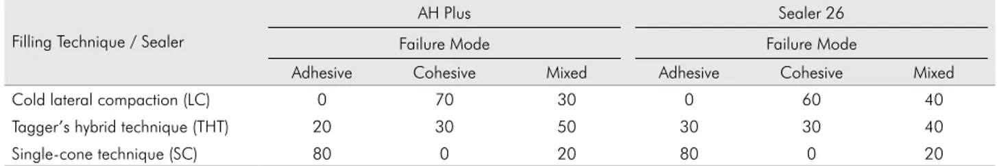
After completion of the push-out test, the specimens were examined with a stereomicroscope (Stemi 2000-C; Carl Zeiss, Jena, Germany) at 25 × magniication and the failure modes (adhesive, cohesive, or mixed) that occurred because of displacement of the sealer from the specimen were evaluated. Failure was considered adhesive if the sealer was totally separated from dentine (dentine surface without sealer), cohesive if the
fracture occurred within the sealer (dentine surface totally covered by the sealer), and mixed, when a combination of adhesive and cohesive modes (dentine surface partially covered by the sealer) occurred.
Results
When considering the root canal illing techniques, the bond strength (MPa) of LC (1.80 ± 0.98) (p < 0.05) was signiicantly higher than that of THT (1.16 ± 0.50) and SC (0.92 ± 0.25). Tukey’s test demonstrated that AH Plus/LC (2.26 ± 1.15) showed the highest values for the interaction between techniques and sealers and was statistically different from the others (p< 0.05). Sealer 26/LC (1.34 ± 0.42) showed intermediate values and was similar (p > 0.05) to AH Plus/THT
(1.32 ± 0.61), which in turn was similar to Sealer 26/THT (1.00 ± 0.27) (Table 1). The SC technique exhibited the lowest values for bond strength with AH Plus (0.48 ± 0.13) and Sealer 26 (0.30 ± 0.12), which were similar to each other (p > 0.05). The comparison between the root canal thirds revealed a signiicantly higher bond strength in the coronal third (1.45 ± 1.14) (p< 0.05) than in the middle (1.20 ± 0.72) and apical thirds (0.92 ± 0.81).
The analysis of the failure modes (Table 2) showed that, regardless of the sealer used, the specimens
Table 1. Bond strength mean values and standard deviations (in MPa) according to the filling technique and sealer used.
Filling technique Sealer Material Bond Strenght* Cold lateral compaction (LC) AH Plus 2.26 ± 1.15 a Sealer 26 1.34 ± 0.42 b Tagger’s hybrid technique (THT) AH Plus 1.32 ± 0.61 b Sealer 26 1.00 ± 0.27 b Single-cone technique (SC) AH Plus 0.48 ± 0.13 c Sealer 26 0.30 ± 0.12c * Different letters indicate statistical significance (p < 0.05).
Table 2. Distribution of failure modes (%) after the push-out test for each type of root canal filling technique/sealer.
Filling Technique / Sealer
AH Plus Sealer 26
Failure Mode Failure Mode
Adhesive Cohesive Mixed Adhesive Cohesive Mixed
Cold lateral compaction (LC) 0 70 30 0 60 40
Tagger’s hybrid technique (THT) 20 30 50 30 30 40
illed by LC predominantly demonstrated cohesive failure; specimens illed by THT and SC showed mixed failure and adhesive failure, respectively.
Discussion
During obturation, the sealer penetrates into the dentinal tubules and gives rise to a mechanical interlocking between the sealer and dentin.13 The
ability of root canal sealers to penetrate to dentinal tubules is related to their physical and/or chemical properties.14,15 Therefore, various techniques have
been studied in order to identify the optimal root canal obturation system, of which LC, SC, and THT present good standards for clinical use.
The bond strength of the filling material was evaluated using the push-out test. This test represents a reliable and reproducible method that simulates clinical conditions, i.e., the failure occurs parallel to the dentin/material interface.16,17 In the present study,
to evaluate the material dislocation resistance, three points with different diameters were used. The point’s diameter was slightly smaller than each root canal third slice, which allowed the point to be itted to the illing material, leading to more accurate results. The epoxy-based sealers used here were previously shown to have satisfactory physicochemical properties15,18,19
and root canal bond strength.20
In the present study, cold lateral compaction had the highest bond strength. These results could be related to the pressure provided by the spreaders over the master and accessory cones, which can create forces in lateral and apical directions and can favor the sealer interlocking with the dentin irregularities and/or tubules. Alternatively, the single-cone technique probably generates forces mainly in the apical direction. Corroborating this hypothesis, this technique had the lowest bond strength values. Furthermore, the root canal anatomy variations can overlap the iles’ shaping ability, which can negatively affect the cone adaptation.5 However,
the evaluation of this condition wasn’t included in our aims and should be further evaluated with different methods.
The thermoplasticized GP cones negatively affected the root canal bond strength of epoxy-based sealers. The lowability and longer polymerization
time of epoxy resin-based sealers allow them to penetrate deeper into the irregularities,1,6,8 thus
enhancing the mechanical interlocking between the sealer and dentine.11 However, our results showed a
signiicantly lower dentin bond strength with Sealer 26. These results could be related to the sealer’s composition. Sealer 26 has calcium hydroxide, which reduces its lowability21 and the capacity to
ill dentin irregularities,22 and the bond strength as
shown in the present study. Differently, AH Plus has a hard polymeric chain formed by diepoxy molecules and polyamines,18,19which increases its
bond strength to the radicular dentin even upon cold lateral compaction.
Regardless of the sealer type and properties, thermoplastic illing techniques allow the illing of irregularities in the root canal wall with minimal amount of sealer mixed in the illing material.6,23
However, the frictional heat generated during THT alters the physicochemical properties of epoxy-based sealers and resin organic matrix,10,24 thus, negatively
affecting the bond strength of the illing material to the radicular dentin. Additionally, GP cones may cool faster than the sealer, creating gaps in the illing mass and union interface.8 This may have contributed to
the results, unlike what occurred in the LC technique where there is no heating of the illing mass.3
The root thirds may differ structurally with respect to canal geometry and collagen/mineral content.25 In
this study, a higher bond strength was observed in the cervical third than in the middle or apical thirds, regardless of the illing technique and/or sealer used. This could be associated with the higher sealer volume and/or penetration into the less mineralized cervical dentin.25 Our results also showed a direct relation
between the illing technique and the sealer bond strength. Further studies are necessary to elucidate the effects of root canal illing techniques on sealer penetration and its relation to dentin bond strength.
Conclusion
1. Marciano MA, Bramante CM, Duarte MAH, Delgado RJR, Ordinola-Zapata R, Garcia RB. Evaluation of single root canals filled using the lateral compaction, Tagger’s Hybrid, Microseal and Gutta-flow techniques. Braz Dent J. 2010;21(5):411-5. doi:10.1590/S0103-64402010000500006
2. Kumar RV, Shruthi C. Evaluation of the sealing ability of resin cement used as a root canal sealer: an in vitro study. J Conserv Dent. 2012;15(3):274-7. doi:10.4103/0972-0707.97958 3. Gade VJ, Belsare LD, Patil S, Bhede R, Gade JR. Evaluation of
push-out bond strength of endosequence BC sealer with lateral condensation and thermoplasticized technique: an in vitro study. J Conserv Dent. 2015;18(2):124-7. doi:10.4103/0972-0707.153075 4. Levitan ME, Himel VT, Luckey JB. The effect of insertion
rates on fill length and adaptation of a thermoplasticized gutta-percha technique. J Endod. 2003;29(8):505-508. doi:10.1097/00004770-200308000-00004
5. Gordon MPJ, Love RM, Chandler NP. An evaluation of 0.06 tapered gutta-percha cones for filling of 0.06 taper prepared curved root canals. Int Endod J. 2005 Feb;38(2):87-96. doi:10.1111/j.1365-2591.2004.00903.x
6. Tagger M, Tamse A, Katz A, Korzen BH. Evaluation of the apical seal produced by a hybrid root canal filling method, combining lateral condensation and thermatic compaction. J Endod. 1984;10(7):299-303. doi:10.1016/S0099-2399(84)80183-1 7. Kardon BP, Kuttler S, Hardigan P, Dom SO. An in vitro
evaluation of the sealing ability of a new root-canal obturation system. J Endod. 2003;29(10):658-61. doi:10.1097/00004770-200310000-00011
8. Carneiro SMBS, Sousa-Neto MD, Rached-Júnior FJA, Miranda CES, Silva SRC, Silva-Sousa YTC. Push-out strength of root fillings with or without thermomechanical compaction. Int Endod J. 2012;45(9):821-8. doi: 10.1111/j.1365-2591.2012.02039.x 9. Lacey S, Pitt Ford TR, Yuan X-F, Sherriff M, Watson T. The
effect of temperature on viscosity of root canal sealers. Int Endod J. 2006;39(11):860-6. doi:10.1111/j.1365-2591.2006.01154.x 10. Viapiana R, Baluci CA, Tanomaru-Filho M, Camilleri
J. Investigation of chemical changes in sealers during application of the warm vertical compaction technique. Int Endod J. 2015;48(1):16-27. doi:10.1111/iej.12271
11. Rached-Júnior FJA, Souza-Gabriel AE, Alfredo E, Miranda CES, Silva-Sousa YTC, Sousa-Neto MD. Bond strength of Epiphany sealer prepared with resinous solvent. J Endod. 2009;35(2):251-5. doi:10.1016/j.joen.2008.10.027
12. Rached-Júnior FJA, Sousa-Neto MD, Souza-Gabriel AE, Duarte MAH, Silva-Sousa YTC. Impact of remaining zinc oxide-eugenol-based sealer on the bond strength of a resinous sealer to dentine after root canal retreatment. Int Endod J. 2014;47(5):463-9. doi:10.1111/iej.12170
13. Haragushiku GA, Sousa-Neto MD, Silva-Sousa YTC, Alfredo E, Silva SC, Silva RG. Adhesion of endodontic sealers to human root dentine submitted to different surface treatments. Photomed Laser Surg. 2010;28(3):405-10. doi:10.1089/pho.2008.2474
14. Marin-Bauza GA, Rached-Júnior FJA, Souza-Gabriel AE, Sousa-Neto MD, Miranda CES, Silva-Sousa YTC. Physicochemical properties of methacrylate resin-based root canal sealers.J Endod. 2010;36(9):1531-6. doi:10.1016/j.joen.2010.05.002
15. Flores DSH, Rached-Júnior FJA, Versiani MA, Guedes DFC, Sousa-Neto MD, Pécora JD. Evaluation of physicochemical properties of four root canal sealers. Int Endod J. 2011;44(2):126-35. doi:10.1111/j.1365-2591.2010.01815.x 16. Gesi A, Raffaelli O, Goracci C, Pashley DH, Tay FR, Ferrari
M. Interfacial strength of Resilon and gutta-percha to intraradicular dentin. J Endod. 2005;31(11):809-13. doi:10.1097/01.don.0000158230.15853.b7
17. Costa JA, Rached-Júnior FJA, Souza-Gabriel AE, Silva-Sousa YTC, Sousa-Neto MD. Push-out strength of methacrylate resin-based sealers to root canal walls. Int Endod J. 2010;43(8):698-706. doi:10.1111/j.1365-2591.2010.01766.x 18. Resende LM, Rached-Junior FJA, Versiani MA, Souza-Gabriel
AE, Miranda CE, Silva-Sousa YTC, et al. A comparative study of physicochemical properties of AH Plus, Epiphany, and Epiphany SE root canal sealers. Int Endod J. 2009;42(9):785-93. doi:10.1111/j.1365-2591.2009.01584.x
19. Borges RP, Sousa-Neto MD, Versiani MA, Rached-Júnior FJA, De-Deus G, Miranda CES, et al. Changes in the surface of four calcium silicate-containing endodontic materials and an epoxy resin-based sealer after a solubility test. Int Endod J. 2012;45(5):419-8. doi: 10.1111/j.1365-2591.2011.01992.x 20. Vilanova WV, Carvalho-Junior JR, Alfredo E, Sousa-Neto MD,
Silva-Sousa YTC. Effect of intracanal irrigants on the bond strength of epoxy resin-based and methacrylate resin-based sealers to root canal walls. Int Endod J. 2012;45(1):42-8. doi:10.1111/j.1365-2591.2011.01945.x
21. Bernardes RA, Campelo AA, Silva Junior DS, Pereira LO, Duarte MAH, Moraes IG, et al. Evaluation of the flow rate of 3 endodontic sealers: Sealer 26, AH Plus, and MTA Obtura. Oral Surg Oral Med Oral Pathol Oral Radiol Endod. 2010;109(1):e47-9. doi:10.1016/j.tripleo.2009.08.038
22. Wu MK, Fan B, Wesselink PR. Leakage along apical root fillings in curved root canals. Part I: effects of apical transportation on seal of root fillings. Endod. 2000;26(4):210-6. doi:10.1097/00004770-200004000-00003
23. Gambarini G, Tagger M. Sealing ability of a new hydroxyapatite-containing endodontic sealer using lateral condensation and thermatic compaction of gutta-percha, in vitro. J Endod. 1996;22(4):165-7. doi:10.1016/S0099-2399(96)80093-8
24. Rose N, Le Bras M, Bourbigot S, Delobel R. Thermal oxidative degradation of epoxy resins: evaluation of their heat resistance using invariant kinetic parameters. Polym Degrad Stabil. 1994;45(3):387-97. doi:10.1016/0141-3910(94)90209-7 25. Mjör IA, Smith MR, Ferrari M, Mannocci F. The structure