Fernanda Cristina Mendes de Santana GIONGO(a)
Bruna MUA(a)
Clarissa Cavalcanti Fatturi PAROLO(a)
Anette CARLÉN(b)
Marisa MALTZ(a)
(a)Department of Social and Preventive
Dentistry, Faculty of Dentistry, Universidade Federal do Rio Grande do Sul – UFRGS, Porto Alegre, RS, Brazil.
(b)Department of Oral Microbiology and
Immunology, Institute of Odontology, University of Gothenburg, Gothenburg, Sweden.
Effects of lactose-containing stevioside
sweeteners on dental biofilm
acidogenicity
Abstract: The aim of this study was to evaluate the effect of a com-mercial lactose-containing stevioside sweetener on biofilm acido-genicity in vivo. Nine volunteers refrained from brushing their teeth for 3 days in five phases. On the 4th day of each phase, the pH of the biofilm was measured by the “Strip method”. Interproximal plaque pH was measured before and up to 60 minutes after a 10 mL mouthrinse for 1 minute with the test solutions: I - sweetener with 93% lactose and 7% stevioside; II - sweetener with 6.8% saccharin, 13.6% cyclamate, and 0.82% stevioside; III - 18% sucrose solution (positive control); IV - mineral water (negative control); and V- 93% lactose solution. The results revealed that the most pronounced pH fall was found with sucrose (positive control), followed by the 93% lactose solution, the sweetener with lactose + stevioside, the sweet-ener with saccharin + cyclamate + stevioside, and finally water (negative control). According to the area under the curve, the two sweeteners containing stevioside were significantly different, and the sweetener with lactose + stevioside was significantly different from water but not from sucrose. The critical pH for dentin
demin-eralization (pH ≤ 6.5) was reached by all volunteers after rinsing
with sucrose solution, lactose solution, and the stevioside + lactose sweetener. Analysis of the data suggests that lactose-containing stevioside sweeteners may be cariogenic, especially to dentin.
Keywords: Cariogenic Agents; Sweetening Agents; Dental Plaque.
Introduction
Sucrose is considered the most cariogenic carbohydrate.1 Searches for alternatives to sucrose have resulted in the development of artii -cial sweeteners, many of which are considered safe for teeth, such as aspartame, saccharin, cyclamate, xylitol, and manitol.2,3,4,5 These sweet-eners have been used as sugar substitutes for caries-active patients. Non-cariogenic natural products of plant origin have been discovered and accepted for general use to sweeten foods and beverages. Stevio-side, a natural sweetener extracted from Stevia rebaudiana, offers par-ticular advantages over other non-caloric sucrose substitutes in being heat-stable, resistant to acid hydrolysis, and non-fermentable.6 Addi-tionally, a caries-preventive action of stevioside extract can be related
Declaration of Interests: The authors certify that they have no commercial or associative interest that represents a conflict of interest in connection with the manuscript.
Corresponding Author: Marisa Maltz
E-mail: [email protected]
DOI: 10.1590/1807-3107BOR-2014.vol28.0026 Epub XXX XX, 2014
Submitted: Nov 19, 2013
to its antimicrobial properties. In vitro studies have
shown reduced numbers of bioilm-viable cells and
polysaccharide formation.7,8,9,10
However, commercial products contain several
components and formulations that might inluence
their cariogenicity. These sugars and their deriva-tives have been added to sweeteners as excipient agents. There is evidence that saccharin, cyclamate, and xylitol are not cariogenic,11 while lactose shows some cariogenic properties.12 Commercial products containing lactose plus aspartame show decreased surface microhardness similar to that with lactose solution.13 A recent in vitro study also showed that a commercial aspartame sweetener containing
lac-tose promotes signiicantly higher bioilm acido -genicity and higher counts of viable S. mutans cells than a commercial stevioside sweetener containing lactose.9 The antimicrobial effect of stevioside could inluence the cariogenic properties of commercial
sweetening products. There is no in vivo study ana-lyzing the cariogenic potential of commercial stevio-side products containing lactose. Therefore, the aim of this study was to evaluate the effect of commercial
stevioside sweeteners on dental bioilm acidogenic -ity. Our hypothesis was that commercial sweeten-ers containing lactose plus stevioside cause less pH drop than with lactose solution.
Methodology
Study population
Nine dentists or dental students from the Uni-versidade Federal do Rio Grande do Sul (ages 20-31 yrs) participated in the study. Inclusion criteria were that they were in good general and dental health and had not used antibiotics for the 2 months preceding the beginning of the study. The protocol for the current study was approved by the Ethics Committee of the Faculty of Dentistry from the Universidade Federal do Rio Grande do Sul (process n˚ 300/08). Informed and
written consent was obtained from all individuals.
Study design
The experiment involved a randomized
cross-over, double-blind design performed in 5 phases. All
participants and the examiner were blinded to the rinses used in each phase. Both crossover and
blind-ing procedures were performed by an operator not involved in the experimental protocol. The rinsing solutions were: (I) sweetener containing 93% lactose, 7% stevioside, and silicon dioxide as an anti-wetting agent (SóStevia, Lowçucar®, Marialva, Brazil), dis-solved in deionized distilled water at the ratio of 0.8
g (content of a packet) per 50 mL; (II) sweetener con -taining 6.8% saccharin, 13.6% cyclamate, and 0.82% stevioside, water, and sodium benzoate preservative (Steva Plus, Lowçucar®, Marialva, Brasil), dissolved in deionized distilled water at the ratio of 6 drops
per 50 mL; (III) 18% sucrose (positive control), with 2 teaspoons of sugar dissolved in 50 mL deionized
water; (IV) mineral water (negative control); and (V)
93% lactose, which corresponds to 46.5 g of lactose diluted in 50 mL of water. These ratios correspond
to a sweetness power of two teaspoons of sucrose
used to sweeten a volume of 50 mL (Brazilian small
cup of coffee). All participants had refrained from brushing their posterior teeth for 3 days before the experimental rinsing (solutions I to V) but main-tained normal oral hygiene for anterior teeth with a conventional dentifrice (> 1000 ppm F). The partici-pants did not receive dietary instructions, since the study followed a crossover design, and all partici-pated in all steps. The participants were instructed not to eat or drink anything at least 1 hour prior to each test session (4th day). A 7-day washout period was performed. During the washout periods, the participants brushed their teeth with a conventional dentifrice (> 1000 ppm F). They were not instructed about brushing frequency during the washout period and before the experimental rinsing.
Calibration
One examiner performed all pH measurements. Intra-examiner calibration of the pH indicator strips was performed with standard solutions. The pH val-ues corresponding to the pH scale of the strips (pH
4.0, 5.0, 6.0, 7.0) were prepared with an ion-speciic
The measurements were performed blinded (pre-coded solutions).
Test session and plaque pH measurement
At the 4th day, the plaque pH and its response to the respective treatment solutions were determined. The pH measurement was performed with the new so-called “Strip method”14 based on the usage of pH indicator strips (Merck®, Darmstadt, Germany – pH
4.0-7.0). Briely, the strips were cut into 4 slices (± 2
mm in width) and inserted into approximal sites dur-ing 10 s. The pH value (one decimal) was assessed by comparison of the color of the strip with the color index guide supplied by the manufacturer. The rest-ing pH (0 minute) and the pH curve were measured at 2 approximal sites, between the 2nd pre-molar and the 1st molar on the right and left sides of the upper jaw.15 Plaque pH was measured at baseline (0
min-ute) and at 5, 15, 20, 30, and 60 minutes. Rinsing with solution (10 mL) was performed during the irst min -ute. Relative isolation was performed to avoid saliva bias in the plaque pH.
Statistical analyses
Intra-examiner ability to interpret the pH indica-tor strips was analyzed by the Intra-Class Correlation
Coeficient. The mean pH values of the two approxi
-mal sites (right and left sides) were calculated. The mean pH curves, resting pH, minimum pH, and time-point of maximum pH decrease were also calculated.
The areas under the curve for pH 6.5 (AUC 6.5) were determined with the UTHSCA Image Tool computer program(UTHSCSA©, San Antonio, USA). Data from
the time-point of maximum pH decrease and AUC6.5 were analyzed by the Kruskal-Wallis test, followed
by the Mann-Whitney test. Data from resting, inal,
and minimum pH were analyzed by ANOVA. The
signiicance limit was set at 5%. The analyses were
performed with the statistical package SPSS 17.0 for Windows (SPSS Inc., Chicago, USA). Power analysis was calculated with the Web site www.openepi.com,
and the results of area under the curve at pH 6.5 were
compared between the two sweeteners.
Results
Intra-examiner ability to reveal the pH of the stan-dard solutions by means of the pH indicator strips was
very good (Intra-Class Correlation Coeficient 0.7). The mean pH curves of the dental bioilm and the
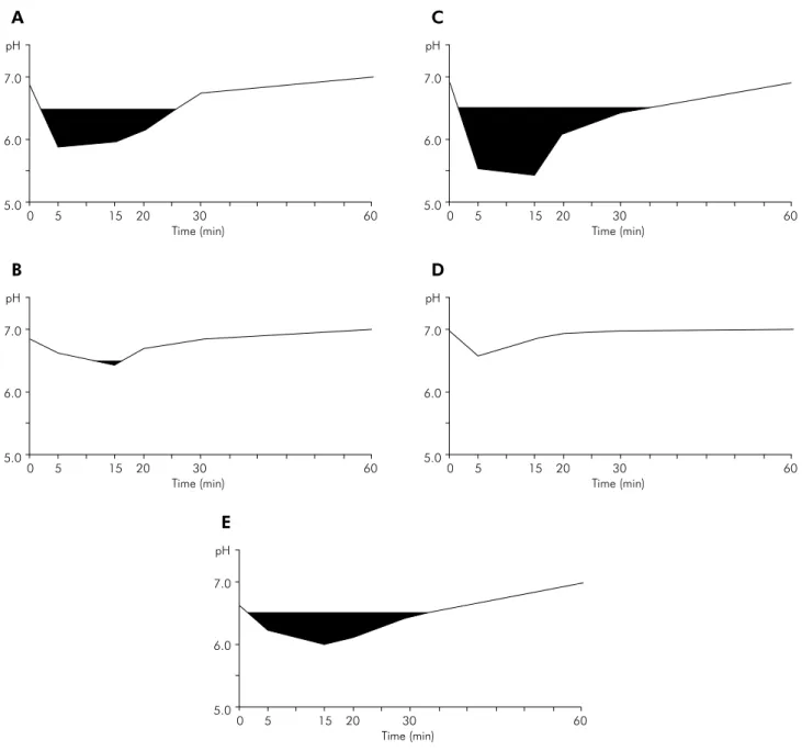
AUC6.5 for the 9 volunteers, obtained with the 5 test solutions, are shown in Figure 1. The most pronounced pH fall was found with sucrose (positive control), followed by the lactose solution, the sweetener with lactose + stevioside, the sweetener with saccharin
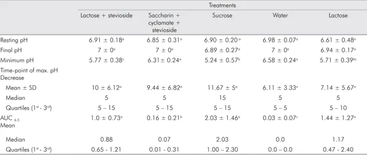
Table 1. Mean ± SD value of resting pH, final pH, minimum pH, time-point of maximum pH decrease, and areas under the curve for pH 6.5 (AUC 6.5) at the approximal sites in 9 individuals.
Treatments Lactose + stevioside Saccharin +
cyclamate + stevioside
Sucrose Water Lactose
Resting pH 6.91 ± 0.18a 6.85 ± 0.31a 6.90 ± 0.20 a 6.98 ± 0.07a 6.61 ± 0.48a
Final pH 7 ± 0a 7 ± 0a 6.89 ± 0.27a 7 ± 0a 6.94 ± 0.17a
Minimum pH 5.77 ± 0.38c 6.31± 0.24a 5.24 ± 0.57b 6.58 ± 0.24a 5.71 ± 0.39bc
Time-point of max. pH Decrease
Mean ± SD 10 ± 6.12a 9.44 ± 6.82a 11.67 ± 5a 6.11 ± 3.33a 7.14 ± 5.67a
Median 5 5 15 5 5
Quartiles (1st - 3rd) 5 – 15 5 – 15 5 – 15 5 – 5 5 – 10
AUC 6.5
Mean
1.0 ± 0.73a 0.16 ± 0.21b 2.03 ± 1.46a 0.03 ± 0.07c 1.44 ± 1.27a
Median 0.88 0.07 2.03 0.0 1.17
Quartiles (1st - 3rd) 0.65 - 1.21 0.01 - 0.31 1.00 – 2.30 0.0 – 0.0 0.47 - 2.40
+ cyclamate + stevioside, and, inally, water (nega -tive control). Accordingly, the area under the curve (AUC6.5) showed the greatest value with sucrose and the smallest with water (p < 0.001; AUC6.5,) (Table 1). The AUC6.5 values for the two sweeteners containing
stevioside were signiicantly different (p = 0.005), and
the sweetener with saccharin + cyclamate +
stevio-side was also signiicantly different from both lactose
(p = 0.037) and sucrose (p = 0.001). The AUC6.5 for the sweetener with lactose + stevioside was not differ-ent from the AUC6,5 of sucrose (p = 0.145) and lactose (p = 0.626). Water was signiicantly different from
all other groups (p < 0.05). The statistical power for
comparison of the results of the area under the curve
at pH 6.5 between the two sweeteners was 91.27%.
The mean time-point of maximal pH fall varied from 6.11 to 11.67 minutes among the test solutions. For all solutions, pH had returned to the baseline level after 60 minutes (Table 1).
The majority of the volunteers showed a
pro-nounced pH fall (pH ≤ 5.5) after the sucrose rinse.
Two of them presented pH falls below the critical pH for enamel demineralization after rinsing with the stevioside + lactose sweetener, and three showed similar pH falls after rinsing with the lactose
sweet-ener. No drop to pH ≤ 5.5 was observed with the ste -vioside + saccharin + cyclamate sweetener. Regard-ing pH drop equal to or lower than pH 6, only one participant reached these values after rinsing with saccharin + cyclamate + stevioside, while almost all participants reached these values after rinsing with lactose and lactose + stevioside.
Discussion
In the present study, it was found that commer-cial stevioside sweeteners containing lactose as the excipient agent showed an AUC6.5 greater than that of those containing non-fermentable sweeteners such as saccharin and cyclamate. The mean AUC6.5 value from the stevioside sweeteners containing lactose was similar to that reached after participants rinsed with 18% sucrose and 93% lactose solutions. These
indings reject our hypothesis, showing that the com -mercial sweetener containing lactose plus stevioside does not cause less pH drop than the lactose solution.
The pH measurements were performed by the newly introduced “strip method” for plaque-pH examina-tions. This method has been shown to give approxi-mal plaque-pH values equal to those obtained with the microtouch method in individuals after rinsing with 10% sucrose.14 The fact that both the strip method and the microtouch method disrupt the bioilm and may remove some bioilm from the interproximal
site could be a limitation. However, Lingström et al.15 evaluated plaque pH measurements by the telemetric method, which uses a pH electrode under an
undis-rupted bioilm, and by the microtouch method and
found a similar trend in the pH curves between the
two methods. To minimize possible bioilm disruption,
pH measurements were limited to 6 time-points. The pH strip used in this study (pH 4.0 to 7.0) has a smaller color shift for pH intervals > 6 than in the pH 4 to 6 intervals.14 This could result in a less precise assess-ment for higher pH values. However, in this study,
the cutoff point for AUC analysis was 6.5 (Figure 1).
Stevioside is heat-stable, resistant to acid hydroly-sis, and non-fermentable for oral bacteria.16 Therefore, it is considered to be a safe product for teeth.6 How-ever, due to the fact that this intense sweetener is ≥
300 times sweeter than sucrose, it has been used in very small quantities and mixed with other sweeten-ers or their derivatives in foods.11 In the present study, the commercial product containing stevioside mixed
with the artiicial substitutes saccharin and cycla -mate showed pH falls almost similar to those with
the negative control (water). The small but signiicant
difference observed in the AUC between these two solutions could be due to other components present in the commercial formulation, such as the preserva-tive sodium benzoate, which has bacteriostatic and fungistatic properties under acidic conditions.11,17
Lactose is a fermentable carbohydrate, although it is less cariogenic than sucrose.2,13,18 Our indings conirmed that sucrose can cause pronounced pH
drops in the dental plaque,19 reaching the critical pH for both dentin (6.5) and enamel (5.5). Similar
results have been observed in a study with a lactose-containing nitroglycerin tablet that also produced a marked pH drop in dental plaque.18
and demineralize dentin in situ.13 This is consis-tent with our findings of a more pronounced pH fall (AUC6.5) for the lactose-containing sweetener compared with that for the stevioside sweetener containing saccharin and cyclamate. The stevio-side extract present in the commercial steviostevio-side sweetener containing lactose was unable to pro-mote reduced biofilm acidogenicity after a 1-min-ute rise in vivo, since no difference in pH drop was found between this commercial sweetener and the
93% lactose solution. A previous in vitro study has also shown that a commercial stevioside sweet-ener containing lactose promoted significantly less biofilm acidogenicity and fewer viable cells than a 10% sucrose solution.9 However, the pres-ent in vivo study found that the commercial ste-vioside sweetener containing lactose promoted dental biofilm acidogenicity (AUC6.5) similar to that of the 18% sucrose solution, despite differ-ences in the minimum pH values.
Figure 1. Mean pH drop of the following solutions: (A) sweetener with 93% lactose and 7% stevioside; (B) sweetener with 6.8% saccharin, 13.6% cyclamate, and 0.82% stevioside; (C) 18% sucrose solution; (D) water; and (E) 93% lactose solution. Dark areas represent AUC6,5.
Time (min) pH
7.0
6.0
5.0
0 5 15 20 30 60
A
Time (min) pH
7.0
6.0
5.0
0 5 15 20 30 60
D
Time (min) pH
7.0
6.0
5.0
0 5 15 20 30 60
B
Time (min) pH
7.0
6.0
5.0
0 5 15 20 30 60
C
Time (min) pH
7.0
6.0
5.0
0 5 15 20 30 60
Conclusion
Analysis of the data showed that the cario-genic potential of different commercial stevioside sweeteners differs. The commercial stevioside sweetener containing 93% lactose may be cario-genic, especially to dentin.
Acknowledgements
The authors are not professionally afiliated with
any corporation. We thank Lowçucar®, the sweet-ener company, for product donations, the Univer-sidade Federal do Rio Grande do Sul (UFRGS), and all volunteers for their dedication.
1. Zero DT. Sugars - the arch criminal?. Caries Res. 2004
May-Jun;38(3):277-85.
2. Frostell G. Effects of mouth rinses with sucrose, glucose, fructose, lactose, sorbitol and Lycasin on the pH of dental plaque. Odontol Revy. 1973 Jul-Sep;24(3):217-26.
3. Scheinin A, Mäkinen KK, Tammisalo E, Rekola M. Turku sugar studies XVIII. Incidence of dental caries in relation to 1-year consumption of xylitol chewing gum. Acta Odontol
Scand. 1975 Sep-Oct;33(5):269-78.
4. Loesche WJ. The rationale for caries prevention through the
use of sugar substitutes. Int Dent J. 1985 Mar;35(1):1-8.
5. Park KK, Schemehorn BR, Stookey GK, Butchko HH, Sanders
PG. Acidogenicity of high-intensity sweeteners and polyols.
Am J Dent. 1995 Feb;8(1):23-6.
6. Das S, Das AK, Murphy RA, Punwani IC, Nasution MP, King-horn AD. Evaluation of the cariogenic potential of the intense natural sweeteners stevioside and rebaudioside A. Caries
Res. 1992 Sep-Oct;26(5):363-6.
7. Mohammadi-Sichani M, Karbasizadeh V, Aghai F, Mofid MR. Effect of different extracts ofStevia rebaudiana leaves on Streptococcus mutans growth. J Med Plants Res. 2012 Aug;6(32):4731-4.
8. Brambilla E, Cagetti MG, Ionescu A, Campus G, Lingström P. An in vitro and in vivo comparison of the effect of Stevia rebaudiana extracts on different caries-related variables: a randomized controlled trial pilot study. Caries Res. 2014 Jan;48(1):19-23.
9. Giacaman RA, Campos P, Muñoz-Sandoval C, Castro RJ. Cariogenic potential of commercial sweeteners in an ex-perimental biofilm caries model on enamel. Arch Oral Biol.
2013 Sep;58(9):1116-22.
References
10. Gamboa F, Chaves M. Antimicrobial potential of extracts from Stevia rebaudiana leaves against bacteria of impor-tance in dental caries. Acta Odontol Latinoam. 2012 May-
Aug;25(2):171-5.
11. Imfeld T. Efficacy of sweeteners and sugar substitutes in
caries prevention. Caries Res. 1993;27 Suppl 1:50-5.
12. Bowen WH, Pearson SK, VanWuyckhuyse BC, Tabak LA. Influence of milk, lactose-reduced milk, and lactose on
car-ies in desalivated rats. Carcar-ies Res. 1991 Jul-Aug;25(4):283-6.
13. Aires CP, Tabchoury CP, Del Bel Cury AA, Cury JA. Effect of a lactose-containing sweetener on root dentine demineraliza-tion in situ. Caries Res. 2002 May-Jun;36(3):167-9.
14. Carlén A, Hassan H, Lingström P. The ‘strip method’: a simple method for plaque pH assessment. Caries Res. 2010 Sep;44(4):341-4.
15. Lingström P, Birkhed D, Granfeldt Y, Björck I. pH
measure-ments of human dental plaque after consumption of starchy foods using the microtouch and the sampling method. Caries
Res. 1993 Sep-Oct;27(5):394-401.
16. Kinghorn AD, Soejarto DD. Intensely sweet compounds of
natural origin. Med Res Rev. 1989 Jan-Mar;9(1):91-115.
17. Bowen WH, Pearson SK. Residual effects of fluoride on a severe cariogenic challenge in rats. Caries Res. 1994
Jun-Aug;28(4):246-50.
18. Lingström P, Birkhed D. Effect of buccal administration of a lactose-containing nitroglycerin tablet (Suscard) on plaque pH. Scand J Dent Res. 1994 Dec;102(6):324-8.
19. Stephan RM. Two factors of possible importance in relation to the etiology and treatment of dental caries and other dental