287
ABSTRACT
BACKGROUND AND OBJECTIVES: Among electro agents
ultrasound is one of the most common, however, there is in-suicient evidence of the beneicial efects with the parameters currently used. he aim of this study was to compare the efect of continuous and pulsed ultrasound therapy on experimental hyperalgesia and edema in knees of Wistar rats.
METHODS: 18 rats were divided into three groups: CG- con-trol group; GUP - treated with pulsed ultrasound 50%; and GUC - continuous ultrasound. To accomplish the lesion, ani-mals were manually restrained and 100µL of 5% formalin solu-tion were injected into the right tibiofemoral space. For assess-ment of nociception digital Von Frey ilaassess-ment was used on the medial side of the joint, until clinching. Edema was evaluated with mid-lateral knee caliper. Assessments occurred in the pre-injury (EV1), after 15 (EV2), 30 (EV3) and 60 (EV4) minutes of the injury. After EV2, treatment was initiated with ultrasound with 0.4W/cm2 (SATA), pulsed or continuous.
RESULTS: he CG had hypernociception, with no return to baseline. GUP has returned to baseline as from EV3 and for continuous ultrasound in EV4. All three groups showed similar behavior for edema, with onset in EV2, without reduction. CONCLUSION: herapeutic ultrasound was efective to de-crease nociception, and the pulsed form showed early results, however, both forms of application had no efect on the forma-tion and maintenance of acute edema.
Keywords: Edema, Pain measurement, Ultrasound therapy.
Comparison of continuous and pulsed ultrasound therapy in knee
hyperalgesia of Wistar rats*
Comparação do ultrassom terapêutico contínuo e pulsado na hiperalgesia de joelho de ratos Wistar
Gladson Ricardo Flor Bertolini1, Josinéia Gresele Coradini2, Regina Inês Kunz2, Bruno Pogorzelski Rocha2, Lígia Inez da Silva3
*Received from State University of Western Paraná, Cascavel, PR, Brazil.
1. University of São Paulo, School of Medicine of Ribeirão Preto, Ribeirão Preto, SP, Brazil. 2. State University of Western Paraná, Department of Physiotherapy, Cascavel, PR, Brazil. 3. Federal University of Paraná, Department of Physiotherapy, Curitiba, PR, Brazil.
Submitted in September 16, 2014.
Accepted for publication in November 06, 2014. Conlict of interests: none – Sponsoring sources: none.
Correspondence to: Gladson Ricardo Flor Bertolini
Rua Universitária, 2069 – Jardim Universitário 85819-110 Cascavel, PR, Brasil.
E-mail: [email protected]
© Sociedade Brasileira para o Estudo da Dor
RESUMO
JUSTIFICATIVA E OBJETIVOS: Dentre os agentes eletrotér-micos o ultrassom é um dos mais comuns, contudo, há insuici-ente evidência dos efeitos benéicos com os parâmetros corrinsuici-ente- corrente-mente utilizados. Assim, o objetivo deste estudo foi comparar o efeito do ultrassom terapêutico contínuo e pulsado sobre hiperal-gesia e edema experimentais em joelhos de ratos Wistar. MÉTODOS: Foram utilizados 18 ratos, divididos em três gru-pos: GC - grupo controle; GUP - tratado com ultrassom pul-sado 50%; e, GUC - ultrassom contínuo. Para realizar a lesão, os animais foram contidos manualmente e 100µL de solução de formalina a 5% foram injetados no espaço tíbio-femoral direito. Para avaliação da nocicepção foi utilizado o ilamento de Von Frey digital, na face medial da articulação, até a retirada do mem-bro. A avaliação do edema foi realizada com paquimetria médio-lateral ao joelho. As avaliações ocorreram no momento pré-lesão (AV1), após 15 (AV2), 30 (AV3) e 60 (AV4) minutos da lesão. Após AV2, foi iniciado o tratamento com ultrassom com 0,4W/ cm² (SATA), de forma pulsada ou contínua.
RESULTADOS: Para GC, houve a presença de hipernocicep-ção, sem retorno aos valores basais. Para GUP houve retorno aos valores basais a partir de AV3 e para o ultrassom contínuo em AV4. Para o edema, os três grupos apresentaram comportamento semelhante, com formação em AV2, sem redução posterior. CONCLUSÃO: O ultrassom terapêutico mostrou-se eicaz para redução do quadro nociceptivo, sendo que a forma pulsada mostrou resultados precocemente ao contínuo, contudo, ambas as formas de aplicação não tiveram efeito sobre a formação e ma-nutenção do edema agudo.
Descritores:Edema, Mensuração da dor, Terapia por ultrassom.
INTRODUCTION
Among electrothermal agents used in physiotherapy, ultra-sound is one of the most common therapies and equipment is available and frequent in the clinical setting. However, in spite of its intensive use, review studies have shown that there is not enough evidence to support beneficial ultra-sound effects with current parameters used in the clinical practice1,2.
Anti-inlammatory efects are credited to this resource, which afterward would be responsible for analgesia, due to increased local temperature. However, important non-thermal efects
Rev Dor. São Paulo, 2014 oct-dec;15(4):287-9 ORIGINAL ARTICLE
288
Bertolini GR, Coradini JG, Kunz RI, Rocha BP and Silva LI Rev Dor. São Paulo, 2014 oct-dec;15(4):287-9
have been currently presented, which would be mediated by cell sodium and calcium concentrations, which would pro-duce direct analgesic actions by changing cell depolarization and activation thresholds, even being able to be the stimula-tion basis for local endogenous opioids release3.
Studies have contradictory results of therapeutic ultrasound in different types of injuries, having among studied variables pain, such as impingement syndromes4,5, ankle torsions6,
low back pain7,8 and knee osteoarthritis9. However, the
di-versity of both clinical and research parameters is extreme2,
thus being useful controlled experiments to test different ultrasound treatment parameters, such as different doses, frequencies and working cycles, as well as continuous and pulsed ultrasound, which have presented different results, even when the same mean temporal and spatial intensity (SATA) is used10.
So, this study aimed at evaluating and comparing the efects of therapeutic ultrasound in diferent presentations on hyper-algesia and edema induced by 5% formalin injection in the knees of Wistar rats.
METHODS
Eighteen Wistar rats, weighing 436.0±33.0g, were kept in polypropylene cages with free access to water and food, with controlled 12h light/dark cycle and controlled room tem-perature (24±1º C). Animals were randomly divided in three groups:
• CG – animals submitted to right knee hyperesthesia induc-tion and not treated;
• GUP – animals submitted to right knee hyperesthesia in-duction and treated with pulsed therapeutic ultrasound 5:5;
• GUC – animals submitted to right knee hyperesthesia in-duction and treated with continuous therapeutic ultrasound. Animals were manually restrained and 100µL of 5% formalin were injected in the right tibio-femoral joint space, aiming at inducing synovitis, with hyperesthesia and edema.
Nociception evaluation
Insight® Von Frey digital ilament was used to evaluate
noci-ception. Test was carried out with animals manually restrained and ilament applied to the medial face of right hind paw tibio-femoral joint. Filament polypropylene tip was applied perpendicularly to the area, with gradual pressure increase and test was interrupted at clinching for clinching threshold recording. Before test, animals were trained with the equip-ment, for three days, aiming at their familiarization. he day after the last training, clinching threshold values were col-lected in moments pre-injury (EV1), 15 (EV2), 30 (EV3) and 60 (EV4) minutes after chemical irritation.
Edema evaluation
Right knees diameter was evaluated with caliper positioned medio-laterally at the joint interline region. his evaluation was performed in moments similar to clinching threshold moments.
Treatment protocols
Treatment was started after the second evaluation, that is, 15 minutes after hyperalgesia induction. CG has not sufered any therapeutic intervention, just simulation. Treatment con-sisted in transcutaneous use of ultrasound (Ibramed®) with
frequency of 1MHz, 0.4V/cm2 (SATA), on the knee joint
in-terline, with slow and circular movements. GUP has received pulsed ultrasound (0.8W/cm2 – SATP), with modulation of
5ms on 5ms of, that is, 50% of active cycle; and GUC has received continuous ultrasound. After last evaluation, animals were euthanized.
Statistical analysis
Data normality was checked with Kolmogorov-Smirnov test. hen, data were analyzed by unidirectional ANOVA (for comparison among groups) and ANOVA with repeated mea-sures (comparison within groups). In all cases, signiicance level was 5%.
his study was carried out according to international animal experiment ethics rules, after approval by the Ethics Commit-tee for Animal Use, UNIOESTE, under opinion 4412/2012.
RESULTS
Results have shown that there has been hypernociception in GC without returning to baseline values, diferently from what was observed for treated groups. GUP has returned to baseline levels as from EV3 and GUC as from EV4 (Table 1). here have been no signiicant diferences when comparing among groups.
Table 1. Values in grams, obtained with digital Von Frey ilament, for the three groups, in different evaluation moments
EV1 EV2 EV3 EV4
CG 344.6±53.0a 246.0±38.6b 205.0±47.2bc 155.1±53.5c
GUP 304.9±87.1a 157.8±66.7b 182.2±38.8ab 257.9±64.1ab
GUC 354.6±108.3a 234.0±100.5b 202.3±55.9b 248.6±128.0ab
CG: control group; GUP: pulsed therapeutic ultrasound group; GUC: conti-nuous therapeutic ultrasound group.
Different small letters indicate signiicant differences within the same group.
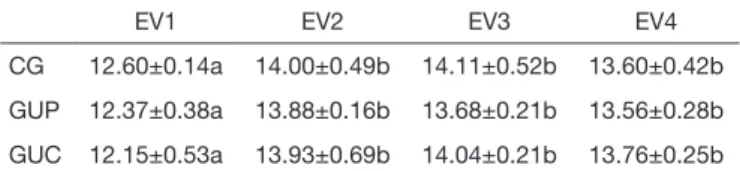
For edema, the three groups had similar behavior, with edema formation in EV2 without posterior reduction (Table 2).
Table 2. Values in millimeters, obtained with the caliper for the three groups in different evaluation moments.
EV1 EV2 EV3 EV4
CG 12.60±0.14a 14.00±0.49b 14.11±0.52b 13.60±0.42b
GUP 12.37±0.38a 13.88±0.16b 13.68±0.21b 13.56±0.28b
GUC 12.15±0.53a 13.93±0.69b 14.04±0.21b 13.76±0.25b
CG: control group; GUP: pulsed therapeutic ultrasound group; GUC: conti-nuous therapeutic ultrasound group.
289
Comparison of continuous and pulsed ultrasound therapy in knee hyperalgesia of Wistar rats
Rev Dor. São Paulo, 2014 oct-dec;15(4):287-9
DISCUSSION
Ultrasound is a very popular tool to treat musculoskeletal problems, however there is the need for further studies about its beneicial efects. A survey with 207 North-American physiotherapists specialized in orthopedic physiotherapy, has shown that it is primarily used for inlammatory processes, also aiming at decreasing pain. It was observed that 75% of answers have pointed to continuous ultrasound for pain re-lief, with just 17.1% using 50% pulse cycles, being that most doses were between 1 and 2W/cm2 2, in spite of hints
indicat-ing the usefulness of lower intensities for the treatment1, that
is, there is the need for studies evaluating lower intensities and diferent ultrasound release ways.
In our study, the irritation model has as major feature two nociceptive behavior stages separated by a quiescence stage around the ifth to the tenth minute after formalin injection11.
So, we decided to carry out the irst evaluation 15 minutes af-ter injury, avoiding evaluating in the quiescent period, with new evaluation 30 and 60 minutes after, aiming at observing nociceptive and edema behavior along 1h after injury. Evaluation with Von Frey ilament provides sensitive, objec-tive and quantiiable nocicepobjec-tive measures12, and edema
for-mation measurement using the caliper is also presented by the literature13. Considering that evaluators were experienced and
animals were trained before evaluations, presented data are re-liable, showing that there have been no changes in edema for-mation with the use of ultrasound, producing neither increase nor decrease, since it has been used during acute irritation stage. his is diferent from what has been observed by a study with tendon trauma in rats, where there has been increased edema evaluated 2 and 8h after trauma, with signiicant de-creases 24 hours later. hey also mention that nociception evaluated by clinching time, has shown hypernociception decrease 8 hours after for GUP and just 24 hours after for GUC14. Although being diferent times, pattern was similar
to our study where the two therapies used have produced no-ciceptive threshold decrease 20 minutes after for pulsed and 60 minutes after for continuous ultrasound.
In a diferent study using types of injury and evaluation simi-lar to our study, it was observed that for continuous ultra-sound animals had increased nociceptive threshold 2 hours after chemical irritation, which was changed in the group receiving naloxone before the injury, showing that a possible route for ultrasound-mediated analgesic efect could be the release of endogenous opioids after chemical irritation3. It is
also stressed that with parameters used, similar to the con-tinuous group in our study, thermal ultrasound efects may be discarded, that is, the so called non-thermal efects were responsible for changes in pain threshold in both studies. A diferent study, with evaluation, injury and ultrasound treatment similar to our study, aiming at evaluating
cumula-tive efects of low-level laser, has observed that isolated appli-cation of this resource was better than laser or the association of techniques15. In a previous study comparing pulsed and
continuous ultrasound, in animals submitted to experimental sciatica model, ultrasound was efective to decrease pain eval-uated by clinching time and, similarly to our study, pulsed ultrasound had faster analgesic results10. hat is, aiming at
analgesic efects, not only inal dose seems to be important but, since pulsed ultrasound had earlier positive results, tem-poral peak (SATP) may inluence the action of this resource and should be the focus of future investigations. Limitation of this study was the lack of inlammatory process molecular evaluation, which also suggests further studies.
CONCLUSION
herapeutic ultrasound was efective to decrease nociception and pulsed ultrasound had earlier results as compared to con-tinuous ultrasound; however, both types of applications had no efect on acute edema formation and maintenance.
REFERENCES
1. Warden SJ. A new direction for ultrasound therapy in sports medicine. Sports Med. 2003;33(2):95-107.
2. Wong RA, Schumann B, Townsend R, Phelps CA. A survey of therapeutic ultra-sound use by physical therapists who are orthopaedic certiied specialists. Phys her. 2007;87(8):986-94.
3. Martignano CC, Silva LI, Meireles A, Rocha BP, Rosa CT, Bertolini GR. Avaliação do ultrassom sobre a hiperalgesia e o edema em joelhos de rato Wistar e interferências de um inibidor de opioides endógenos. Fisioter Bras. 2013;14(4):289-93.
4. Yildirim MA, Ones K, Celik EC. Comparison of ultrasound therapy of various du-rations in the treatment of subacromial impingement syndrome. J Phys her Sci. 2013;25(9):1151-4.
5. Alexander LD, Gilman DR, Brown DR, Brown JL, Houghton PE. Exposure to low amounts of ultrasound energy does not improve soft tissue shoulder pathology: a sys-tematic review. Phys her. 2010;90(1):14-25.
6. van den Bekerom MP, van der Windt DA, Ter Riet G, van der Heijden GJ, Bou-ter LM. herapeutic ultrasound for acute ankle sprains. Eur J Phys Rehabil Med. 2012;48(2):325-34.
7. Fiore P, Panza F, Cassatella G, Russo A, Frisardi V, Solfrizzi V, et al. Short-term efects of high-intensity laser therapy versus ultrasound therapy in the treatment of low back pain: a randomized controlled trial. Eur J Phys Rehabil Med. 2011;47(3):367-73. 8. Licciardone JC, Minotti DE, Gatchel RJ, Kearns CM, Singh KP. Osteopathic manual
treatment and ultrasound therapy for chronic low back pain: a randomized controlled trial. Ann Fam Med. 2013;11(2):122-9.
9. Luksurapan W, Boonhong J. Efects of phonophoresis of piroxicam and ultrasound on symptomatic knee osteoarthritis. Arch Phys Med Rehabil. 2013;94(2):250-5. 10. Ciena AP, Cunha NB, Moesch J, Mallmann JS, Carvalho AR, Moura PJ de, et al.
Efeitos do ultrassom terapêutico em modelo experimental de ciatalgia. Rev Bras Med Esporte. 2009;15(6):424-7.
11. Martins MA, de Castro Bastos L, Tonussi CR. Formalin injection into knee joints of rats: pharmacologic characterization of a deep somatic nociceptive model. J Pain. 2006;7(2):100-7.
12. Vivancos GG, Verri WA Jr, Cunha TM, Schivo IR, Parada CA, Cunha FQ, et al. An electronic pressure-meter nociception paw test for rats. Braz J Med Biol Res. 2004;37(3):391-9.
13. Gould D, Yousaf N, Fatah R, Subang MC, Chernajovsky Y. Gene therapy with an improved doxycycline-regulated plasmid encoding a tumour necrosis factor-alpha inhibitor in experimental arthritis. Arthritis Res her. 2007;9(1):R7.
14. Bertolini GR, Silva TS, Ciena AP, Artifon EL. Comparação do ultrassom pulsado e contínuo no reparo tendíneo de ratos. Fisioter Pesq. 2012;19(3):242-7.