Avaliação de 100 pacientes com nefrite
lúpica acompanhados por dois anos
Analysis of 100 patients with lupus
nephritis followed up for 2 years
Ana Karla Guedes de Melo(1), Alessandra Barbosa Avelar(2), Flávia Kamy Marciel Maegawa(3), Branca Dias Batista de Souza(4)
Recebido em (Received on) 01/11/2007. Aprovado (Approved), após revisão, em 21/10/08. Declaramos a inexistência de conflitos de interesse (We declare no conflict of interest).
Trabalho desenvolvido no Departamento de Medicina da Irmandade da Santa Casa de Misericórdia de São Paulo - SP Study carried out at Department of Medicine of Irmandade da Santa Casa de Misericórdia de São Paulo-SP
1. Médica especialista em Reumatologia e pós-graduanda da Faculdade de Ciências Médicas da Santa Casa de Misericórdia de São Paulo-SP 1. Rheumatologist. Postgraduate student of Medical Sciences School at Santa Casa de Misericórdia de São Paulo-SP
2. Médica especialista em Reumatologia e pós-graduanda da Faculdade de Ciências Médicas da Santa Casa de Misericórdia de São Paulo-SP 2. Rheumatologist. Postgraduate student of Medical Sciences School at Santa Casa de Misericórdia de São Paulo-SP
3. Médica especialista em Reumatologia 3. Rheumatologist
4. Chefe do Serviço de Reumatologia da Irmandade da Santa Casa de Misericórdia de São Paulo e chefe da disciplina de Reumatologia da Faculdade de Ciências Médicas da Santa Casa de Misericórdia de São Paulo – SP
4. Chief of Rheumatology Department at Irmandade da Santa Casa de Misericórdia de São Paulo; Head of the Rheumatology Department of Medical Sciences School at Santa Casa de Misericórdia de São Paulo-SP
Endereço para correspondência (Correspondence to): Branca Dias Batista de Souza - Departamento de Medicina/Reumatologia (Department of Medicine/ Rheumatology). Rua Dr. Cesário Motta Júnior, 112 – Vila Buarque CEP (Zip Code): 01221-000 Tel/Fax: 55 (11) 2176-7281.
RESUMO
Objetivos: Determinar a frequência de remissão total e parcial no tra-tamento da nefrite lúpica aos 12 e 24 meses de seguimento. Comparar esses subgrupos aos 12 meses, correlacionando as variáveis renais iniciais com a resposta ao tratamento. Analisar e comparar os resultados terapêuticos do subgrupo com glomerulonefrite proliferativa difusa por correlação clínico-patológica (“classe IV clínica”) com aqueles de “classe IV histológica”, isto é, com biópsia renal comprovada pela Organização Mundial da Saúde. Material e métodos: Foram estudados 100 pacientes consecutivos com diagnóstico de lúpus eritematoso sis-têmico (LES) e nefrite, atendidos no Serviço de Reumatologia da Santa Casa de Misericórdia de São Paulo e acompanhados por dois anos. Os portadores de comorbidades que comprometem os rins foram excluí-dos. Foram analisadas as variáveis demográicas, clínicas, laboratoriais e o índice de atividade da doença (SLEDAI). Os pacientes com classe histológica III, IV ou V receberam corticosteroide e ciclofosfamida como tratamento de indução da nefrite lúpica e aqueles com classe II receberam apenas corticosteroide. Resultados: A idade média ao diagnóstico de LES foi de 24,71 + 10,14 anos, com predomínio do sexo feminino (88%). O SLEDAI calculado ao diagnóstico foi de 16,09 ± 6,48. Em relação às variáveis renais iniciais, a creatinina média foi de 1,02 + 0,49 mg/dL, a proteinúria de 24 horas média foi de 2,57 + 2,39 g e o anticorpo anti-dsDNA foi encontrado em 66% dos casos. Todos os pacientes receberam corticosteroide e 75% utilizaram a ciclofosfa-mida. Cinquenta e seis pacientes foram submetidos à biópsia renal. Os subtipos II e IV foram os mais prevalentes (33,9% e 32,2%,
respec-ABSTRACT
Este trabalho foi desenvolvido para demonstrar a expe-riência de um serviço terciário no tratamento da indução e manutenção da remissão da nefrite lúpica. Sabe-se que esse é um dos temas de maior relevância na Reumatologia, apesar do pequeno número de publicações sobre o assunto em nosso meio, somado às controvérsias quanto à indicação de biópsia renal nesses pacientes. Os objetivos principais deste estudo são determinar a frequência de remissão total e parcial da nefrite aos 12 e 24 meses de acompanhamento; comparar esses subgrupos aos 12 meses, correlacionando as variáveis renais iniciais com a resposta terapêutica e demonstrando a experiência de um serviço brasileiro no manejo das manifes-tações renais do LES. Outro objetivo é comparar os resultados terapêuticos dos subgrupos de pacientes com glomerulonefrite proliferativa difusa por correlação clínico-patológica (“classe IV clínica”) daqueles com “classe IV histológica”, ou seja, com biópsia renal.
MATERIAL E MÉTODOS
Trata-se de uma análise retrospectiva de prontuários de pa-cientes com diagnóstico de LES e nefrite, segundo os critérios
de classiicação do ACR de 1997, atendidos no Serviço de
Reumatologia da Santa Casa de Misericórdia de São Paulo,
no período de 1990 a 2003. A nefrite lúpica é deinida pela
presença de proteinúria persistente (> 0,5g nas 24 horas) ou maior que 3+ no EAS ou pela cilindrúria (cilindros hemáticos, tubulares, granulosos ou mistos).
INTRODUÇÃO
O lúpus eritematoso sistêmico (LES) é uma doença inlamató -ria crônica, de natureza autoimune, que pode afetar múltiplos sistemas orgânicos. O envolvimento renal no LES é uma causa
signiicativa de morbidade e mortalidade devido à possibilidade de progressão para insuiciência renal e a complicações rela -cionadas ao tratamento. Pelos critérios do Colégio Americano de Reumatologia (American College of Rheumatology, ACR
– 1997), a nefrite lúpica é deinida pela presença de proteinúria
persistente (> 0,5g nas 24 horas) ou maior que 3+ ou pela cilin-drúria (cilindros hemáticos, tubulares, granulosos ou mistos). Entre os padrões histológicos elaborados pela Organização Mundial da Saúde (OMS), a glomerulonefrite proliferativa difusa (classe IV) representa o subtipo mais prevalente e o de pior prognóstico.1 A nefrite constitui a principal causa de internações e mortalidade entre os pacientes com LES.2
Estudos randomizados anteriores demonstraram que o uso da ciclofosfamida para o tratamento da nefrite lúpica se correla-ciona com maiores taxas de preservação da função renal a longo prazo e com menor incidência de doença renal terminal.3
Existem controvérsias quanto à indicação de biópsia renal em todos os pacientes com LES e nefrite. Esdaile et al. estabe-leceram uma correlação entre os achados clínico-laboratoriais
e a classiicação histológica da nefrite lúpica, com o intuito de
orientar a conduta terapêutica e racionalizar a realização de procedimentos invasivos nessa população4 (Tabela 1). tivamente). Após 12 meses de acompanhamento, todos os pacientes apresentaram redução signiicativa da proteinúria de 24 horas, melhora do sedimento urinário e dos valores das frações do complemento (C3, C4, CH50). A frequência de remissão total aos 12 meses foi 72,7% e, aos 24 meses, 85,7% (p = 0,013). A remissão parcial ocorreu em 27,3% dos doentes aos 12 meses e em 14,3% aos 24 meses. O sexo masculino apresentou menor frequência de remissão total comparado ao feminino aos 12 meses de acompanhamento (45,5% versus 81,6%, p = 0,007). Dentre as diferentes variáveis estudadas, nenhuma se correlacionou com remissão total ou parcial aos 12 meses. O subgrupo “classe IV clínica” apresentou maior frequência de remissão total que o subgrupo “classe IV histológica”. Conclusão: Com o esquema terapêutico usado em nosso serviço, veriicou-se um excelente desfecho em dois anos. Não foram observadas correlações entre variáveis clínico-laboratoriais e remissão total ou parcial. O sexo masculino apresentou menores taxas de remissão total comparado ao feminino. Apesar do pequeno número de pacientes estudados e das controvérsias quanto à biópsia renal, a taxa de remissão total foi maior nos pacientes com “classe IV clínica” em relação àqueles com “classe IV histológica”.
Palavras-chave: lúpus eritematoso sistêmico, nefrite, remissão total, biópsia renal.
After 12 months, all the patients had signiicant reduction in the
24-hour urinary protein level, improvement in the urinary sediment and increase in the fractional values of the complement (C3, C4, CH50). The frequency of total remission at 12 months was 72.7% and, at 24 months, 85.7% (p =0.013). Partial remission occurred in 27.3% at 12 months and, in 14.3%, at 24 months. Male gender presented a lower rate of total remission compared with women at 12 months (45.5% versus 81.6%, p = 0.007). Among several factors studied, none was correlated with total or partial remission at 12 months. The subgroup “clinical class IV” presented a higher frequency of total remission than the subgroup “histological class IV”. Conclusion: Our therapeutic approach achieved an excellent outcome in two years. Correlations between clinical and laboratory variables and total or partial remission were not observed. Male gender presented a lower rate of total remission compared with women. Despite the small number of patients studied and the controversies of renal biopsy, the rate of total remission was higher in patients with “clinical class IV” than in those with “histological class IV”.
com hipertensão arterial no início da doença, com o anti-DNA positivo e com o uso de ciclofosfamida. Possíveis explicações para esses fatos discrepantes decorrem, principalmente, de diferenças metodológicas das séries: critérios de inclusão e seleção de pacientes, estudos retrospectivos, tempos de
acom-panhamento, tratamentos realizados, deinição dos desfechos
clínicos, tamanho da amostra, entre outros.
O sexo masculino apresentou menores taxas de remissão total aos 12 meses comparado ao feminino, o que poderia suge-rir pior prognóstico da nefrite lúpica entre os homens na nossa casuística. Esse dado difere da literatura em que se demonstra que o sexo do indivíduo não apresenta valor prognóstico na evolução da nefrite lúpica.16
Apesar do pequeno número de pacientes estudados e das controvérsias quanto à biópsia renal inicial, as taxas de remissão nos pacientes com “classe IV clínica” e “classe IV histológica” foram altas aos 12 meses. Um balanço entre os ganhos potenciais de informações que podem guiar um esque-ma terapêutico e as complicações inerentes ao procedimento invasivo sempre deve ser levado em consideração antes de indicar a biópsia renal. Estudos prévios demonstraram que parâmetros clínico-laboratoriais são capazes de predizer com acurácia o prognóstico da nefrite lúpica, sem a necessidade de biópsia em todos os casos com envolvimento renal.17,18 Já outros estudos advogam que os dados histológicos oferecem informações adicionais aos parâmetros laboratoriais e podem guiar, de maneira mais precisa, a escolha terapêutica.19 Como pode ser percebido, o tema permanece controverso. Na expe-riência do nosso serviço, a biópsia renal é individualizada e indicada, sobretudo, nas seguintes situações: dúvida quanto à correlação clínico-histológica, presença de síndrome nefrótica, ou em casos que não responderam à terapia de indução com ciclofosfamida. Pelo presente estudo, a frequência de remissão
total aos 12 meses foi signiicativamente mais alta nos pacien -tes com “classe IV clínica”, sem a necessidade de informações adicionais pelo exame histológico. Na casuística apresentada, as variáveis renais e do LES, bem como a dose cumulativa da ciclofosfamida não diferiram entre os pacientes com “classe IV histológica” ou “clínica”, isto é, com e sem biópsia renal.
Portanto, concluímos que, neste estudo, a realização de biópsia renal para a determinação do subtipo histológico não
inluenciou na resposta ao tratamento em pacientes com nefrite
lúpica acompanhados por dois anos. Os dados apresentados demonstraram que a correlação clínico-histológica pode ajudar na decisão terapêutica desses pacientes, reservando a biópsia renal apenas para situações especiais.
Analysis of 100 patients with lupus
nephritis followed up for 2 years
INTRODUCTION
Systemic lupus erythematosus (SLE) is an autoimmune chronic
inlammatory disease that may affect multiple systems. Renal involvement in SLE is a signiicant cause of morbidity and
mortality due to the possibility of progression to end stage renal disease (ESRD) and treatment-related complications. By the criteria of the American College of Rheumatology
(ACR – 1997), lupus nephritis is deined by the presence of
persistent proteinuria (> 0,5g in 24 hours), or higher than 3+, or by cylindruria (hematic, tubular, granular or mix cylinder). Among histological standards created by World Health Or-ganization (WHO), diffuse proliferative glomerulonephritis (class IV) represents the most prevalent and worst prognostic subtype.1 Nephritis is the main cause of hospitalization and mortality among SLE patients.2
Controlled trials have shown that the use of cyclophos-phamide for lupus nephritis treatment is correlated with hi-gher rates of long term renal function preservation and lower incidence of ESRD.3
There are controversies about indicating renal biopsy to every lupus patients with nephritis. Esdaile et al. established
a correlation between clinical and laboratory indings and histological classiication of lupus nephritis, in order to guide
therapeutic approach and rationalize the performance of inva-sive procedures in such population.4 (Table 1)
MATERIAL AND METHODS
Retrospective chart analysis was done for patients with SLE and nephritis who attended the Rheumatology Department at Santa Casa de Misericórdia de São Paulo from 1990 to 2003.
Lupus nephritis is deined according the ACR criteria as per -sistent proteinuria (> 0,5 g in 24 hours) or higher than 3+ or by cylindruria (hematic, tubular, granular or mix cylinders).
Patients who had been submitted to initial renal biopsy or
not have been included. Classiication of nephritis had been
clinically established according to Esdaile4 or by histopatho-logy. Renal biopsy was individually assigned based, mainly,
on the following situations: dificulty to establish a clinicopa -thological correlation (doubtful cases), presence of nephrotic syndrome and/or to support therapeutic decision.
Patients had been followed up for two years since nephritis was diagnosed and therapy started, based on clinicopatho-logical correlation or on renal biopsy data. Our therapeutic approach included high doses of corticosteroids (prednisone 1-2 mg/kg/day orally) in every patient or 1g/day doses of me-thylprednisolone intravenously, during 3-5 consecutive days, in more severe cases. Intravenous (IV) monthly cyclophospha-mide was used in 0.5 to 1 g/m2 doses of body surface area as a
irst-choice of immunosuppressive agent for induction of class
III, IV or V nephritis remission. Azathioprine or mycophenolate mofetil (MMF) were used for nephritis maintenance treatment after 6-9 months of cyclophosphamide IV, or in case of failure or intolerance to the approach described before. Patients with mesangial glomerulonephritis were treated only with high doses of glucocorticoid.
Therapeutic response was evaluated after 12 and 24 mon-ths. Boumpas & Balow (1998) criteria were used to classify
therapeutic response as complete, partial and no response.5
According to these authors, complete remission is deined
as stabilization or normalization of serum creatinine, urinary
sediment with less than ive erythrocytes per ield and 24-hour
proteinuria lower than 1.0 g for, at least, six months. Partial remission is characterized by stabilization or improvement of
serum creatinine, hematuria lower than ive cells per ield and
persistent 24-hour proteinuria reduction (if nephrotic, reduction higher than or equal to 50%, but with value lower than 3,0 g/24 hours; if non nephrotic, reduction higher than or equal to 50%, but with value higher than 1.0 g/24 hours). When presenting with renal function deterioration (excluding other causes, like sepsis, nephrotoxic drugs, renal vein thrombosis), proteinuria increment or reduction not meeting criteria above, patients were considered as no response.
Patients with comorbidities that compromise the kidneys were excluded from this study. The demographic, clinical and laboratory variables and the Systemic Lupus Erythematosus Disease Activity Index (SLEDAI) were analyzed. In order to
verify the inluence of different factors in nephritis prognosis,
patients were divided into groups of complete and partial re-mission, “clinical class IV” and “histological class IV”.
Data were expressed as mean ± standard deviation. The program used for statistical analysis was SPSS version 13.0. Comparison between means was performed using “t” Student test, and comparison between frequencies was done with Mc-Nemar and chi-square tests. When p < 0,05, difference was
considered signiicant.
This study had been previously approved by the Research Ethics Committee of the institution.
Table 1
Correlation between clinical and laboratory indings and histological classiication of lupus nephritis
Class II Class III Class IV Class V
Arterial hypertension None Rare Frequent Rare
Proteinuria (grams/24hours) < 1 < 2 1-20 3,5-20
Hematuria
Erythrocytes/per field) 5-15 5-15 > 15 None
Leucocyturia/leucocytes per field) 5-15 5-15 > 15 None
Creatinine (mg/dl) Normal Normal or
incremented Frequently incremented Normal
Glomerular filtration rate (ml/min) Normal 60 - 80 < 60 Normal
CH50 ↓ ↓ ↓↓ Normal
C3 ↓ ↓ ↓↓ Normal
Anti-dsdna ↑ ↑ ↑↑ Normal
RESULTS
Records of 100 patients with lupus nephritis were analyzed during two years. The average age at SLE diagnosis was 24.71 ± 10.14 years, ranging from 5 to 46 years old. The average age at lupus nephritis diagnosis was 26.78 ± 10.95 years, ranging from 5 to 53 years. Period of time between SLE and nephritis diagnosis was 2.32 ± 4.70 years. In this trial, 88% were female and predominantly caucasian (Table 2).
The main extra-renal SLE manifestations were joints, mucous-cutaneous, hematologic and serositis, found in 81%, 79%, 67% and 24% of the patients. Neuropsychiatric lupus occurred in 11% of patients. The average SLEDAI value at diagnosis was 16.09 ± 6.48, expressing disease in considerable activity, that is, SLEDAI ≥ 4.
Baseline average serum creatinine was 1.02 ± 0.49 mg/dL, ranging from 0.4 to 3.6 mg/dL, and average proteinuria was 2.57 +2.39 grams/24 hours. At the time of nephritis diagnosis, 33% of patients were hypertensive according to criteria pro-posed by World Health Organization6 and 66% had high titers of antibody anti-dsDNA (Table 3).
Fifty-six patients had been submitted to percutaneous renal biopsy. The most frequent histological subtypes were WHO class II and IV (33.9% and 32.2%, respectively). Glomerulo-nephritis class III was observed in 11 of 56 patients (19.6%) and only eight patients presented class V (14.3%). Out of 44 patients who had not been submitted to renal biopsy, 6 had clinical and laboratory data compatible to mesangial glome-rulonephritis, 11 with focal proliferative glomeglome-rulonephritis, 23 with diffuse proliferative and 4 with membranous.
Regarding the treatment, all patients received glucocorti-coid for nephritis, and prednisone had been used in 100% of the cases. Out of 100 patients, 75% used IV cyclophosphamide, since they presented glomerulonephritis class III, IV or V, due to clinical- histological correlation or renal biopsy; with cumu-lative doses of 9.44 ± 4.22 grams. Fifteen patients received IV cyclophosphamide and methylprednisolone. Among patients using cyclophosphamide, 60 (80%) completed remission induction period and, then, received 2-3 mg/kg/day doses of azathioprine for maintenance treatment. Fifteen patients discontinued cyclophosphamide due to intolerance or adver-se effects, such as gastrointestinal effects (nauadver-sea and vomit in 8 patients), infections (5 patients) and severe leucopenia (< 2000 cells/mm³ in 4 individuals). In case of cyclophospha-mide discontinuation during induction phase, azathioprine was administered in 10 patients, and mycophenolate mofetil was used in 5 cases, in a 2 g/day doses, taken twice a day. Most patients with cutaneous and articular manifestations (85%)
were taking antimalarials (6 mg/kg/day of hydroxychloroquine or 4 mg/kg/day of chloroquine diphosphate).
Renal variables analyzed were: Serum creatinine, 24-hour proteinuria, changes in urinary sediment and fractional com-plement. Comparing such variable results at the beginning and after 12 months of follow-up, it was found that all patients
showed signiicant reduction in the 24-hour urinary protein
level, improvement in the urinary sediment and increase in the fractional values of the complement (Table 4). During follow-up, there was a trend to increment serum creatinine, without,
however, reaching statistic signiicance.
Complete remission frequency at 12 month was 72.7%, and at 24 months, 85.7% (p = 0,013). Partial remission was achieved in 27.3% of patients at 12 months, and 14.3% at 24 months (Figure 1). Comparison among subgroups with complete and partial remission at 12 months and the baseline parameters of SLE and nephritis showed that no variable had
correlation with therapeutic response (Table 5). Renal lare
during nephritis treatment was observed in 40% of cases, most of them (80%) during maintenance phase. In such cases, glucorticoid and immunosuppressive doses were optimized, according to doses intervals described above.
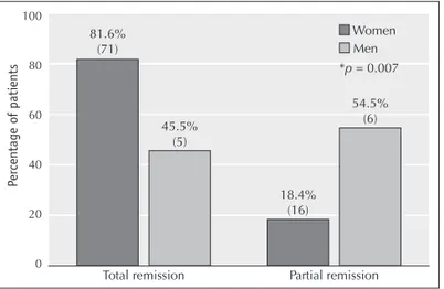
Male patients presented a lower rate of complete remis-sion when compared with female at 12 months (45.5% versus 81.6%, respectively, p = 0.007), which could suggest a less favorable outcome of lupus nephritis among men of this
po-pulation. (Figure 2). As may be veriied by the presented data,
men who did not achieve complete remission at 12 months did accomplish partial response to the established treatment.
Comparative analysis of “clinical class IV”,“histological class IV”, and initial variables of nephritis and SLE, showed that there was no statistical difference among SLEDAI values, serum creatinine and 24-hour proteinuria, as well as in the urinary se-diment, the fraction values of complement and the cumulative doses of cyclophosphamide in both groups (Table 6). The rate of complete remission was higher in patients with “clinical class IV” than in those with “histological class IV”. (Figure 3)
DISCUSSION
In this study, we report the evolution of 100 patients with lupus nephritis followed up for two years. The treatment regimen of
al.6, patients with diffuse proliferative lupus nephritis who had received intravenous cyclophosphamide for a short period of time, followed by MMF or azathioprine maintenance therapy, demonstrate more effective results and more safety than the prolonged use of cyclophosphamide. Houssiau corroborated that IV cyclophosphamide is the only therapy able to reduce Table 2
Analysis of 100 patients with lupus nephritis followed up for 2 years. Demographic data.
Age at SLE diagnosis in years
(M ± SD) 24.71 ± 10.14(5-46)
Age at nephritis diagnosis in years
(M ± SD) 26.78 ± 10.95(5-53)
Interval between SLE diagnosis and nephritis diagnosis in years (M ± SD)
2.32 ± 4.70 (0-24)
Women (%) 88 (88%)
Race (%) White Others Non registered
57 (57%) 29 (29%) 14 (14%)
SLE = Systemic Lupus Erythematosus; M = mean; SD = standard deviation.
Table 3
Initial clinical and laboratory data of 100 patients with lupus nephritis
Creatinine in mg/dL
(M ± SD/variation) 1.02 ± 0.49 (0.4-3.6)
Proteinuria in g/24h
(M ± SD/variation) 2.57 ± 2.39 (0-10.3)
SAH (%) 37 (37%)
Anti-dsDNA positivity (%) 66 (66%)
M = mean; SD = standard deviation; SAH = Systemic Arterial Hypertension; Anti-dsDNA = double-stranded-dsDNA.
Table 4
Comparison of variables related to nephritis at baseline (T0) and after 12 months (T12) in 100 patients with lupus nephritis
T0 T12 P*
Creatinine in mg/dL
(M ± SD) 0.99 ± 0.42 1.03 ± 0.94 0.663
Proteinuria in g/24h
(M ± SD) 2.58 ± 2.42 1.26 ± 1.83 0.000
Hematuria in cell p/f
(M ± SD) 22.99 ± 23.42 13.06 ± 18.64 0.000
Leucocituria in cell p/f
(M ± SD) 14.61 ± 16.77 10.10 ± 15.34 0.037
CH50 (M ± SD) 172.50 ± 73.42 190.67 ± 89.83 0.031
C4 (M ± SD) 16.96 ± 8.04 21.48 ± 9.86 0.024
C3 (M ± SD) 82.44 ± 39.36 100.12 ± 33.49 0.026
* t de student test
M = mean; DP = standard deviation; Cel p/f = cells per field;
CH50 = total serum hemolytic complement; C4 = complement 4; C3 = complement 3
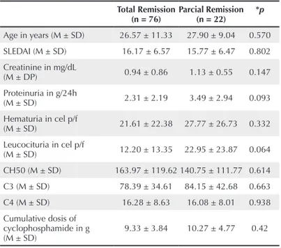
Table 5
Comparison among subgroups with total and partial remission at 12 months and SLE and nephritis variables at baseline: Prognostic factors.
Total Remission
(n = 76) Parcial Remission(n = 22) *p
Age in years (M ± SD) 26.57 ± 11.33 27.90 ± 9.04 0.570 SLEDAI (M ± SD) 16.17 ± 6.57 15.77 ± 6.47 0.802 Creatinine in mg/dL
(M ± DP) 0.94 ± 0.86 1.13 ± 0.55 0.147
Proteinuria in g/24h
(M ± SD) 2.31 ± 2.19 3.49 ± 2.94 0.093
Hematuria in cel p/f
(M ± SD) 21.61 ± 22.38 27.77 ± 26.73 0.332
Leucocituria in cel p/f
(M ± SD) 12.20 ± 13.35 22.95 ± 23.87 0.064
CH50 (M ± SD) 163.97 ± 119.62 140.75 ± 111.77 0.614
C3 (M ± SD) 78.39 ± 34.61 84.15 ± 42.68 0.663
C4 (M ± SD) 16.28 ± 8.63 16.08 ± 8.01 0.938
Cumulative dosis of cyclophosphamide in g
(M ± SD) 9.33 ± 3.84 10.27 ± 4.77 0.42
* t de student test
SLE = Systemic Lupus Erythematosus, M = mean, SD = standard deviation; SLEDAI = Systemic Lupus Erythematosus Disease Activity Index; Cel p/f = cells per field; CH50 = total serum hemolytic complement; C4 = complement 4; = C3 complement 3.
Figure 1. Comparison among subgroups with total and partial remission at 12
months and 24 months of follow-up (n = 77).
*Mc Nemar test
Total remission
12m Partial remission12m Total remission24m Partial remission24m 80
60
40
20
0
Number of patients
56 (72.7%)
21 (27.3%)
12
9 2 54
66 (87.7%)
11 (14.3%)
CH50). The frequency of complete remission at 12 months was 72.7% and, at 24 months, 85.7% (p = 0,013). According to Urowitz et al., patients with active lupus nephritis treated with cyclophosphamide or other immunosuppressive achieve complete remission in approximately 70% of cases.8
According to our data, no patient presented clinical and laboratory criteria for lack of therapeutic response by the end of 2-year of follow-up. However, these data should be carefully interpreted, since, during the study period, there was
recurrence of active renal disease (lare) in approximately 40%
of the patients, but after treatment optimization (increment of corticosteroids and/or immunosuppressive doses) these patients responded in a satisfactory way.
Comparison among subgroups with complete and partial remission at 12 months and the baseline variables of SLE and nephritis showed that no variable had correlation with thera-peutic response. Previous studies demonstrated that variables like age, proteinuria, hematuria and arterial hypertension at diagnosis time had a questionable prognostic value in lupus nephritis outcome.9,10,11,12 Other authors concluded in their studies that high creatinine and hypoalbuminaemia represen-ted factors of predicting worst renal prognosis.13,14 In a study published in Brazil,15 it was prooved that patients with SLE onset after 16 years old, the worse outcomes had occurred in those with nephritis, revealed clinically at the beginning or in the evolution of the disease, with arterial hypertension in the beginning, with positive anti-DNA and those with cyclophos-phamide use. Possible explanations for these contradictory facts derive mainly from methodological differences of the series: criteria of patient inclusion and selection, retrospective Table 6
Comparison among “clinical class IV” and “histological class IV” subgroups
Class IV
*p Histological
(n = 18) (n = 23)Clinical
SLEDAI (M ± SD) 16.22 ± 5.26 17.83 ± 5.67 0.356 Creatinine in mg/dL
(M ± SD) 1.13 ± 0.33 1.09 ± 0.37 0.707
Proteinuria in g/24h
(M ± SD) 2.67 ± 2.12 3.37 ± 2.70 0.360
Hematuria in cel p/f
(M ± SD) 37.33 ± 25.22 34.96 ± 26.33 0.771
Leucocituria in cel p/f
(M ± SD) 15.61 ± 12.77 19.78 ± 23.44 0.472
CH50 (M ± SD) 135.71 ± 119.62 124.22 ± 83.65 0.833
C3 (M ± SD) 86.76 ± 46.82 58.53 ± 19.17 0.117
C4 (M ± SD) 15.00 ± 6.61 13.07 ± 6.90 0.511
Cumulative dosis of cyclophosphamide in g
(M ± SD) 8.72 ± 3.79 10.31 ± 4.48 0.227
* t de student test
M = mean, SD = standard deviation; SLEDAI = Systemic Lupus Erythematosus Disease Activity Index; Cel p/f = cells per field; CH50 = total serum hemolytic complement; C4 = complement 4; = C3 complement 3.
Figure 2. Comparison among men and women subgroups referring to total and
partial remission at 12 months.
* chi-square test
Total remission Partial remission
Per
cen
tag
e o
f pati
en
ts
100
80
60
40
20
0
81.6% (71)
45.5% (5)
54.5% (6)
18.4% (16)
Women Men
Figure 3. Comparison among subgroups with “clinical class IV” and “histological
class IV” total and partial remission at 12 months of follow-up.
* chi-square test
Total remission Partial remission
Per
cen
tag
e o
f pati
en
ts
100
80
60
40
20
0
50% (9)
82.6% (19)
17.4% (4) 50%
(9)
Class IV histological Class IV clinical
development into ESRD in these patients based in studies with long period of follow-up.7
Our data demonstrated satisfactory outcomes in all patients at the end of the 12-month follow-up. Among renal variables
analyzed, there was a signiicant reduction in the 24-hour uri -nary protein level, improvement in the uri-nary sediment and increase in the fractional values of the complement ( C3, C4,
studies, follow-up periods, treatment regimen, clinical outcome
deinition, sample size and others.
Male patients presented a lower rate of complete remission compared to female at 12 months, what could suggest worse prognosis of lupus nephritis among men in our population. This data differ from that in literature, where gender has no prognostic value in lupus nephritis outcome.16
Despite the small number of patients studied, and the controversies of renal biopsy, the rates of complete remission was higher in patients with “clinical class IV” than in those with “histological class IV” at 12 months. A balance between potential gain of information that can guide a therapeutic approach and complications associated with invasive pro-cedure must always be considered before recommending a renal biopsy. Previous studies demonstrated that clinical and laboratory parameters are able to predict with accuracy the lupus nephritis prognosis, and biopsy is not required in every case with renal involvement.17,18 Other studies defend that histological data provide additional information to laboratory parameters and may guide, in a more accurate way, therapeutic selection.19 As may be noticed, this topic remains controvertial. In our experience, renal biopsy is individual and assigned, mainly, in the following situations: Question about clinical and histological correlation, presence of nephrotic syndrome or, failure to cyclophosphamide induction therapy. For this study, the frequency of complete remission at 12 months
was signiicantly higher in patients with “clinical class IV”,
without the need for additional information by histological examination. In our data, renal variables and SLE, as well as cumulative cyclophosphamide doses were not different among patients with “clinical class IV” and “histological class IV”, that is, with renal biopsy.
In conclusion, in this study, renal biopsy to determine the
histological subtype did not inluenced on therapeutic response
in lupus nephritis patients during two years of follow-up. Our data demonstrated that clinical and histological correlation may help therapeutic decision of these patients, leaving renal biopsy only for special situations.
REFERÊNCIAS BIBLIOGRÁFICAS
REFERENCES
1. Appel GB, Valeri A. The course and treatment of lupus nephritis. Ann Rev Med 1994;45:525-37.
2. Cervera R, Khamashta MA, Font J, Sebastiani GD, Gil A, Lavilla P,
et al. Morbidity and mortality in systemic lupus erythematous during a 10-year period: a comparison of early and late manifestations in a cohort of 1,000 patients. Medicine. 2003;82:299-308.
3. Steinberg AD, Steinberg SC. Long-term preservation of renal functions in patients with lupus nephritis receiving treatment that includes cyclophosphamide versus those treated with prednisone only. Arthritis Rheum. 1991;34:945-50.
4. Esdaile JM, Federgreen W, Quintal H, Suissa A, Hayslett JP, Kashgarian M. Predictors of one year outcome in lupus nephritis: The importance of renal biopsy. Q J Med. 1991;81:907-18
5. Boumpas DT, Balow JE. Outcome criteria for lupus nephritis trials: a critical overview. Lupus. 1998;7:622-9.
6. Contreras G, Pardo V, Leclercq B, Lenz O, Tozman E, O’Nan P, et al. Sequential therapies for proliferative lupus nephritis. 2004 ;350: 971-80.
7. Houssiau FA, Vasconcelos C, D’Cruz D, Sebastiani GD, Garrido Ed Ede R, Danieli MG, et al. Immunosuppressive therapy in lupus nephritis: the Euro-Lupus Nephritis Trial, a randomized trial of low-dose versus high-dose intravenous cyclophosphamide. Arthritis Rheum. 2002;46:2121-31.
8. Urowitz MB, Ibañez D, Ali Y, Gladman DD. Outcomes in patients with active lupus nephritis requiring immunosupressives who never received cyclophosphamide. J Rheumatol. 2007;34:1491-96.
9. Donadio Jr JV, Hart GM, Bergstralh EJ. Prognostic determinants in lupus nephritis: a long-term clinicopathologic study. Lupus. 1995;4: 109-15.
10. Bakir AA, Levy PS, Dunea G. The prognosis of lupus nephritis in African-Americans: A retrospective analysis. Am J Kidney Dis. 1994;24:159-71.
11. Appel GB, Cohen DJ, Pirani CL, Meltzer JL, Estes D. Long- term follow-up of patients with lupus nephritis. A study based on the classiication of the World Health Organization. Am J Med. 1987;83:877-85.
12. Baqi N, Moazami S, Singh A, Ahmad H, Balachandra S, Tejani A. Lupus nephritis in children: A longitudinal study of prognostic factors and therapy. J Am Soc Nephrol. 1996;7:924-9.
13. Seedat YK, Parag KB, Ramsaroop R. Systemic lupus erythematous and renal involvement. A South African experience. Nephron. 1994;66:426-30.
14. Villas Boas ML, Nakayama E, Carvalho, MFC, Delino Filho J, Neiva SLA, Habermann F, et al. Lupus eritematoso sistêmico: estudo de 48 pacientes com ênfase no comprometimento renal. Rev Ass Med Bras. 1988;34:165-73.
15. Costallat LTL, Appenzeller S, Marini R. Evolução e fatores prognósticos do lúpus eritematoso sistêmico em relação com a idade de início. Rev Bras Reumatol. 2002;42(2):91-8.
16. Leaker B, Fairley KF, Dowling J, Kincaid-Smith P. Lupus nephritis: clinical and pathological correlation. Q J Med. 1987;62:163-79.
17. Whiting-O’Keefe Q, Henke JE, Shearn MA, Hopper J Jr, Biava CG, Epstein WV.The information content from renal biopsy in systemic lupus erythematosus. Ann Intern Med. 1982;96(part 1):718-23.
18. Austin HA, Muenz LR, Joyce KM, Antonovych TA, Kullick ME, Klippel JH, et al. Prognostic factors in lupus nephritis. Contribution of renal histologic data. Am J Med. 1983;75:382-91.