UPDATE ARTICLE
Mitochondria and the central nervous system: searching
for a pathophysiological basis of psychiatric disorders
Emilio L. Streck,
1,2,3Cinara L. Gonc¸alves,
1,2,3Camila B. Furlanetto,
1,2,3Giselli Scaini,
1,2,3Felipe Dal-Pizzol,
1,2,3Joa˜o Quevedo
2,3,41Bioenergetics Laboratory, Graduate Program in Health Sciences, Universidade do Extremo Sul Catarinense (UNESC), Criciu´ma, SC, Brazil. 2National Science and Technology Institute for Translational Medicine (INCT-TM), Porto Alegre, RS, Brazil.3Center of Excellence in Applied
Neurosciences of Santa Catarina (NENASC), Floriano´polis, SC, Brazil.4Neuroscience Laboratory, Graduate Program in Health Sciences, UNESC, Criciu´ma, SC, Brazil.
Introduction: Mitochondrial dysfunction has been postulated to participate in the development of many neuropsychiatric disorders, but there is no consensus as to its role. The aim of this paper is to review recent studies and to outline the current understanding of the association between mitochondrial dysfunction and psychiatric disorders.
Methodology: We reviewed articles that evaluated mitochondrial dysfunction and psychiatric disorders, with a particular focus on depression, bipolar disorder, anxiety disorders, obsessive-compulsive disorder, and autism spectrum disorder, and the association between mitochondrial dysfunction and development of these disorders.
Results:Evidence suggests that alterations in mitochondrial morphology, brain energy metabolism, and mitochondrial enzyme activity may be involved in the pathophysiology of different neurop-sychiatric disorders, given their key role in energy metabolism in the cell.
Conclusions:Understanding the interactions between mitochondrial dysfunction and development of psychiatric disorders may help establish more effective therapeutic strategies for these disorders and thus lead to better outcomes for affected subjects.
Keywords: Mitochondria; central nervous system; neuroplasticity; cell death
Introduction
Biological systems cannot be described as random molecules commanded by physical and chemical laws of diffusion and casual interactions.1 It is essential to understand biological phenomena as part of a large system; thus, the cell is now understood as an assembly of molecular machines made of proteins that interact to preserve their functions.2 The same occurs with the structure and function of mitochondria, and the traditional belief that mitochondria are autonomous organelles is changing. The current challenge is to understand the structural and functional cooperation of mitochondria with the rest of the cell, their relation to the endoplasmic reticulum,3 and the cross-talk between nuclear and mitochondrial genetic machinery.4
Mitochondria are essential for the life of the cell. They produce most of the adenosine triphosphate (ATP) by oxidative phosphorylation. Mitochondria have two mem-branes (outer and inner), an intermembrane space, and an internal matrix. The inner mitochondrial mem-brane contains the electron transport chain (ETC), the
molecular machinery for energy production.5Five protein complexes form the ETC. Of these, three (I, III, and IV) pump protons (H+) through the inner membrane, gen-erating a H+gradient required for the synthesis of ATP at complex V (ATP synthase). The mitochondrial genome codes for 13 of the ETC proteins. The cell nucleus encodes other mitochondrial proteins (more than 1,000), which mediate processes such as the regulation of ion homeostasis, stress responses, cell survival, and signal transduction.5
Neuronal energy supplies are completely dependent on mitochondrial oxidative phosphorylation. Neurons have limited capacity to obtain energy through glycolysis when oxidative phosphorylation is compromised,6which makes them particularly vulnerable to mitochondrial dysfunction. The aim of this paper is to review recent findings and outline the current understanding of the association between mitochondrial dysfunction and psychiatric dis-orders, with a particular focus on depression, bipolar disorder (BD), anxiety disorders, obsessive-compulsive disorder, and autism spectrum disorders.
More than a power station
It is generally assumed that the mitochondrion is the energy-providing organelle of the cell, but it processes several other compounds as well.7Neurotransmitters and neurotrophic factors control mitochondrial dynamics Correspondence: Prof. Emilio Luiz Streck, Laborato´rio de
Bio-energe´tica, Universidade do Extremo Sul Catarinense, Av. Universita´ria, 1105, Universita´rio, CEP 88806-000, Criciu´ma, SC, Brazil.
E-mail: [email protected]
Submitted Aug 01 2013, accepted Oct 03 2013. 156–167 ß2014 Associac¸a˜o Brasileira de Psiquiatria
through their influence upon neuronal energy metabo-lism, Ca2+ homeostasis, and dendritic and axonal motility.5
Many investigations have focused on the role of mitochondria in neurogenesis, especially during neuronal differentiation. The amount of this organelle per cell increases, while the velocity at which mitochondria move decreases, as neurite outgrowth slows and synaptogen-esis takes place.8 A study by Vayssie`re et al.9showed that treatment with chloramphenicol (an inhibitor of mitochondrial protein synthesis) prevents cell differentia-tion, whereas oligomycin (an inhibitor of the mitochondrial ATP synthase) does not, suggesting that increased mitochondrial mass (but not ATP production) is required for neuronal differentiation. Additionally, the focal applica-tion of nerve growth factor (NGF) to growing axons results in an accumulation of mitochondria near the site of NGF stimulation by a mechanism that involves docking interactions with the actin cytoskeleton, suggesting a role for mitochondria in facilitating growth cone responses to neurotrophic factors.10
Presynaptic terminals characteristically contain multi-ple mitochondria, because the majority of the ATP produced is required to maintain synaptic ion home-ostasis and phosphorylation reactions.5Looking beyond the obvious function of this organelle, recent findings from experimental models in which the motility and function of mitochondria were visualized suggest that mitochondria play active roles in synaptic plasticity.5
Interestingly, the movement of mitochondria into dendritic protrusions during synaptogenesis correlates with the development and morphological plasticity of spines; when dendritic mitochondrial content is increased, the number and plasticity of spines and synapses enhances greatly.11 It remains to be determined if and how recruitment of mitochondria within active synapses contributes to long-term changes in synaptic strength.5 Besides, it is known that changes in mitochondrial functions, such as Ca2+ regulation, energy metabolism, and oxyradical production, play a role in synaptic plasticity as well.5
Mitochondria: the crucial signaling
Studies have showed emerging roles of mitochondria in neuroplasticity.11-13 Several prominent signaling path-ways that stimulate mitochondrial biogenesis and energy metabolism, while simultaneously regulating neuroplasti-city, have been demonstrated.13 Mitochondria are dis-tributed throughout the length of axons and presynaptic terminals. In addition, they are found in dendrite shafts and associated with dendritic spines.14,15 Mitochondria also participate in the metabolism of reactive oxygen species (ROS), calcium signaling and apoptosis.5
Among many specialized cell types in the body, neurons are particularly remarkable. They have tree-like shapes, are electrically excitable, and act in signal detection, integration, and storage, as well as in the generation of adaptive responses.13Through the use of imaging and molecular biology techniques for the study of
mitochondria, several surprising properties and functions of mitochondria in neuroplasticity have been revealed. Mitochondria move rapidly within and between subcel-lular compartments16; undergo fission and fusion17; respond (e.g., move, change their energy output, take up or release calcium) to electrical activity and activation of neurotransmitters and growth factor receptors18; and function as signaling outposts that contain kinases, deacetylases, and other signal transduction enzymes.19
Indeed, events in learning and memory processes, such as long-term potentiation (LTP), have been asso-ciated with changes in mitochondria.20-22 Other studies suggest that mitochondria work as mediators of some of the effects of glutamate and brain-derived neurotro-phic factor (BDNF) on synaptic plasticity. BDNF is known to promote synaptic plasticity, partly because it enhances mitochondrial energy production23 and increases mitochondrial respiratory coupling at complex I.24 Furthermore, BDNF expression and signaling is increased in response to environmental factors –– such as exercise and cognitive stimulation –– that increase cellular energy demand.25
Although changes in the location of mitochondria within axons and dendrites may play roles in synaptic plasticity, rapid functional changes in mitochondria are increasingly being implicated in synaptic plasticity. These changes may include mitochondrial Ca2+ uptake or release, production of superoxide and other ROS, and release of proteins and other factors.13
Apoptosis
Apoptosis is the prototypical form of programmed cell death (PCD) in neurons during development and adult cell turnover, and it may also occur in a range of neurodegenerative conditions. Under normal circum-stances, apoptosis is suppressed, as a result of the rigorous compartmentalization of catabolic enzymes and their activators. Morphologically, it is characterized by cell shrinkage, membrane blebbing, and karyorrhexis.5
apoptosis of the cell.27 The intrinsic pathway, which predominates in neurons, is triggered by signals as trophic factor withdrawal, moderate overactivation of glutamate receptors, oxidative stress, and DNA damage.5 These triggers activate kinases and transcription factors that induce mitochondrial translocation of Bax and Bak, which form pores in the outer mitochondrial membrane.5 Mitochondria also participate in death-regulating bio-chemical signals. For example, cytochromecis restricted to the mitochondrial intermembrane space, which pre-vents its interaction with apoptotic-protease-activating factor 1 (Apaf-1), a cytosolic protein. When this mem-brane is compromised, cytochrome c binds to Apaf-1, leading to allosteric activation of pro-caspase-9, which in turn activates caspase-3.28 In the same way, Smac/ DIABLO and Omi/HtrA2, two intermembrane proteins, are usually separated from cytosolic inhibitors of apopto-sis proteins (IAPs). Once mitochondrial membrane permeabilization (MMP) occurs, Smac/DIABLO and Omi/HtrA2 inactivate IAP, preventing caspase 3 and -9 inhibition. Two DNAses, apoptosis inducer factor (AIF) and endonuclease G, are also normally confined to the mitochondrial intermembrane space, but following MMP they can move to the nucleus and mediate chromatinolysis.29
MMP is a key event in physiological as well as pathological cell death30 and it is regulated at multiple levels. Many proapoptotic agents can induce MMP, including Ca2+, ROS, lipid messengers (e.g., ceramide and ganglioside GD3), and stress kinases. Briefly, MMP formation is facilitated by proapoptotic proteins from the Bcl-2 family, and is inhibited by antiapoptotic Bcl-2-like proteins.30
In this regard, several studies have sought to understand the role of each of the proteins involved in the process. While caspase inhibition can prevent cell death, it does not prevent cytochromecrelease, compromising mitochondrial ATP generation and increasing superoxide production. These alterations can make neurons vulnerable to necro-sis.5This is similar to AIF and HtrA2, which could impair mitochondrial function when released into the cytosol.31,32 In addition, studies have shown that mitochondria can themselves release controlled amounts of cytochromec, Smac, HtrA2, and AIF, by a still-unclear mechanism.33,34 Inhibitors of caspase -1 and -3 modify LTP of synaptic transmission in hippocampal synapses,35,36 indicating functions for apoptotic cascades in synaptic plasticity.
Reactive oxygen species
Mitochondria are the main intracellular source of ROS5,37; ROS production contributes to mitochondrial damage in a range of pathologies and is also important in redox signaling from the organelle to the cell.38,39
Each complex of the mitochondrial respiratory chain has a singular function and works in association with the others. A fault at any part of the chain can disturb energy supply. In the absence of ADP, the movement of H+ through ATP synthase ceases and electron flow slows down, decreasing the speed of the respiratory chain
(State IV respiration), and O2–Nformation increases.40In Complex I, the primary source of O2–Nappears to be one of the iron-sulfur clusters.41-43In Complex III, most of the O2–Nappears to be formed as a result of the autoxidation of ubisemiquinone.44,45 In addition, O2–N interacts with nitric oxide to generate peroxynitrite. Peroxynitrite nitra-tion of tyrosine residues impairs the formanitra-tion of monoaminergic neurotransmitters and other aminergic compounds.46
On the other hand, the mitochondrion possesses various antioxidant defenses that detoxify O2–N and H2O2. Superoxide is enzymatically converted to H2O2 by a family of metalloenzymes called superoxide dis-mutases (SOD).47 H
2O2 can diffuse to the cytosol and then be converted to H2O by glutathione peroxidase and catalase. Moreover, mitochondria use several antioxidant molecules, such as coenzyme Q10 (ubiquinone), crea-tine, nicotinamide, and glutathione, to interrupt or mini-mize oxidative processes.5
Despite their potential to cause damage, ROS act on signaling functions in physiological processes, including synaptic plasticity and learning and memory.48
Ca2+signaling
Mitochondria also play a role in calcium homeostasis.18,49 Calcium (Ca2+) is the principal second messenger that contributes to the regulation of both neurotransmission and short- and long-term neuronal plasticity in the brain. The role of Ca2+ signals in apoptosis has been further reinforced by the demonstration that antiapoptotic proteins (such as Bcl-2) decrease Ca2+ levels in the endoplasmic reticulum (ER) and reduce cytosolic and mitochondrial Ca2+ responses to extracellular stimuli by increasing the leak of Ca2+from the ER.50-53Proapoptotic proteins, on the other hand, exert the opposite effect.54
The fine spatial and temporal organization of intracel-lular calcium signals is essential to central nervous system (CNS) function.55Signals are conveyed through-out the CNS by local changes in calcium concentration ([Ca2+]c).55Thus, the Ca2+signals that are essential for synaptic transmission and therefore for transmission of information throughout the CNS are transmitted to the mitochondria, where it is assumed that Ca2+ modulates mitochondrial metabolism as described elsewhere –– with upregulation of the TCA cycle, ATP synthase, and the aspartate carrier,56 and presumably with a consequent increase in the supply of ATP.
Alternatively, local changes in intracellular Ca2+ con-centration ([Ca2+]c) can diffuse across the cell, leading to an effect at a distant site. Indeed, mitochondrial Ca2+ overload has long been associated with necrosis in heart ischemia-reperfusion injury and excitotoxicity.57
Choi58showed that prolonged exposure to high concen-trations of glutamate leads to Ca2+-dependent cell death in neuronal culture.
Activation of NMDA receptors by glutamate results in an increase in [Ca2+]c.59,60 In addition, depolarization induces opening of voltage-gated Ca2+ channels, while the activity of the plasma membrane Na+/Ca2+ exchan-gers is reversed. During this increase in [Ca2+]c, mitochondria accumulate and retain Ca2+ to buffer the cytosolic loading. However, this increase in [Ca2+]c caused by glutamate promotes extensive accumulation of Ca2+ for several hours. In this context, it has been shown that necrosis is initiated by this delayed Ca2+ influx, which is independent of Ca2+ release from mitochondria, but dependent on declining activity of cytoplasmic Ca2+ clearing mechanisms (for example, calpain-mediated cleavage of Na+/Ca2+ exchanger). Following the initiation of necrosis, mitochondria are overloaded with Ca2+, the electrochemical proton gradi-ent collapses, and necrotic cell death is induced.61Thus, it seems that alteration of this cellular response (for example, by a tumor or viral proteins)51,54,62,63 plays a role in the pathogenesis of human disorders. Prolonged permeability transition pore opening leads to a complete collapse of the membrane potential and Ca2+ release, which results in the complete loss of mitochondrial function and necrotic cell death.
Mitochondrial impairment and psychiatric disorders
Mitochondrial dysfunction has been studied in patients with brain diseases, including neurodegenerative dis-eases and psychiatric disorders.64,65 Evidence that patients with psychiatric disorders (depression, BD, and schizophrenia) exhibit mitochondrial abnormalities at the structural, molecular, and functional levels has been reviewed66(Figure 1).
These findings suggest that a mitochondrial deficit is sufficient to trigger one or more psychiatric disorders. Mitochondrial deficits in psychiatric disorders are sug-gested by positron emission tomography (PET) analysis of brain energy metabolism.5In addition, data suggesting a role for mitochondrial alterations in psychiatric disorders are only correlations; therefore, it remains to be deter-mined whether these alterations contribute to the disease process or are just epiphenomena.5
Interestingly, psychiatric symptoms have been observed in subjects with mitochondrial diseases. Fattal et al.67 identified 19 confirmed cases of mitochondrial disease with psychiatric complications, including BD, major depressive disorder, psychosis, anxiety disorders, and personality changes. Indeed, symptoms of mental illness have been previously documented in subjects affected by mitochondrial cytopathies.68 In addition, a case report of mitochondrial encephalomyopathy, lactic acidosis, and stroke-like episodes (MELAS), a typical mitochondrial encephalopathy, which had presented as mania prior to diagnosis, supports the role of mtDNA mutations in the etiology of psychiatric disorders.69
Taken together, the reviewed lines of evidence, including ultrastructural, neuroradiological, biochemical, and genetic data, seem to point to a possible role of mitochondrial dysfunction in the pathological mechanism of some psychiatric disorders. However, the exact mechanisms by which deficits of energy metabolism occur in the brain of subjects affected with psychiatric disorders are not completely understood.68
Mitochondrial dysfunctions (leading to decreased ATP production, oxidative stress, and apoptosis) occur in the early stages of different neurodegenerative diseases associated with mood disorders. Findings from genetic, postmortem brain, brain-imaging, and biomarker studies in humans with psychiatric disorders and rodent models of such disorders have confirmed this hypothesis.
Depression
Several lines of evidence suggest that mitochondrial dysfunction is an important component of the neurobiol-ogy of depression. Patients with depression have reduced glucose utilization in the prefrontal cortex, anterior cingulate gyrus, and caudate nucleus.70 The energy metabolism deficits observed in patients with depression may be widespread, as suggested by data that demonstrates reduced mitochondrial ATP production and increased mitochondrial DNA deletions as compared with control subjects.71
Magarinos et al.72found that stress did not affect the number of neuronal mitochondria; however, the total mitochondrial area increased after a stress paradigm, suggesting that longer duration of stress could compro-mise ATP synthesis. Gardner et al.73showed a significant decrease of mitochondrial ATP production and mitochon-drial enzyme content in muscle of patients with major depressive disorder. Madrigal et al.74 reported that complexes I-III and II-III of the mitochondrial respiratory chain were inhibited in the rat brain after chronic stress, as was brain Na+, K+-ATPase.75 Rezin et al.76reported that mitochondrial respiratory chain complexes I, III and IV were inhibited in the cerebral cortex and cerebellum of rats after 40 days of chronic mild stress (CMS), and this was reversed by administration of ketamine.77 Gong et al.78showed that exposure to CMS inhibited mitochon-drial respiration and dissipated mitochonmitochon-drial membrane potential. In addition, the mitochondrial ultrastructure was altered in brains of mice exposed to CMS.78
Genetic evidence points to a role for mitochondrial impairment in depression. Postmortem brain tissue from a patient with severe depression was found to have more mtDNA deletions than postmortem muscle tissues from the same patient, suggesting that the accumulation of mtDNA deletions in the brain might play a role in the pathophysiology of depression.79
Bipolar disorder
Several investigators have proposed that mitochondrial dysfunction is related to the pathophysiology of BD. Brain magnetic resonance spectroscopy has demonstrated decreased levels of N-acetyl-aspartate (a marker of mitochondrial energy production) in the prefrontal cortex of patients with BD as compared with healthy controls, indicating neurodevelopmental alterations in the former.80 Stork & Renshaw81proposed a hypothesis of mitochon-drial dysfunction in BD that involves impaired oxidative phosphorylation, a shift toward glycolytic energy produc-tion, a decrease in total energy production and/or substrate availability, and altered phospholipid metabo-lism. Postmortem studies have reported changes in mitochondrial-related gene expression in BD as well.82,83 In addition, manic-like hyperactivity induced by d-amphetamine, which is considered an animal model of mania, is associated with oxidative stress in the rat brain.84-86 Correˆa et al.87 showed that citrate synthase activity was inhibited in the rat hippocampus after mania induced by amphetamine, and this was reversed by valproate (VPA) and lithium (Li) administration. In contrast, Streck et al.88demonstrated that amphetamine inhibited creatine kinase activity in rat brains, but VPA and Li were not able to prevent this. Zugno et al.89 showed that amphetamine increased Na+, K+-ATPase activity in rat brains, and that VPA or Li reversed this effect. Moreover, VPA and Li did not alter Na+, K+ -ATPase activity. Valvassori et al.90 showed that AMPH inhibited mitochondrial respiratory chain activity in rat brains, and VPA, but not Li, reversed this. Feier et al.91 showed that methamphetamine inhibited the activities of
Krebs cycle enzymes and complexes of the mitochondrial respiratory chain, and Li and VPA reversed methamphe-tamine-induced energy metabolism dysfunction.
Bachmann et al.92 have demonstrated that long-term treatment with Li and VPA enhanced cell respiration rate, mitochondrial membrane potential, and mitochondrial oxidation in SH-SY5Y cells. Additionally, methampheta-mine reduces mitochondrial cytochrome c, mitochondrial antiapoptotic Bcl-2/Bax ratio, and mitochondrial cyto-chrome oxidase (COX) activity, and treatment with Li or VPA prevents these alterations. Treatment with Li or VPA prevented Bcl-2 attenuation of apoptosis by sequestering pro-caspases; preventing the release of mitochondrial apoptogenic factors, such as calcium, cytochromec, and apoptosis-inducing factor, into the cytoplasm; and enhan-cing mitochondrial calcium uptake.93,94 Studies showed that upregulation of Bcl-2 is a result of the activation of extracellular signal-regulated kinase (ERK) and phosphoi-nositide 3-kinase (PI3K) pathways by mood stabilizers.95
Patients with BD exhibit impaired brain energy meta-bolism, reduced levels of mitochondrial proteins involved in energy metabolism, and increased mtDNA muta-tions.96 A study using Southern blot analysis did not demonstrate mtDNA deletions in postmortem cerebral cortex samples of patients with mood disorders. When a highly sensitive quantitative PCR method was used, more mtDNA deletions were found in the brains of patients with BD than in control brains.97 Konradi et al.98 reported decreased expression of nuclear genes coding for the enzyme complexes responsible for oxidative phosphor-ylation and reduced expression of nuclear genes related to proteasome degradation in the hippocampus of nine subjects with BD. MacDonald et al.99 showed that creatine kinase mtRNA was decreased in patients with BD, mainly in the hippocampus. The inhibition of creatine kinase activity by amphetamine reinforces the hypothesis that metabolism impairment is involved in the pathophy-siology of BD.
Anxiety disorders
Recent evidence demonstrates that impairment of cellular plasticity and resilience may underlie the pathophysiology of anxiety disorders, and that antidepressants have major effects on the signaling pathways that regulate neuro-plasticity and cell survival.100,101
It has been suggested that mitochondrial Ca2+ seques-tration plays a key role in modulating the tone of synaptic plasticity in a variety of neuroanatomical regions, includ-ing those implicated in the pathophysiology of anxiety disorders.102A study about the relative roles of mitochon-drial and ER Ca2+ buffering showed that dendritic mitochondria rapidly accumulate Ca2+, while the ER displays a more delayed increase in Ca2+ during high-frequency stimulation.103 Thus, is possible that the regulation of mitochondrial function plays an important role in regulating the synaptic strength of the neuronal circuitry mediating complex behaviors.
improve mitochondrial function.104 The activation of mitochondrial benzodiazepine receptors reduced stress and anxiety in rats, and neurosteroids that had been widely recognized as having anxiolytic properties have specific binding sites on mitochondria and have been confirmed to modulate mitochondrial Ca2+ efflux and increase mitochondrial resilience.105-111
Murphy et al.112have shown in isolated mitochondria that Bcl-2 overexpression increases mitochondrial Ca2+ uptake capacity, increasing the resistance of mitochon-dria to Ca2+-induced inhibition of respiration. Einat et al.,113using BCL2-heterozygous mice, demonstrated an increase in anxiety-like behavior with reduced mito-chondrial Bcl-2 levels, suggesting that mitomito-chondrial function, modulated by Bcl-2, may be related to the regulation of anxiety behaviors, thus playing a critical role in the etiology of anxiety disorders.
Obsessive-compulsive disorder
Obsessive-compulsive disorder (OCD) is a common psychiatric disorder defined by the presence of obsessive thoughts and repetitive compulsive actions, and it often includes anxiety and depressive symptoms.114Although the etiology of OCD remains unknown, the results of twin studies, familial studies, and segregation analysis have provided evidence that OCD has a strong genetic component.115-119
Some investigators have also found markers of oxidative stress in the brain tissues of patients with OCD.120-122 Moreover, Kuloglu et al.120 showed signifi-cantly lower levels of vitamin E and C, and higher levels of malondialdehyde (MDA) in patients with OCD com-pared to controls, suggesting that OCD is linked to oxidative stress. Free radicals in the brain are mainly produced by catecholamine metabolism, and this increase in catecholaminergic metabolism seems to be associated with increased tissue damage.123,124Depleted levels of glutathione (GSH) have also been found in postmortem prefrontal cortex samples of patients with psychiatric disorders.125 Diminished GSH levels affect mitochondrial function and inhibit the activity of the mitochondrial complexes, especially complex I.126
Although no molecules that induce OCD symptoms have been identified, several neurotransmitters, including serotonin, dopamine, glutamate and gamma-aminobutyric acid (GABA), have been suggested to play regulatory roles in OCD. Neurotransmitters are highly redox-reactive molecules, and produce ROS during normal neurotrans-mission. Alterations in these neurotransmitter pathways may increase the oxidative burden in the brain.127On the other hand, some investigators have suggested that dopamine also plays an important role in the pathogenesis of OCD.128,129
In addition, a multivariate logistic regression analysis demonstrated that MnSOD, UCP2 I/D genotypes, and GSH had significant impacts on OCD. Prevalence differences between the genders of OCD patients have been previously reported.130-132Yamada et al.133showed that mitochondrial uncoupling protein 2 (UCP-2) regulates
neurotransmission and that overexpression of UCP2
prevents dopamine transmission in the CNS. Knock-down of the UCP2 gene increases mitochondrial mem-brane potential and ROS production in murine endothelial cells.134 However, in transgenic mice, UCP-2 protects cells against apoptosis and oxidative stress. Walder et al.135have suggested that a 45 bp insertion inUCP2
affects the amount and activity of UCP-2 protein by influencing mRNA stability, translation, and posttransla-tional modification. De Bilbao et al.136 suggested that resistance to cerebral ischemic injury inUCP2knockout mice is regulated by mitochondrial GSH levels in microglia.
Although the pathology of mitochondrial disorders in OCD has not yet been identified, it is well known that mitochondrial dysfunction related to mutations of mito-chondrial DNA or of nuclear-encoded genes linked to mitochondrially based oxidative phosphorylation leads to impaired energy metabolism, perturbs calcium home-ostasis, and increases ROS and apoptosis,137-139 changes that might influence on neurotransmitter release, leading to OCD, since synaptic transmission requires high levels of ATP.
Autism spectrum disorder
Clinical, genetic, and biochemical evidence suggests that mitochondrial dysfunction in autism spectrum disorder (ASD) is more common than expected. Some patients with ASD phenotypes clearly have genetic-based primary mitochondrial disease.140
ASD encompass severe developmental disorders characterized by variable degrees of impairment in language, communication, and social skills, as well as by repetitive and stereotypic patterns of behavior. Substantial percentages of autistic patients display peripheral markers of mitochondrial energy metabolism dysfunction, such as elevated lactate, pyruvate, and alanine levels in blood, urine, and/or cerebrospinal fluid, serum carnitine deficiency, and/or enhanced oxidative stress. These biochemical abnormalities are accompa-nied by highly heterogeneous clinical presentations, which generally encompass neurological and systemic symptoms that are relatively unusual in idiopathic autistic disorder. In some patients, these abnormalities have been successfully explained by the presence of specific mutations or rearrangements in their mitochondrial or nuclear DNA. However, in most cases, abnormal energy metabolism cannot be immediately linked to specific genetic or genomic defects. Recent evidence from postmortem studies of autistic brains points to abnorm-alities in the mitochondrial function as possible down-stream consequences of dysreactive immunity and altered Ca2+signaling.141
processes are present in the autistic brain by showing that transforming growth factor (TGF-b1), interleukin (IL)-6 and IL-10 are increased in the brain of autistic patients. A number of studies have also shown that inflammatory cytokines, including tumor necrosis factor (TNF)-a, inter-feron (IFN)-c, IL-1, IL-6, IL-8, and IL-12, are ele-vated in blood mononuclear cells, serum, plasma, and cerebrospinal fluid (CSF) of autistic subjects.144-150 Additionally, El-Ansary & Al-Ayadhi142 have shown that autistic patients have remarkably higher plasma HSP70, TGF-b2, caspase-7, and INF-c levels as compared with age- and gender-matched controls, suggesting that these parameters confirm the role of neuroinflammation and apoptosis mechanisms in the etiology of autism, as well as the possibility of using these parameters as predictive biomarkers.
Mitochondria as a pharmacological target for psychiatric disorders
Mitochondria are an ideal target for therapeutic modifica-tion, because they are key regulators of energy producmodifica-tion, ROS production, and apoptosis. Mitochondria-targeted drugs are therapeutic agents that can directly target mitochondria to address perturbed cellular bioenergetics, oxidative stress, mtDNA mutations, impaired mitochon-drial Ca2+-handling capacity, and mitochondrial-mediated apoptosis.151-153 However, mitochondria-targeted drugs
can interact with mitochondria in a secondary manner, where primary targets are other cellular locations.154
As mitochondria are also involved in initiating apoptotic cell decay, they are vulnerable targets for experimental and/or pharmacological interventions. Mood stabilizers also promote Bcl-2-related neuronal processes such as neurite outgrowth, adult hippocampal neuronal neurogen-esis, and neuronal protection against a variety of insults.155,156 Thus, the members of the Bcl-2 protein family may mediate some of the behavioral effects of antidepressants, and pharmacological modulation of Bcl-2 function might produce antidepressant-like behavioral effects. Studies have shown that mice overexpressing Bcl-2-associated athanogene (Bag1) or Bax inhibitor 1 and mice that received brain infusions of the BID inhibitor BI-11A7 had a protective phenotype in several tests of depression-like behavior.154,157,158 Furthermore, mice overexpressing Bax inhibitor 1 displayed resilience to monoamine depletion-induced anhedonia-like symptoms, and Bag1-transgenic mice showed resilience to amphe-tamine-induced hyperlocomotion.157,158 Besides, Zhu et al.159demonstrated that mitochondrial Ca2+depletion promotes apoptosis. In animal models of depression, inositol trisphosphate receptor inhibitors and other com-pounds that regulate mitochondrial Ca2+ influx produced behavioral effects similar to those produced by antide-pressants, lithium, and ketamine.160,161 Additionally, nifedipine and nimodipine have produced promising results in treating and stabilizing mood in both animal
models of depression and in humans with BD or other types of depressive disorder.162-165
Considering that the mitochondrial permeability transi-tion pore (mPTP) opens upon collapse of the mitochon-drial membrane potential (Dy), an mPTP inhibitor would be a good candidate for further testing. Kubota et al.166 showed that NIM811, an mPTP inhibitor, counteracted the behavioral phenotypes associated with BD in Polg1-transgenic mice. In addition, aminopropyl carbazole (P7C3) exerts its pro-neurogenic activity by protecting newborn neurons from apoptosis and inhibiting mPTP, and an mPTP inhibitor has demonstrated neuroprotective effects in vitro.167,168
Some psychiatric medications already exhibit antiox-idant activity.169-172To protect mitochondria and the cell against such damaging effects, several measures can be applied. One is to increase the intracellular glutathione content. This can be done by supplying precursors for glutathione synthesis, such as N-acetylcysteine.173 A double-blind, placebo-controlled adjunctive study showed that the antioxidant N-acetylcysteine improved function-ing and depressive symptoms in patients with BD after 24 weeks, compared with placebo treatment.174 Other compounds acting as general intracellular antioxidants include ascorbic acid (vitamin C), a-tocopherol (vitamin E), b-carotene, and a-lipoic acid.175,176 All these com-pounds are naturally present in the cell, but their levels can be increased by exogenous supplementation.
Conclusions
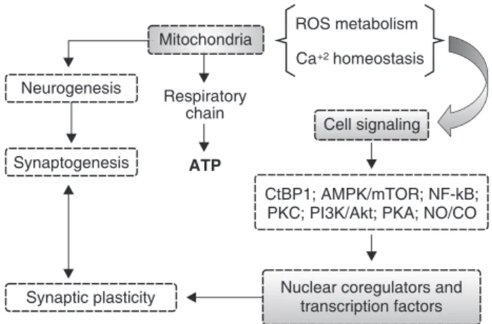
Mitochondria play a pivotal role in cellular energy metabolism. The final product of this process is ATP, used as a source of chemical energy. Besides this major role, mitochondria have been shown to be involved in other functions, such as signaling, calcium homeostasis, cellular differentiation, and cell death, as well as control of the cell cycle and of cell growth (Figure 2). Thus, understanding the interactions between mitochondrial dysfunction and development of psychiatric disorders, such as depression, BD, anxiety disorders, OCD, and ASDs, may help establish more effective therapeutic strategies for these psychiatric conditions and, consequently, enable better outcomes for affected subjects. Moreover, this evidence highlights the role of mitochondrial dysfunction in the pathophysiology of psychiatric disorders, which represents an interesting research prospect.
Acknowledgements
This study was supported by grants from the Graduate Program in Health Sciences at Universidade do Extremo Sul Catarinense (UNESC), Coordenac¸a˜o de Aperfei-c¸oamento de Pessoal de Nı´vel Superior (CAPES), and Conselho Nacional de Desenvolvimento Cientı´fico e Tecnolo´gico (CNPq).
Disclosure
The authors report no conflicts of interest.
References
1 Lenaz G, Genova ML. Structure and organization of mitochondrial respiratory complexes: a new understanding of an old subject. Antioxid Redox Signal. 2010;12:961-1008.
2 Alberts B. The cell as a collection of protein machines: pre-paring the next generation of molecular biologists. Cell. 1998;92: 291-4.
3 Pinton P, Giorgi C, Siviero R, Zecchini E, Rizzuto R. Calcium and apoptosis: ER-mitochondria Ca2+ transfer in the control of apoptosis. Oncogene. 2008;27:6407-18.
4 Ryan MT, Hoogenraad NJ. Mitochondrial-nuclear communications. Annu Rev Biochem. 2007;76:701-22.
5 Mattson MP, Gleichmann M, Cheng A. Mitochondria in neuroplas-ticity and neurological disorders. Neuron. 2008;60:748-66. 6 Herrero-Mendez A, Almeida A, Fernandez E, Maestre C, Moncada
S, Bolanos JP. The bioenergetic and antioxidant status of neurons is controlled by continuous degradation of a key glycolytic enzyme by APC/C-Cdh1. Nat Cell Biol. 2009;11:747-52.
7 Brie`re JJ, Chre´tien D, Be´nit P, Rustin P. Respiratory chain defects: what do we know for sure about their consequences in vivo? Biochim Biophys Acta. 2004;1659:172-7.
8 Chang DT, Reynolds IJ. Differences in mitochondrial movement and morphology in young and mature primary cortical neurons in culture. Neuroscience. 2006;141:727-36.
9 Vayssie`re JL, Cordeau-Lossouarn L, Larcher JC, Basseville M, Gros F, Croizat B. Participation of the mitochondrial genome in the differentiation of neuroblastoma cells. In Vitro Cell Dev Biol. 1992;28A:763-72.
10 Chada SR, Hollenbeck PJ. Nerve growth factor signaling regulates motility and docking of axonal mitochondria. Curr Biol. 2004;14: 1272-6.
11 Li Z, Okamoto K, Hayashi Y, Sheng M. The importance of dendritic mitochondria in the morphogenesis and plasticity of spines and synapses. Cell. 2004;119:873-87.
12 Levy M, Faas GC, Saggau P, Craigen WJ, Sweatt JD. Mitochondrial regulation of synaptic plasticity in the hippocampus. J Biol Chem. 2003;278:17727-34.
13 Cheng A, Hou Y, Mattson MP. Mitochondria and neuroplasticity. ASN Neuro. 2010;2:e00045.
14 Cameron HA, Kaliszewski CK, Greer CA. Organization of mito-chondria in olfactory bulb granule cell dendritic spines. Synapse. 1991;8:107-18.
15 Popov V, Medvedev NI, Davies HA, Stewart MG. Mitochondria form a filamentous reticular network in hippocampal dendrites but are present as discrete bodies in axons: a three-dimensional ultra-structural study. J Comp Neurol. 2005;492:50-5.
16 Zinsmaier KE, Babic M, Russo GJ. Mitochondrial transport dynamics in axons and dendrites. Results Probl Cell Differ. 2009;48:107-39.
17 Liesa M, Palacı´n M, Zorzano A. Mitochondrial dynamics in mammalian health and disease. Physiol Rev. 2009;89:799-845. 18 MacAskill AF, Atkin TA, Kittler JT. Mitochondrial trafficking and the
provision of energy and calcium buffering at excitatory synapses. Eur J Neurosci. 2010;32:231-40.
19 Stowe DF, Camara AK. Mitochondrial reactive oxygen species production in excitable cells: modulators of mitochondrial and cell function. Antioxid Redox Signal. 2009;11:1373-414.
20 Williams JM, Thompson VL, Mason-Parker SE, Abraham WC, Tate WP. Synaptic activity-dependent modulation of mitochondrial gene expression in the rat hippocampus. Brain Res Mol Brain Res. 1998;60:50-6.
21 Calabresi P, Gubellini P, Picconi B, Centonze D, Pisani A, Bonsi P, et al. Inhibition of mitochondrial complex II induces a long-term potentiation of NMDA-mediated synaptic excitation in the striatum requiring endogenous dopamine. J Neurosci. 2001;21: 5110-20.
22 Li Z, Jo J, Jia JM, Lo SC, Whitcomb DJ, Jiao S, et al. Caspase- 3 activation via mitochondria is required for long-term depression and AMPA receptor internalization. Cell. 2010;141:859-71.
24 Markham A, Cameron I, Franklin P, Spedding M. BDNF increases rat brain mitochondrial respiratory coupling at complex I, but not complex II. Eur J Neurosci. 2004;20:1189-96.
25 Mattson MP. Pathways towards and away from Alzheimer’s disease. Nature. 2004;430:631-9.
26 Stefanis L. Caspase-dependent and -independent neuronal death: two distinct pathways to neuronal injury. Neuroscientist. 2005; 11:50-62.
27 Bender T, Martinou JC. Where killers meet-permeabilization of the outer mitochondrial membrane during apoptosis. Cold Spring Harb Perspect Biol. 2013;5:a011106.
28 Wang X. The expanding role of mitochondria in apoptosis. Genes Dev. 2001;15:2922-33.
29 Penninger JM, Kroemer G. Mitochondria, AIF and caspases: rivaling for cell death execution. Nat Cell Biol. 2003;5:97-9. 30 Zamzami N, Kroemer G. Apoptosis: mitocondrial membrane
permeabilization –– the (w)hole story? Curr Biol. 2003;13:R71-3. 31 Cheung EC, Joza N, Steenaart NA, McClellan KA, Neuspiel M,
McNamara S, et al. Dissociating the dual roles of apoptosis-inducing factor in maintaining mitochondrial structure and apopto-sis. EMBO J. 2006;25:4061-73.
32 Martins LM, Morrison A, Klupsch K, Fedele V, Moisoi N, Teismann P, et al. Neuroprotective role of the Reaper-related serine protease HtrA2/Omi revealed by targeted deletion in mice. Mol Cell Biol. 2004;24:9848-62.
33 Huang X, Zhai D, Huang Y. Dependence of permeability transition pore opening and cytochrome C release from mitochondria on mitochondria energetic status. Mol Cell Biochem. 2001;224:1-7. 34 Wang H, Yu SW, Koh DW, Lew J, Coombs C, Bowers W, et al.
Apoptosis-inducing factor substitutes for caspase executioners in NMDA-triggered excitotoxic neuronal death. J Neurosci. 2004;24:10963-73.
35 Lu C, Wang Y, Furukawa K, Fu W, Ouyang X, Mattson MP. Evidence that caspase-1 is a negative regulator of AMPA receptor-mediated long-term potentiation at hippocampal synapses. J Neurochem. 2006;97:1104-10.
36 Gulyaeva NV, Kudryashov IE, Kudryashova IV. Caspase activity is essential for long-term potentiation. J Neurosci Res. 2003;73:853-64.
37 Massaad CA, Klann E. Reactive oxygen species in the regulation of synaptic plasticity and memory. Antioxid Redox Signal. 2011;14:2013-54.
38 Balaban RS, Nemoto S, Finkel T. Mitochondria, oxidants, and aging. Cell. 2005;120:483-95.
39 Droge W. Free radicals in the physiological control of cell function. Physiol Rev. 2002;82:47-95.
40 Boveris A, Oshino N, Chance B. The cellular production of hydrogen peroxide. Biochem J. 1972;128:617-30.
41 Barja G, Herrero AJ. Localization at complex I and mechanism of the higher free radical production of brain nonsynaptic mitochondria in the short-lived rat than in the longevous pigeon. J Bioenerg Biomembr. 1998;30:235-43.
42 Genova ML, Ventura B, Giuliano G, Bovina C, Formiggini G, Parenti Castelli G, et al. The site of production of superoxide radical in mitochondrial Complex I is not a bound ubisemiquinone but presumably iron-sulfur cluster N2. FEBS Lett. 2001;505:364-68. 43 Kushnareva Y, Murphy AN, Andreyev A. Complex I-mediated
reactive oxygen species generation: modulation by cytochrome c and NAD(P)+ oxidation-reduction state. Biochem J. 2002;368: 545-53.
44 Lesnefsky EJ, Gudz TI, Migita CT, Ikeda-Saito M, Hassan MO, Turkaly PJ, et al. Ischemic injury to mitochondrial electron transport in the aging heart: damage to the iron-sulfur protein subunit of electron transport complex III. Arch Biochem Biophys. 2001; 385:117-28.
45 St-Pierre J, Buckingham JA, Roebuck SJ, Brand MD. Topology of superoxide production from different sites in the mitochondrial electron transport chain. J Biol Chem. 2002;277:44784-90. 46 Beckman JS, Koppenol WH. Nitric oxide superoxide and
peroxyni-trite: the good the bad and the ugly. Am J Physiol. 1996;271: C1424-37.
47 Fridovich I. Superoxide radical and superoxide dismutases. Annu Rev Biochem. 1995;64:97-112.
48 Kishida KT, Klann E. Sources and targets of reactive oxygen species in synaptic plasticity and memory. Antioxid Redox Signal. 2007;9:233-44.
49 Gleichmann M, Mattson MP. Neuronal calcium homeostasis and dysregulation. Antioxid Redox Signal. 2011;14:1261-73.
50 Pinton P, Ferrari D, Magalha˜es P, Schulze-Osthoff K, Di Virgilio F, Pozzan T, et al. Reduced loading of intracellular Ca2+ stores and downregulation of capacitative Ca2+ influx in Bcl-2-overexpressing cells. J Cell Biol. 2000;148:857-62.
51 Pinton P, Ferrari D, Rapizzi E, Di Virgilio F, Pozzan T, Rizzuto R. The Ca2+ concentration of the endoplasmic reticulum is a key determinant of ceramide-induced apoptosis: significance for the molecular mechanism of Bcl-2 action. EMBO J. 2001;20:2690-701. 52 Palmer AE, Jin C, Reed JC, Tsien RY. Bcl-2-mediated alterations in endoplasmic reticulum Ca2+ analyzed with an improved genetically encoded fluorescent sensor. Proc Natl Acad Sci USA. 2004; 101:17404-9.
53 Foyouzi-Youssefi R, Arnaudeau S, Borner C, Kelley WL, Tschopp J, Lew DP, et al. Bcl-2 decreases the free Ca2+ concentration within the endoplasmic reticulum. Proc Natl Acad Sci USA. 2000;97:5723-8.
54 Scorrano L, Oakes SA, Opferman JT, Cheng EH, Sorcinelli MD, Pozzan T, et al. BAX and BAK regulation of endoplasmic reticulum Ca2+: a control point for apoptosis. Science. 2003;300:135-9. 55 Duchen MR. Mitochondria, calcium-dependent neuronal death and
neurodegenerative disease. Pflugers Arch. 2012;464:111-21. 56 Pardo B, Contreras L, Serrano A, Ramos M, Kobayashi K, Iijima M,
et al. Essential role of aralar in the transduction of small Ca2+ signals to neuronal mitochondria. J Biol Chem. 2006;281:1039-47. 57 Orrenius S, Zhivotovsky B, Nicotera P. Regulation of cell death: the
calcium-apoptosis link. Nat Rev Mol Cell Biol. 2003;4:552-65. 58 Choi DW. Glutamate neurotoxicity in cortical cell culture is calcium
dependent. Neurosci Lett. 1985;58:293-7.
59 Nicholls DG. Mitochondrial calcium function and dysfunction in the central nervous system. Biochim Biophys Acta. 2009;1787: 1416-24.
60 Pivovarova NB, Andrews SB. Calcium-dependent mitochondrial function and dysfunction in neurons. FEBS J. 2010;277:3622-36. 61 Bano D, Young KW, Guerin CJ, Lefeuvre R, Rothwell NJ, Naldini L,
et al. Cleavage of the plasma membrane Na+/Ca2+ exchanger in excitotoxicity. Cell. 2005;120:275-85.
62 Campanella M, de Jong AS, Lanke KW, Melchers WJ, Willems PH, Pinton P, et al. The coxsackievirus 2B protein suppresses apoptotic host cell responses by manipulating intracellular Ca2+ home-ostasis. J Biol Chem. 2004;279:18440-50.
63 Chami M, Ferrari D, Nicotera P, Paterlini-Brechot P, Rizzuto R. Caspase-dependent alterations of Ca2+ signaling in the induction of apoptosis by hepatitis B virus X protein. J Biol Chem. 2003;278: 31745-55.
64 Jhons DR. The other human genome: mitochondrial DNA and disease. Nat Med. 1996;2:1065-8.
65 Schon EA, Manfredi G. Neuronal degeneration and mitochondrial dysfunction. J Clin Invest. 2003;111:303-12.
66 Shao L, Martin MV, Watson SJ, Schatzberg A, Akil H, Myers RM, et al. Mitochondrial involvement in psychiatric disorders. Ann Med. 2008;40:281-95.
67 Fattal O, Budur K, Vaughan AJ, Franco K. Review of the literature on major mental disorders in adult patients with mitochondrial diseases. Psychosomatics. 2006;47:1-7.
68 Scaglia F. The role of mitochondrial dysfunction in psychiatric disease. Dev Disabil Res Rev. 2010;16:136-43.
69 Grover S, Padhy SK, DAS CP, Vasishta RK, Sharan P, Chakrabarti S. Mania as a first presentation in mitochondrial myopathy. Psychiatry Clin Neurosci. 2006;60:774-85.
70 Videbech P. PET measurements of brain glucose metabolism and blood flow in major depressive disorder: a critical review. Acta Psychiatr Scand. 2000;101:11-20.
71 Gardner A, Johansson A, Wibom R, Nennesmo I, von Do¨beln U, Hagenfeldt L, et al. Alterations of mitochondrial function and correlations with personality traits in selected major depressive disorder patients. J Affect Disord. 2003;76:55-68.
CA3 pyramidal neurons after chronic stress. Eur J Pharmacol. 1999;371:113-22.
73 Gardner A, Salmaso D, Nardo D, Micucci F, Nobili F, Sanchez-Crespo A, et al. Mitochondrial function is related to alterations at brain SPECT in depressed patients. CNS Spectr. 2008;13:805-14. 74 Madrigal JL, Olivenza R, Moro MA, Lizasoain I, Lorenzo P, Rodrigo J, et al. Glutathione depletion, lipid peroxidation and mitochondrial dysfunction are induced by chronic stress in rat brain. Neuropsychopharmacology. 2001;24:420-9.
75 Gamaro GD, Streck EL, Matte´ C, Prediger ME, Wyse AT, Dalmaz C. Reduction of hippocampal Na+, K+-ATPase activity in rats subjected to an experimental model of depression. Neurochem Res. 2003;28:1339-44.
76 Rezin GT, Cardoso MR, Gonc¸alves CL, Scaini G, Fraga DB, Riegel RE, et al. Inhibition of mitochondrial respiratory chain in brain of rats subjected to an experimental model of depression. Neurochem Int. 2008;53:395-400.
77 Rezin GT, Gonc¸alves CL, Daufenbach JF, Fraga DB, Santos PM, Ferreira GK, et al. Acute administration of ketamine reverses the inhibition of mitochondrial respiratory chain induced by chronic mild stress. Brain Res Bull. 2009;79:418-21.
78 Gong Y, Chai Y, Ding JH, Sun XL, Hu G. Chronic mild stress damages mitochondrial ultrastructure and function in mouse brain. Neurosci Lett. 2011;488:76-80.
79 Suomalainen A, Majander A, Haltia M, Somer H, Lo¨nnqvist J, Savontaus ML, et al. Multiple deletions of mitochondrial DNA in several tissues of a patient with severe retarded depression and familial progressive external ophthalmoplegia. J Clin Invest. 1992;90:61-6.
80 Caetano SC, Olvera RL, Hatch JP, Sanches M, Chen HH, Nicoletti M, et al. Lower N-acetyl-aspartate levels in prefrontal cortices in pediatric bipolar disorder: a1H magnetic resonance spectroscopy study. J Am Acad Child Adolesc Psychiatry. 2011;50:85-94. 81 Stork C, Renshaw PF. Mitochondrial dysfunction in bipolar disorder:
evidence from magnetic resonance spectroscopy research. Mol Psychiatry. 2005;10:900-19.
82 Iwamoto K, Bundo M, Kato T. Altered expression of mitochondria-related genes in postmortem brains of patients with bipolar disorder or schizophrenia, as revealed by large-scale DNA microarray analysis. Hum Mol Genet. 2005;14:241-53.
83 Munakata K, Iwamoto K, Bundo M, Kato T. Mitochondrial DNA 3243A.G mutation and increased expression of LARS2 gene in the brains of patients with bipolar disorder and schizophrenia. Biol Psychiatry. 2005;57:525-32.
84 Frey BN, Martins MR, Petronilho FC, Dal-Pizzol F, Quevedo J, Kapczinski F. Increased oxidative stress after repeated ampheta-mine exposure: possible relevance as a model of mania. Bipolar Disord. 2006;8:275-80.
85 Frey BN, Valvassori SS, Gomes KM, Martins MR, Dal-Pizzol F, Kapczinski F, et al. Increased oxidative stress in submitochondrial particles after chronic amphetamine exposure. Brain Res. 2006;1097:224-9.
86 Frey BN, Valvassori SS, Re´us GZ, Martins MR, Petronilho FC, Bardini K, et al. Effects of lithium and valproate on amphetamine-induced oxidative stress generation in an animal model of mania. J Psychiatry Neurosci. 2006;31:326-32.
87 Correˆa C, Amboni G, Assis LC, Martins MR, Kapczinski F, Streck EL, et al. Effects of lithium and valproate on hippocampus citrate synthase activity in an animal model of mania. Prog Neuropsychopharmacol Biol Psychiatry. 2007;31:887-91. 88 Streck EL, Amboni G, Scaini G, Di-Pietro PB, Rezin GT, Valvassori
SS, et al. Brain creatine kinase activity in an animal model of mania. Life Sci. 2008;82:424-9.
89 Zugno AI, Valvassori SS, Scherer EB, Mattos C, Matte´ C, Ferreira CL, et al. Na+,K+-ATPase activity in an animal model of mania. J Neural Transm. 2009;116:431-6.
90 Valvassori SS, Rezin GT, Ferreira CL, Moretti M, Gonc¸alves CL, Cardoso MR, et al. Effects of mood stabilizers on mitochon-drial respiratory chain activity in brain of rats treated with d-amphetamine. J Psychiatr Res. 2010;44:903-9.
91 Feier G, Valvassori SS, Varela RB, Resende WR, Bavaresco DV, Morais MO, et al. Lithium and valproate modulate energy metabolism in an animal model of mania induced by methamphe-tamine. Pharmacol Biochem Behav. 2013;103:589-96.
92 Bachmann RF, Wang Y, Yuan P, Zhou R, Li X, Alesci S, et al. Common effects of lithium and valproate on mitochondrial functions: protection against methamphetamineinduced mitochon-drial damage. Int J Neuropsychopharmacol. 2009;12:805-22. 93 Adams JM, Cory S. The Bcl-2 protein family: arbiters of cell survival.
Science. 1998;281:1322-6.
94 Bruckheimer EM, Cho SH, Sarkiss M, Hermann J, McDonnell TJ. The Bcl-2 gene family and apoptosis. Adv Biochem Eng Biotechnol. 1998;62:75-105.
95 Chen G, Henter ID, Manji HK. Translational research in bipolar disorder: emerging insights from genetically based models. Mol Psychiatry. 2010;15:883-95.
96 Kato T. Mitochondrial dysfunction as the molecular basis of bipolar disorder: therapeutic implications. CNS Drugs. 2007;21:1-11. 97 Kato T, Stine OC, McMahon FJ, Crowe RR. Increased levels of a
mitochondrial DNA deletion in the brain of patients with bipolar disorder. Biol Psychiatry. 1997;42:871-5.
98 Konradi C, Eaton M, MacDonald ML, Walsh J, Benes FM, Heckers S. Molecular evidence for mitochondrial dysfunction in bipolar disorder. Arch Gen Psychiatry. 2004;61:300-8.
99 MacDonald ML, Naydenov A, Chu M, Matzilevich D, Konradi C. Decrease in creatine kinase messenger RNA expression in the hippocampus and dorsolateral prefrontal cortex in bipolar disorder. Bipolar Disord. 2006;8:255-64.
100 Coyle JT, Duman RS. Finding the intracellular signaling pathways affected by mood disorder treatments. Neuron. 2003;38:157-60. 101 Duman RS. Pathophysiology of depression: the concept of synaptic
plasticity. Eur Psychiatry. 2002;17:306-10.
102 Schweitzer N. Pegging pathology on mitochondrial dysfunction. Scientist. 2004;18:28.
103 Pivovarova NB, Pozzo-Miller LD, Hongpaisan J, Andrews SB. Correlated calcium uptake and release by mitochondria and endoplasmic reticulum of CA3 hippocampal dendrites after afferent synaptic stimulation. J Neurosci. 2002;22:10653-61.
104 Thiffault C, Quirion R, Poirier J. The effect of L-deprenyl, D-deprenyl and MDL72974 on mitochondrial respiration: a possible mechanism leading to an adaptive increase in superoxide dismutase activity. Brain Res Mol Brain Res. 1997;49:127-36. 105 Aikey JL, Nyby JG, Anmuth DM, James PJ. Testosterone rapidly
reduces anxiety in male house mice (Mus musculus). Horm Behav. 2002;42:448-60.
106 Bitran D, Foley M, Audette D, Leslie N, Frye CA. Activation of peripheral mitochondrial benzodiazepine receptors in the hippo-campus stimulates allopregnanolone synthesis and produces anxiolytic-like effects in the rat. Psychopharmacology (Berl). 2000;151:64-71.
107 Horvat A, Nikezic´ G, Petrovic´ S, Kanazir DT. Binding of estradiol to synaptosomal mitochondria: physiological significance. Cell Mol Life Sci. 2001;58:636-44.
108 Kaasik A, Safiulina D, Kalda A, Zharkovsky A. Dehydroep-iandrosterone with other neurosteroids preserve neuronal mito-chondria from calcium overload. J Steroid Biochem Mol Biol. 2003;87:97-103.
109 Morin C, Zini R, Simon N, Tillement JP. Dehydroepiandrosterone and alpha-estradiol limit the functional alterations of rat brain mitochondria submitted to different experimental stresses. Neuroscience. 2002;115:415-24.
110 Reddy DS, Kulkarni SK. Differential anxiolytic effects of neuroster-oids in the mirrored chamber behavior test in mice. Brain Res. 1997;752:61-71.
111 Reddy DS, Kulkarni SK. Role of GABA-A and mitochondrial diazepam binding inhibitor receptors in the anti-stress activity of neurosteroids in mice. Psychopharmacology (Berl). 1996;128:280-92.
112 Murphy AN, Bredesen DE, Cortopassi G, Wang E, Fiskum G. Bcl-2 potentiates the maximal calcium uptake capacity of neural cell mitochondria. Proc Natl Acad Sci USA. 1996;93:9893-8. 113 Einat H, Yuan P, Manji HK. Increased anxiety-like behaviors and
mitochondrial dysfunction in mice with targeted mutation of the Bcl-2 gene: further support for the involvement of mitochondrial function in anxiety disorders. Behav Brain Res. 2005;165:172-80. 114 Torres AR, Lima MC. [Epidemiology of obsessive-compulsive
115 Inouye E. Similar and dissimilar manifestations of obsessive-compulsive neuroses in monozygotic twins. Am J Psychiatry. 1965;121:1171-5.
116 do Rosario-Campos MC, Leckman JF, Curi M, Quatrano S, Katsovitch L, Miguel EC, et al. A family study of early-onset obsessive-compulsive disorder. Am J Med Genet B Neuropsychiatr Genet. 2005;136B:92-7.
117 Hanna GL, Himle JA, Curtis GC, Gillespie BW. A family study of obsessivecompulsive disorder with pediatric probands. Am J Med Genet B Neuropsychiatr Genet. 2005;134B:13-9.
118 van Grootheest DS, Cath DC, Beekman AT, Boomsma DI. Twin studies on obsessive-compulsive disorder: a review. Twin Res Hum Genet. 2005;8:450-8.
119 Pauls DL. The genetics of obsessive compulsive disorder: a review of the evidence. Am J Med Genet C Semin Med Genet. 2008;148C:133-9.
120 Kuloglu M, Atmaca M, Tezcan E, Gecici O, Tunckol H, Ustundag B. Antioxidant enzyme activities and malondialdehyde levels in patients with obsessive-compulsive disorder. Neuropsychobiology. 2002;46:27-32.
121 Selek S, Herken H, Bulut M, Ceylan MF, Celik H, Savas HA, et al. Oxidative imbalance in obsessive compulsive disorder patients: a total evaluation of oxidant-antioxidant status. Prog Neuropsycho-pharmacol Biol Psychiatry. 2008;32:487-91.
122 Chakraborty S, Dasgupta A, Das HN, Singh OP, Mandal AK, Mandal N. Study of oxidative stress in obsessive compulsive disorder in response to treatment with Fluoxetine. Indian J Clin Biochem. 2009;24:194-7.
123 Jesberger JA, Richardson JS. Oxygen free radicals and brain dysfunction. Int J Neurosci. 1991;57:1-17.
124 Weber GF. The pathophysiology of reactive oxygen intermediates in the central nervous system. Med Hypothesis. 1994;43:223-30. 125 Gawryluk JW, Wang JF, Andreazza AC, Shao L, Young LT.
Decreased levels of glutathione, the major brain antioxidant, in post-mortem prefrontal cortex from patients with psychiatric disorders. Int J Neuropsychopharmacol. 2011;14:123-30. 126 Bharath S, Hsu M, Kaur D, Rajagopalan S, Andersen JK.
Glutathione, iron and Parkinson’s disease. Biochem Pharmacol. 2002;64:1037-48.
127 Dean OM, van den Buuse M, Bush AI, Copolov DL, Ng F, Dodd S, et al. A role for glutathione in the pathophysiology of bipolar disorder and schizophrenia? Animal models and relevance to clinical practice. Curr Med Chem. 2009;16:2965-76.
128 Alexander GE, Crutcher MD. Functional architecture of basal ganglia circuits: neural substrates of parallel processing. Trends Neurosci. 1990;13:266-71.
129 Brambilla F, Bellodi L, Perna G, Arancio C, Bertani A. Dopamine function in obsessive compulsive disorder: growth hormone response to apomorphine stimulation. Biol Psychiatry. 1997;42: 889-97.
130 Lochner C, Hemmings SM, Kinnear CJ, Moolman-Smook JC, Corfield VA, Knowles JA, et al. Gender in obsessive-compulsive disorder: clinical and genetic findings. Eur Neuropsychopharmacol. 2004;14:105-13.
131 Labad J, Menchon JM, Alonso P, Segalas C, Jimenez S, Jaurrieta N, et al. Gender differences in obsessive-compulsive symptom dimensions. Depress Anxiety. 2008;25:832-8.
132 Wang Y, Samuels JF, Chang YC, Grados MA, Greenberg BD, Knowles JA, et al. Gender differences in genetic linkage and association on 11p15 in obsessive-compulsive disorder families. Am J Med Genet B Neuropsychiatr Genet. 2009;150B:33-40. 133 Yamada S, Isojima Y, Yamatodani A, Nagai K. Uncoupling protein 2
influences dopamine secretion in PC12 cells. J Neurochem. 2003;87:461-9.
134 Duval C, Ne`gre-Salvayre A, Dogilo A, Salvayre R, Pe´nicaud L, Casteilla L. Increased reactive oxygen species production with antisense oligonucleotides directed against uncoupling protein 2 in murine endothelial cells. Biochem Cell Biol. 2002;80:757-64. 135 Walder K, Norman RA, Hanson RL, Schrauwen P, Neverova M,
Jenkinson CP, et al. Association between uncoupling protein polymorphisms (UCP2-UCP3) and energy metabolism/obesity in Pima indians. Hum Mol Genet. 1998;7:1431-5.
136 de Bilbao F, Arsenijevic D, Vallet P, Hjelle OP, Ottersen OP, Bouras C, et al. Resistance to cerebral ischemic injury in UCP2 knockout
mice: evidence for a role of UCP2 as a regulator of mitochondrial glutathione levels. J Neurochem. 2004;89:1283-92.
137 Calabrese V, Lodi R, Tonon C, D’Agata V, Sapienza M, Scapagnini G, et al. Oxidative stress, mitochondrial dysfunction and cellular stress response in Friedreich’s ataxia. J Neurol Sci. 2005;233: 145-62.
138 Lin Y, Han Y, Xu J, Cao L, Gao J, Xie N, et al. Mitochondrial DNA damage and the involvement of antioxidant defense and repair system in hippocampus of rats with chronic seizures. Cell Mol Neurobiol. 2010;30:947-54.
139 Mancuso C, Scapagini G, Curro` D, Giuffrida Stella AM, De Marco C, Butterfield DA, et al. Mitochondrial dysfunction, free radical generation and cellular stress response in neurodegenerative disorders. Front Biosci. 2007;12:1107-23.
140 Haas RH. Autism and mitochondrial disease. Dev Disabil Res Rev. 2010;16:144-53.
141 Palmieri L, Persico AM. Mitochondrial dysfunction in autism spectrum disorders: cause or effect? Biochim Biophys Acta. 2010;1797:1130-7.
142 El-Ansary A, Al-Ayadhi L. Neuroinflammation in autism spectrum disorders. J Neuroinflammation. 2012;9:265.
143 Vargas DL, Nascimbene C, Krishnan C, Zimmerman AW, Pardo CA. Neuroglial activation and neuroinflammation in the brain of patients with autism. Ann Neurol. 2005;57:67-81.
144 Singh VK. Plasma increase of interleukin-12 and interferon-gamma: pathological significance in autism. J Neuroimmunol. 1996;66: 143-5.
145 Croonenberghs J, Bosmans E, Deboutte D, Kenis G, Maes M. Activation of the inflammatory response system in autism. Neuropsychobiology. 2002;45:1-6.
146 Molloy CA, Morrow AL, Meinzen-Derr J, Schleifer K, Dienger K, ManningCourtney P, et al. Elevated cytokine levels in children with autism spectrum disorder. J Neuroimmunol. 2006;172: 198-205.
147 Ashwood P, Wakefield AJ. Immune activation of peripheral blood and mucosal CD3alymphocyte cytokine profiles in children with autism and gastrointestinal symptoms. J Neuroimmunol. 2006;173: 126-34.
148 Chez MG, Burton Q, Dowling T, Chang M, Khanna P, Kramer C. Memantine as adjunctive therapy in children diagnosed with autistic spectrum disorders: an observation of initial clinical response and maintenance tolerability. J Child Neurol. 2007;22: 574-9.
149 El-Ansary A, Ben Bacha AG, Al-Ayadhi LY. Proinflammatory and proapoptotic markers in relation to mono and di-cations in plasma of autistic patients from Saudi Arabia. J Neuroinflammation. 2011;8:142.
150 Tostes MH, Teixeira HC, Gattaz WF, Branda˜o MA, Raposo NR. Altered neurotrophin, neuropeptide,cytokines and nitric oxide levels in autism. Pharmacopsychiatry. 2012;45:241-3.
151 Burchell VS, Gandhi S, Deas E, Wood NW, Abramov AY, Plun-Favreau H. Targeting mitochondrial dysfunction in neurode-generative disease: Part II. Expert Opin Ther Targets. 2010;14: 497-511.
152 Serviddio G, Romano AD, Cassano T, Bellanti F, Altomare E, Vendemiale G. Principles and therapeutic relevance for targeting mitochondria in aging and neurodegenerative diseases. Curr Pharm Des. 2011;17:2036-55.
153 Camara AK, Lesnefsky EJ, Stowe DF. Potential therapeutic benefits of strategies directed to mitochondria. Antioxid Redox Signal. 2010;13:279-347.
154 Malkesman O, Austin DR, Tragon T, Henter ID, Reed JC, Pellecchia M, et al. Targeting the BH3-interacting domain death agonist to develop mechanistically unique antidepressants. Mol Psychiatry. 2012;17:770-80.
155 Hunsberger JG, Austin DR, Chen G, Manji HK. Cellular mechan-isms underlying affective resiliency: the role of glucocorticoid receptor- and mitochondrially-mediated plasticity. Brain Res. 2009;1293:76-84.
156 Hunsberger JG, Austin DR, Chen G, Manji HK. MicroRNAs in mental health: from biological underpinnings to potential therapies. Neuromolecular Med. 2009;11:173-82.
manic-like and depression-like behavioral impairments. Proc Natl Acad Sci USA. 2008;105:8766-71.
158 Hunsberger JG, Machado-Vieira R, Austin DR, Zarate C, Chuang DM, Chen G, et al. Bax inhibitor 1, a modulator of calcium homeostasis, confers affective resilience. Brain Res. 2011;1403: 19-27.
159 Zhu LP, Yu XD, Ling S, Brown RA, Kuo TH. Mitochondrial Ca(2+)homeostasis in the regulation of apoptotic and necrotic cell deaths. Cell Calcium. 2000;28:107-17.
160 Galeotti N, Bartolini A, Ghelardini C. Blockade of intracellular calcium release induces an antidepressant-like effect in the mouse forced swimming test. Neuropharmacology. 2006;50:309-16. 161 Galeotti N, Vivoli E, Norcini M, Bartolini A, Ghelardini C. An
antidepressant behaviour in mice carrying a gene-specific InsP3R1, InsP3R2 and InsP3R3 protein knockdown. Neuropharmacology. 2008;55:1156-64.
162 Taragano FE, Allegri R, Vicario A, Bagnatti P, Lyketsos CG. A double blind, randomized clinical trial assessing the efficacy and safety of augmenting standard antidepressant therapy with nimodipine in the treatment of ‘vascular depression’. Int J Geriatr Psychiatry. 2011;16:254-60.
163 Brunet G, Cerlich B, Robert P, Dumas S, Souetre E, Darcourt G. Open trial of a calcium antagonist, nimodipine, in acute mania. Clin Neuropharmacol. 1990;13:224-8.
164 Pazzaglia PJ, Post RM, Ketter TA, George MS, Marangell LB. Preliminary controlled trial of nimodipine in ultra-rapid cycling affective dysregulation. Psychiatry Res. 1993;49:257-72. 165 Mogilnicka E, Czyrak A, Maj J. BAY K 8644 enhances immobility in
the mouse behavioral despair test, an effect blocked by nifedipine. Eur J Pharmacol. 1988;151:307-11.
166 Kubota M, Kasahara T, Iwamoto K, Komori A, Ishiwata M, Miyauchi T, et al. Therapeutic implications of down-regulation of cyclophilin D in bipolar disorder. Int J Neuropsychopharmacol. 2010;13: 1355-68.
167 Pieper AA, Xie S, Capota E, Estill SJ, Zhong J, Long JM, et al. Discovery of a proneurogenic, neuroprotective chemical. Cell. 2010;142:39-51.
168 Stavrovskaya IG, Narayanan MV, Zhang W, Krasnikov BF, Heemskerk J, Young SS, et al. Clinically approved heterocyclics act on a mitochondrial target and reduce stroke-induced pathology. J Exp Med. 2004;200:211-22.
169 Khanzode SD, Dakhale GN, Khanzode SS, Saoji A, Palasodkar R. Oxidative damage and major depression: the potential antioxidant action of selective serotonin re-uptake inhibitors. Redox Rep. 2003;8:365-70.
170 Bakare A, Shao L, Cui J, Young LT, Wang JF. Mood stabilizing drugs lamotrigine and olanzapine increase expre-ssion and activity of glutathione S-transferase in primary cultured rat cerebral cortical cells. Neurosci Lett. 2009;455: 70-3.
171 Cui J, Shao L, Young LT, Wang JF. Role of glutathione in neuroprotective effects of mood stabilizing drugs lithium and valproate. Neuroscience. 2007;144:1447-53.
172 Shao L, Cui J, Young LT, Wang JF. The effect of mood stabilizer lithium on expression and activity of glutathione s-transferase isoenzymes. Neuroscience. 2008;151:518-24.
173 Benrahmoune M, The´rond P, Abedinzadeh Z. The reaction of superoxide radical with N-acetylcysteine. Free Radic Biol Med. 2000;29:775-82.
174 Berk M, Copolov DL, Dean O, Lu K, Jeavons S, Schapkaitz I, et al. N-acetyl cysteine for depressive symptoms in bipolar disorder-a double-blind randomized placebo-controlled trial. Biol Psychiatry. 2008;64:468-75.
175 Sies H, Stahl W, Sundquist AR. Antioxidant functions of vitamins. Vitamins E and C, beta-carotene, and other carotenoids. Ann NY Acad Sci. 1992;669:7-20.