G.U. Okyay, et al. Portal-systemic encephalopathy with hypermanganesemia 121
Dicle Tıp Derg / Dicle Med J www.diclemedj.org Cilt / Vol 39, No 1, 121-124
Yazışma Adresi /Correspondence: Dr. Coşkun Yenigün
Dışkapı Eğiim ve Araşırma Hastanesi, 3. Dahiliye Kliniği, Dışkapı, Ankara, Türkiye Email: [email protected] Copyright © Dicle Tıp Dergisi 2012, Her hakkı saklıdır / All rights reserved
Dicle Tıp Dergisi / 2012; 39 (1): 121-124
Dicle Medical Journal doi: 10.5798/diclemedj.0921.2012.01.0109
CASE REPORT / OLGU SUNUMU
Portal-systemic encephalopathy with hypermanganesemia: A case report and review
of the literature
Hipermanganesemili portosistemik ensefalopati: olgu sunumu ve literatürün gözden
geçirilmesi
Gülay Ulusal Okyay1, Ezgi Coşkun Yenigün1, Atakan Pirpir1, Osman Ersoy2, İ. Safa Yıldırım1
1Dıskapı Yıldırım Beyazıt Education and Research Hospital, Department of Internal Medicine, Ankara, Turkey
2Hacettepe University Faculty of Medicine, Department of Gastroenterology, Ankara, Turkey
Geliş Tarihi / Received: 30.06.2011, Kabul Tarihi / Accepted: 01.12.2011
ÖZET
Hepatik ensefalopati (HE), kronik karaciğer hastalarının -da izlenen nöropsikiyatrik bir sendromdur. Amonyak
dü-zeylerine ek olarak beyinde artmış manganez düzeyinin de HE patogenezinde rolü olduğu düşünülmektedir. Kara
-ciğer sirozlu hastalarda Manyetik Resonans incelemenin (MRI) T1 ağırlıklı kesitlerinde globus palliduslarda simet
-rik hiperintens görünüm, manganez depozisyonuna ilişkin karakteristik bir bulgudur. Biz bu makalede porto-sistemik ensefalopatili bir vakayı, tipik MR görüntüsü ve artmış kan manganez düzeyi ile sunmaktayız.
Anahtar kelimeler: Kronik karaciğer hastalığı, manga
-nez, T1 ağırlıklı hiperintensite ABSTRACT
Hepatic encephalopathy (HE) is a neuropsychiatric syn
-drome of patients with chronic liver disease. In addition to ammonia levels, increased manganese levels in the brain are also considered as having role in the pathogenesis of HE. On cranial T1-weighted magnetic resonance im
-aging (MRI), hyperintense and symmetrical globus pal
-lidi linked to the manganese deposition are characteristic for patients with cirrhosis of the liver. We presented here a case of portal-systemic encephalopathy demonstrated with typical MR images and increased blood manganese
concentration.
Key words: Chronic liver disease, manganese, T1-weighted hyperintensity
INTRODUCTION
Hepatic encephalopathy (HE) is a neuropsychiatric syndrome characterized by symptoms varied from mild personality changes to coma. Despite an in-crease in concentrations of more than 20 toxic com-pounds in the blood samples of the patients with liver dysfunction, ammonia and manganese are con-sidered as the leading causes for HE.1 Pallidal signal hyperintensity on T1-weighted magnetic resonance imaging (MRI) in patients with chronic liver dis-ease was attributed to the manganese deposition.
Herein, we reported a case of chronic liver disease with recurrent HE episodes having typical cranial MR images attributed to high serum manga-nese concentrations and we discussed the review of relevant literature.
CASE
A 29-year-old man admitted to our emergency de-partment with the complaints of general weakness, decreased appetite, progressive speech disturbance and sleep tendency developed within two days. In his history, he had diagnosed with chronic liver dis-ease at 6 years of age. Sclerotherapy for the esopha-geal variceal bleedings and splenectomy due to hy-persplenism were also noted. He had no HE episode up to the last 3 months. In this period, however, he developed HE for 5 times.
G.U. Okyay, et al. Portal-systemic encephalopathy with hypermanganesemia 122
Dicle Tıp Derg / Dicle Med J www.diclemedj.org Cilt / Vol 39, No 1, 121-124
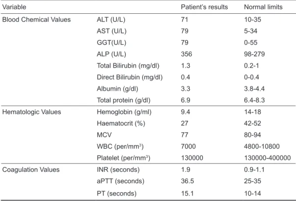
laboratory indings were summarized in table-1. Serum creatinine level and electrolytes were in nor-mal limits. Iron deiciency was detected with serum iron level; 27 µg/dl (50-175), transferrin saturation; 10 % and ferritin; 7.2 ng/ml (18-370). Peripheral blood smear revealed hypochromia and
microcy-tosis. Blood arterial ammonia level was 183µg /dl (45-80). Portal magnetic resonance imaging (MRI) venography showed an increased diameter of portal vein (27 mm), in addition to inferior and superior mesenteric veins. Splenic vein was in connection with renal vein via collateral veins.
Table 1. The results of laboratory tests
Variable Patient’s results Normal limits
Blood Chemical Values ALT (U/L) 71 10-35
AST (U/L) 79 5-34
GGT(U/L) 79 0-55
ALP (U/L) 356 98-279
Total Bilirubin (mg/dl) 1.3 0.2-1 Direct Bilirubin (mg/dl) 0.4 0-0.4
Albumin (g/dl) 3.3 3.8-4.4
Total protein (g/dl) 6.9 6.4-8.3
Hematologic Values Hemoglobin (g/ml) 9.4 14-18
Haematocrit (%) 27 42-52
MCV 77 80-94
WBC (per/mm3) 7000 4800-10800
Platelet (per/mm3) 130000 130000-400000
Coagulation Values INR (seconds) 1.9 0.9-1.1
aPTT (seconds) 36.5 25-35
PT (seconds) 15.1 10-14
Figure 1. Symmetrical hyperintense lesions at bi
-lateral globus pallidi, T1 weighted scan, cranial MRI
Cranial MRI was performed due to repeated encephalopathic states to rule out the organic pa-thologies. MRI demonstrated the symmetric hyper-intense lesions at bilateral globus pallidi which were extending into the mesencephalone on T1 weighted scans (igure-1). Whole blood manganese concen-tration measured by atomic absorption spectro-photometry at admission was 593 nmol/L (72-110 nmol/L). We have started treatment for the HE and iron deiciency anemia. His symptoms improved progressively and during 10 months follow up, HE was not reccured.
DISCUSSION
G.U. Okyay, et al. Portal-systemic encephalopathy with hypermanganesemia 123
Dicle Tıp Derg / Dicle Med J www.diclemedj.org Cilt / Vol 39, No 1, 121-124
in addition to ammonia, levels of manganese were also elevated in the brain 2,3 which may contribute to the pathogenesis of HE.4
Manganese is a trace element, which is primar-ily cleared by the liver. Inadequate elimination of manganese absorbed from the normal diet 5 and in-creased systemic availability due to portal-systemic shunting 2 may lead to manganese overload in the patients with liver disease. Kriger 5 et.al have de-termined the whole blood manganese levels in pa-tients with liver cirrhosis which were signiicantly increased as compared to controls and they have demonstrated the accumulation of manganese in the basal ganglia of patients with end stage liver diseas-es. Spahr and colleagues 1 have also demonstrated increased blood manganese concentrations in cir-rhotic patients, especially with previous portacaval anastomoses or transjugular intrahepatic portosys-temic shunts.
Our patient had the diagnosis of chronic liver disease for 23 years. Cranial MRI revealed bilat-eral symmetric T1 hyperintensities involving glo-bus pallidi and extending into the mesencephalone. This was accepted as the characteristic inding of the manganese deposition.6 Similar patterns of in-creased T1-weighted signal intensities can also be associated with lipid, hemoglobin breakdown prod-ucts, melanoma, neuroibromatosis and calciica-tion, which can be differentiated according to the clinical presentations and radiologic features. Sig-nals induced by calciication can be ruled out by normal cranial computed tomography (CT) ind-ings,7 as performed in our case.
Whole blood manganese level measured via atomic absorption spectrophotometry was 593 nmol/L (72-110 nmol/L). Portal MRI venography established the spontaneous splenorenal shunt. We suggested that increased blood concentration of manganese was a result of impaired clearance of it, due to portal-systemic shunting developed be-cause of hepatocellular dysfunction. In general, the transport of manganese across the intestinal tract is poorly understood. It is thought to occur through mechanisms similar to that regulating non-heme iron uptake.8 Interdependence between manganese and iron on their transport has been demonstrated in some studies.9 Malecki and colleagues 9 found that iron deiciency is an exacerbating factor, for increased intestinal absorption of manganese. In
our patient, iron deiciency anemia due to internal hemoroidal bleeding was detected. We speculated that increased absorption of manganese -in addition to decreased elimination- might have a role in its elevated blood concentration. We added oral iron replacement therapy to the treatment.
The hyperintensity of the T1 signal is related to a high incidence of extrapyramidal dysfunction including rigidity, tremor, akinesia and athetosis in cirrhotic patients.4,10,11 Extrapyramidal symptoms may result from a toxic effect of manganese on bas-al ganglia dopaminergic functions.12 Spahr and col-leagues 1 have reported that there was no signiicant correlation between blood manganese levels and extrapyramidal symptoms. In our patient, although the blood manganese level was elevated signiicant-ly, he only presented with lapping tremor without any other signs of extrapyramidal dysfunction.
Surgical obliteration of portal-systemic shunts or obliteration by interventional radiological tech-niques are fairly effective in reversing intractable portal-systemic encephalopathy, but it is often as-sociated with ascites accumulation and/or forma-tion of esophageal varices.13 In our patient, in spite of the long duration of liver disease, he had never developed HE episode before to the last 3 months. The life threatening risks of intervention were taken into consideration and conservative therapy by re-moving exacerbating factors was initially planned for the patient. The treatment included preventive approaches for bleeding from the hemorrhoids, the iron replacement therapy for the anemia and recom-mendations of the diet poor from the manganese. The patient has remained stable and HE was not noted during 10 months follow up period.
In conclusion, the deposition of manganese in the brain of the patients with chronic liver dis-ease may contribute to the pathogenesis of HE and should be considered especially for the ones having portal-systemic shunts. Manganese overload might be avoided with low manganese diet, removing fac-tors promoting intestinal absorption and probably with chelating agents; which should be addressed in further studies.
REFERENCES
G.U. Okyay, et al. Portal-systemic encephalopathy with hypermanganesemia
124
Dicle Tıp Derg / Dicle Med J www.diclemedj.org Cilt / Vol 39, No 1, 121-124
magnetic resonance signal hyperintensity and neurological symptoms. Hepatology 1996;24(5):1116-20.
2. Riordan S.M, Williams R. Treatment of hepatic encephalopa -thy. N Engl J Med 1997;337 (7):473-9.
3. Choi Y, Park JK, Park NH, et al. Whole blood and red blood cell manganese relected signal intensities of T1-weighted magnetic resonance images better than plasma manganese in liver cirrhotics. J Occup Health 2005;47(1):68-73. 4. Hauser RA, Zesiewicz TA, Rosemurgy AS, Martinez C, Ola
-now CW. Manganese intoxication and chronic liver failure. Ann Neurol 1994;36(6):871-5.
5. Krieger D. Manganese may be of importance in the patho -genesis of chronic hepatic encephalopathy. Biomed Phar -macotherapy 1996;50(2):93-6.
6. Gospe SM Jr, Caruso RD, Clegg MS, et al. Paraparesis, hy -permanganesaemia, and polycythaemia: a novel presenta -tion of cirrhosis. Arch Dis Child 2000;83(5):439-42. 7. Rose C, Butterworth RF, Zayed J, et al. Manganese depo
-sition in basal ganglia structures results from both portal-systemic shunting and liver dysfunction. Gastroenterology 1999;117(3):640-4.
8. Krieger D, Krieger S, Jansen O, Gass P, Theilmann L, Lich -tnecker H. Manganese and chronic hepatic encephalopathy. Lancet 1995;346(8970):270-4.
9. Fitsanakis VA, Zhang N, Garcia S, Aschner M. Manganese (Mn) and iron (Fe): interdependency of transport and regu -lation. Neurotox Res 2010;18:124-31.
10. Weissenborn K, Ehrenheim C, Hori A, Kubicka S, Manns MP. Pallidal lesions in patients with liver cirrhosis: clinical and MRI evaluation. Metab Brain Dis 1995;10(3):219-31. 11. Pomier-Layrargues G, Spahr L, Butterworth RF. Increased
manganese concentrations in pallidum of cirrhotic patients. Lancet 1995;345(8951):735-7.
12. Ikeda S, Yamaguchi Y, Sera Y, et al. Manganese deposition in the globus pallidus in patients with biliary atresia. Trans -plantation 2000; 69(11):2339-43.