Systematic Identification of Cyclic-di-GMP
Binding Proteins in
Vibrio cholerae
Reveals a
Novel Class of Cyclic-di-GMP-Binding ATPases
Associated with Type II Secretion Systems
Kevin G. Roelofs1☯, Christopher J. Jones2☯, Sarah R. Helman1☯, Xiaoran Shang1, Mona W. Orr1, Jonathan R. Goodson1, Michael Y. Galperin3, Fitnat H. Yildiz2, Vincent T. Lee1*
1Department of Cell Biology and Molecular Genetics, University of Maryland at College Park, Maryland, United States of America,2Department of Microbiology and Environmental Toxicology, University of California, Santa Cruz, Santa Cruz, California, United States of America,3National Center for Biotechnology Information, National Library of Medicine, National Institutes of Health, Bethesda, Maryland, United States of America
☯These authors contributed equally to this work. *vtlee@umd.edu
Abstract
Cyclic-di-GMP (c-di-GMP) is a ubiquitous bacterial signaling molecule that regulates a vari-ety of complex processes through a diverse set of c-di-GMP receptor proteins. We have uti-lized a systematic approach to identify c-di-GMP receptors from the pathogenVibrio choleraeusing the Differential Radial Capillary Action of Ligand Assay (DRaCALA). The DRaCALA screen identified a majority of known c-di-GMP binding proteins inV.cholerae
and revealed a novel c-di-GMP binding protein, MshE (VC0405), an ATPase associated with the mannose sensitive hemagglutinin (MSHA) type IV pilus. The known c-di-GMP bind-ing proteins identified by DRaCALA include diguanylate cyclases, phosphodiesterases, PilZ domain proteins and transcription factors VpsT and VpsR, indicating that the DRa-CALA-based screen of open reading frame libraries is a feasible approach to uncover novel receptors of small molecule ligands. Since MshE lacks the canonical c-di-GMP-binding motifs, a truncation analysis was utilized to locate the c-di-GMP binding activity to the N-ter-minal T2SSE_N domain. Alignment of MshE homologs revealed candidate conserved resi-dues responsible for c-di-GMP binding. Site-directed mutagenesis of these candidate residues revealed that the Arg9 residue is required for GMP binding. The ability of c-di-GMP binding to MshE to regulate MSHA dependent processes was evaluated. The R9A allele, in contrast to the wild type MshE, was unable to complement theΔmshEmutant for the production of extracellular MshA to the cell surface, reduction in flagella swimming motil-ity, attachment to surfaces and formation of biofilms. Testing homologs of MshE for binding to c-di-GMP identified the type II secretion ATPase ofPseudomonas aeruginosa
(PA14_29490) as a c-di-GMP receptor, indicating that type II secretion and type IV pili are both regulated by c-di-GMP.
a11111
OPEN ACCESS
Citation:Roelofs KG, Jones CJ, Helman SR, Shang X, Orr MW, Goodson JR, et al. (2015) Systematic Identification of Cyclic-di-GMP Binding Proteins in
Vibrio choleraeReveals a Novel Class of Cyclic-di-GMP-Binding ATPases Associated with Type II Secretion Systems. PLoS Pathog 11(10): e1005232. doi:10.1371/journal.ppat.1005232
Editor:Samuel I Miller, University of Washington School of Medicine, UNITED STATES
Received:June 4, 2015
Accepted:September 25, 2015
Published:October 27, 2015
Copyright:This is an open access article, free of all copyright, and may be freely reproduced, distributed, transmitted, modified, built upon, or otherwise used by anyone for any lawful purpose. The work is made available under theCreative Commons CC0public domain dedication.
Data Availability Statement:All relevant data are within the paper and its Supporting Information files.
Author Summary
Cyclic-di-GMP (c-di-GMP) is a ubiquitous bacterial signaling molecule that regulates important bacterial functions, including virulence, antibiotic resistance, biofilm formation and cell division. The list of known c-di-GMP receptors is clearly incomplete. Here we uti-lized a systematic and unbiased biochemical approach to identify c-di-GMP receptors from the 3,812 genes of theVibrio choleraegenome. Results from this analysis identified most known di-GMP receptors as well as MshE, a protein not known to interact with c-di-GMP. The c-di-GMP binding site was identified at the N-terminus of MshE and requires a conserved arginine residue in the 9thposition. MshE is the ATPase that powers the secretion of the MshA pili onto the surface of the bacteria. We show that c-di-GMP binding to MshE is required for MshA export and the function of the pili in attachment and biofilm formation. ATPases responsible for related processes such as type IV pili and type II secretion were also tested for c-di-GMP binding, which identified theP.aeruginosa ATPase PA14_29490 as another c-di-GMP binding protein. These findings reveal a new class of c-di-GMP receptor and raise the possibility that c-di-GMP regulate membrane complexes through direct interaction with related type II secretion and type IV pili ATPases.
Introduction
Cyclic diguanosine monophosphate (c-di-GMP) is a ubiquitous bacterial nucleotide secondary signaling molecule that regulates cellular processes in response to environmental and cellular stimuli. The elements of this canonical pathway of signal production, signal transduction, altered activity and signal removal were elegantly described by the Benziman lab over twenty-five years ago [1,2]. C-di-GMP is synthesized by diguanylate cyclases (DGCs) via a catalytic GGDEF domain [1,3,4]. Once made in the cell, c-di-GMP binds to macromolecule receptors to allosterically alter their activities. C-di-GMP signaling is terminated through hydrolysis by phosphodiesterases (PDEs) that contain catalytic EAL or HD-GYP domains [5–8]. In the char-acterization of bacterial cellulose synthase inKomagataeibacter xylinus, the Benziman lab dem-onstrated the importance of c-di-GMP in the allosteric activation of the cellulose synthase complex [1,9,10]. Recent structure elucidation of the BcsA-BcsB-c-di-GMP complex validated these early finding and provided a molecular mechanism for c-di-GMP activation of cellulose biosynthesis [11]. Since this initial description of c-di-GMP regulation of cellulose biosynthesis, genome sequencing has revealed genes for DGCs in diverse bacteria indicating that c-di-GMP is a ubiquitous and important signaling molecule in prokaryotes that regulates a variety of phe-notypes [12].
The identification of receptor proteins for c-di-GMP is needed for understanding the regu-lation by this ubiquitous signaling molecule. However, the process of identifying c-di-GMP binding proteins has been challenging for several reasons. First, c-di-GMP simultaneously reg-ulates complex traits including promoting biofilm formation, inhibiting motility and additional pathways [13–15] indicating that there are likely many c-di-GMP receptors in the cell. Second, although there are several defined protein domains that bind c-di-GMP (see below), these domains do not accurately predict c-di-GMP binding proteins. For example, the PilZ domain binds c-di-GMP [16], but the PilZ protein inP.aeruginosa, for which the domain is named, does not bind c-di-GMP [17]. InV.cholerae, there are five PilZ domain proteins, but these five proteins do not fully explain all of the observed c-di-GMP regulated effects [18]. Third, c-di-GMP binds a number of proteins that do not have predicted binding motifs or were predicted
to bind a different ligand. Examples of novel c-di-GMP binding proteins include VpsT [19], VpsR [20] and FlrA [21] inV.choleraeas well as FleQ inP.aeruginosa[22]. In addition, the Clp protein inXanthomoasspecies, a homolog of the cAMP receptor protein (CRP) ofE.coli, binds c-di-GMP rather than cAMP [23,24]. Another example of a c-di-GMP receptor that was not predicted is the BldD ofStreptomyces coelicolorbinds two dimers of c-di-GMP to repress transcription [25]. Together, these studies reveal the diversity of cellular targets of c-di-GMP that mediate complex regulation and the highlight the challenges in identifying the c-di-GMP-binding receptors that are responsible for c-di-GMP regulation.
Many approaches have been utilized to identify c-di-GMP binding proteins. The first pro-teins shown to bind c-di-GMP included the enzymes that make and degrade c-di-GMP. The I-site of DGCs binds c-di-GMP to provide product feedback inhibition of DGC activity [4,26]. Enzymatically active or inactive PDEs are also capable of c-di-GMP binding [5,27–30]. Bioin-formatic studies revealed that the PilZ domain is a c-di-GMP binding domain [16]. In addition, targeted and unbiased approaches have been employed to identify c-di-GMP receptor proteins. In the targeted approach, genes of c-di-GMP regulated processes were tested for c-di-GMP binding [19–25,31,32]. These studies led to the discoveries of the c-di-GMP receptor proteins that have not been predicted by bioinformatics and have motivated identification of novel GMP receptor proteins using systematic approaches. Affinity pull-down assays using c-di-GMP conjugated sepharose resin, biotin, or a tripartite c-di-c-di-GMP capture compound enriched c-di-GMP binding proteins from whole cell lysates, which were subsequently identified by mass spectrometry [33–35]. This approach has also been employed to identify binding proteins of another prokaryotic cyclic dinucleotide, cyclic-di-AMP (c-di-AMP) [36–38]. An alternative unbiased approach utilizes the Differential Radial Capillary Action of Ligand Assay (CALA) to systematically screen protein expression libraries for ligand binding activity. DRa-CALA relies on the differential spreading of bound and unbound radiolabeled ligand when mixed with protein and spotted on a nitrocellulose membrane [39]. In addition, DRaCALA allowed direct detection of c-di-GMP receptors expressed inE.coliwhole cell lysates thus enabling the screening of individual genes from a target genome [39]. This approach has been used onStaphylococcus aureusandEscherichia coliopen reading frame (ORF) libraries to iden-tify c-di-AMP and c-di-GMP binding proteins, respectively [36,40].
Vibrio choleraewas chosen as an organism for a DRaCALA based screen of c-di-GMP bind-ing proteins since an open readbind-ing frame library was available [41] and it is an organisms that extensively utilize c-di-GMP signaling system to regulate motility, biofilm formation, patho-genesis, and survival upon dissemination to environmental reservoirs [42–46]. TheV.cholerae O1 El Tor N16961 genome encodes 62 proteins with domains for c-di-GMP metabolism including 31 GGDEF, 13 EAL, 9 GGDEF+EAL, and 8 HD-GYP domain proteins [47–49]. However, only 5 c-di-GMP receptor proteins have been identified, including 2 PilZ domain proteins PlzC and PlzD [18], and 3 transcription factors VpsT, VpsR, and FlrA [19–21]. From the DRaCALA screen of theV.choleraeopen reading library, a number of predicted c-di-GMP binding proteins were identified. In addition, MshE, an ATPase in the mannose sensitive hem-agglutinin (MSHA) type IV pilus operon, was also revealed as a c-di-GMP receptor. Purified MshE specifically binds c-di-GMP with a high affinity (Kdapproximately 2μM). Screening of
related type II secretion and type IV pili ATPases identified a gene inP.aeruginosa,
type II secretion systems and demonstrate the utility of a DRaCALA screen for identification of c-di-GMP receptor proteins.
Results
DRaCALA screening of a
V
.
cholerae
ORFeome for c-di-GMP binding
activity
We sought to systematically identify protein receptors of c-di-GMP using DRaCALA by indi-vidually testingVibrio choleraeORFs expressed inE.coliwhole cell lysates for32P-c-di-GMP binding activity. The 3,812 unique ORFs from theV.choleraeO1 El Tor N16961 ORFeome pDONR plasmids were recombined into gateway-compatible histidine (His-ORF) or His-malt-ose binding protein (His-MBP-ORF) expression vectors in a single Gateway reaction [50] and selected on agar plates containing either carbenicillin or gentamicin, respectively. For each ORF, multiple transformants were inoculated into a single well of a 96-well microtiter plate to create His-ORF and His-MBP-ORF libraries from which whole cell lysates were generated. Protein expression in whole cell lysates was tested for 348 ORFs by PAGE separation and revealed by staining with Coomassie. A band corresponding to the predicted molecular weight was visualized for 49% of His-ORF and 76% of His-MBP-ORF fusions for a combined coverage of 81% of theV.choleraeORFeome. These results indicate that mostV.choleraeproteins are overexpressed in the His-ORF and His-MBP-ORF libraries, thus enabling a systematic genome-wide DRaCALA screen for c-di-GMP binding proteins.
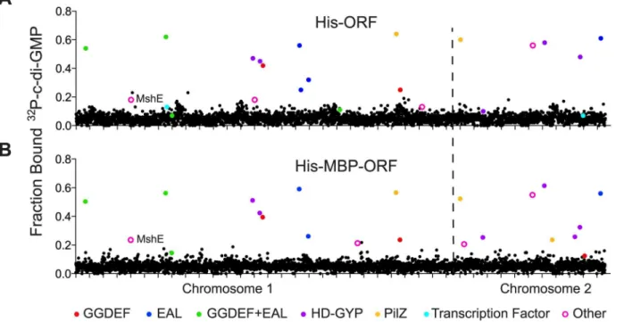
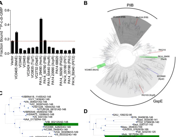
Whole cell lysates from His-ORF and His-MBP-ORF libraries were tested for c-di-GMP binding by DRaCALA using a 96 well pin tool. The fraction bound of32P-c-di-GMP was mea-sured in duplicate for each whole cell lysate and positiveV.choleraeORFs were defined as those having c-di-GMP fraction bound three standard deviations above the mean for both measurements (Fig 1,S1 Table) (seeMaterial and Methods). The positive control expressing PelD, a known c-di-GMP-binding protein, was above the cutoff in each 96-well plate. This pri-mary screen identified 55 His-ORF and 47 His-MBP-ORF proteins that significantly increased the fraction bound of32P-c-di-GMP. A secondary screen was performed to validate these ORFs. In total, 23 His-ORFs and 22 His-MBP-ORFs (28 unique ORFs total) were validated as positive for c-di-GMP binding (Fig 1,Table 1). The specificity for c-di-GMP binding of cell lysates expressing positive ORFs was determined by competition experiments using unlabeled guanosine nucleotides (Table 1). Unlabeled c-di-GMP significantly reduced32P-c-di-GMP binding for ORFs listed inTable 1. In contrast, unlabeled GTP and cGMP did not reduce32 P-c-di-GMP binding, suggesting that the measured binding activity was specific for P-c-di-GMP. Together these results illustrate how sequential high-throughput DRaCALA screens can iden-tify genetic elements that encode proteins with specific ligand binding activity. This screen of theV.choleraeORFeome for GMP binding proteins identified known and candidate c-di-GMP receptors.
Positive ORFs encode c-di-GMP binding proteins
ORFs with GGDEF domains (S1 Fig). Four ORFs (VC0072, VC0658, VC0703 (MbaA), VC1934) were identified with both GGDEF and EAL domains, but lacking the RxxD motif, suggesting that c-di-GMP binding occurs at the EAL domain. Both EAL and HD-GYP
domains can bind c-di-GMP as a substrate for hydrolysis. The screen identified four ORFs con-taining an EAL domain (VC1641, VC1652 (VieA), VC1710, VCA1083) and six ORFs contain-ing a HD-GYP domain (VC1295, VC1348, VCA0210, VCA0681, VCA0895, VCA0931). The V.choleraegenome contains 5 ORFs that encode PilZ domains. While four of these (PlzA, -C, -D, and -E) retain RxxxR and DxSxxG motifs required for c-di-GMP binding, only PlzC (VC2344) and PlzD (VCA0042) have been demonstrated to bind c-di-GMP biochemically [18,
51]. From the DRaCALA-based ORFeome screen, PlzC and PlzD were identified in both ORF and MBP-ORF libraries, while PlzE (VCA0735) was identified only in the His-MBP-ORF library. Previous work demonstrated c-di-GMP binding for His-fusions of PlzC and PlzD but not PlzE, suggesting that a MBP fusion to PlzE may be required for proper fold-ing of the c-di-GMP bindfold-ing site durfold-ing heterologous expression of PlzE [52,53]. Finally, two of three c-di-GMP binding transcription factors, VpsT (VCA0952) [19] and VpsR (VC0665) [20] were identified, but not FlrA (VC2137) [21]. In total, DRaCALA identified 22 of 46 (48%) proteins predicted to bind di-GMP and 6 of 11 (55%) proteins previously shown to bind c-di-GMP (Table 2). These results demonstrate that DRaCALA-based screen can identify all known categories of c-di-GMP binding proteins and represents an unbiased approach to dis-covering receptor proteins of signaling molecules.
Six ORFs were identified which encode proteins that are not known or predicted to bind c-di-GMP, namely VC0405 (MshE), VC1308 (TyrR), VC2066 (FliA), VC2529 (RpoN),
VCA0071 (PstC), and VCA0593 (Table 1). These ORFs represent potentially novel types of c-di-GMP binding proteins. To determine if these proteins bind c-c-di-GMP directly, His-Fig 1. Primary DRaCALA screen ofVibrio choleraeORF libraries.Average fraction bound32P-c-di-GMP vs. individualV.cholerae(A) His-ORFs and (B) His-MBP-ORFs overexpressed inE.coliwhole cell lysates. ORFs are arranged by VC gene number along the X-axis. ORFs validated as positive in the secondary screen are indicated by color and classified by type of c-di-GMP-binding protein.“Other”refers to proteins that have not been predicted or demonstrated to bind c-di-GMP.
MBP-ORF fusions were purified and assayed for c-di-GMP binding activity by DRaCALA. C-di-GMP binding was detected for purified MshE, but not for RpoN, FliA, TyrR, or VCA0593
(S2 Fig). We were unable to purify PstC. These results suggest that heterologous expression of
RpoN, FliA, TyrR, or VCA0593 can induce the expression of c-di-GMP binding proteins encoded within theE.coligenome. In the remainder of the manuscript, we characterize the c-di-GMP binding properties of MshE.
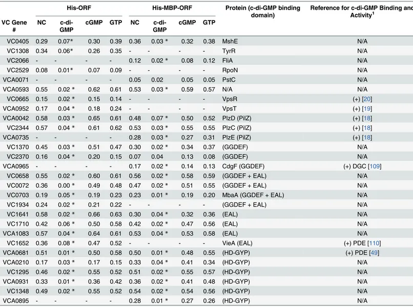
Table 1. Specific binding of validated ORFs to c-di-GMP.
His-ORF His-MBP-ORF Protein (c-di-GMP binding domain)
Reference for c-di-GMP Binding and Activity1
VC Gene #
NC c-di-GMP
cGMP GTP NC c-di-GMP
cGMP GTP
VC0405 0.29 0.07* 0.30 0.39 0.36 0.03* 0.32 0.38 MshE N/A
VC1308 0.34 0.06* 0.26 0.35 - - - - TyrR N/A
VC2066 - - - - 0.12 0.02* 0.08 0.12 FliA N/A
VC2529 0.08 0.01* 0.07 0.09 - - - - RpoN N/A
VCA0071 - - - - 0.05 0.02 0.05 0.05 PstC N/A
VCA0593 0.55 0.02* 0.62 0.61 0.53 0.03* 0.59 0.57 N/A N/A
VC0665 0.15 0.02* 0.15 0.14 - - - - VpsR (+) [20]
VCA0952 0.17 0.04* 0.18 0.24 - - - - VpsT (+) [19]
VCA0042 0.58 0.03* 0.65 0.61 0.48 0.07* 0.50 0.52 PlzD (PilZ) (+) [18]
VC2344 0.57 0.04* 0.61 0.62 0.53 0.03* 0.55 0.55 PlzC (PilZ) (+) [18]
VCA0735 - - - - 0.28 0.03* 0.27 0.31 PlzE (PilZ) (+) [18]
VC1370 0.45 0.03* 0.51 0.47 0.30 0.02* 0.34 0.37 (GGDEF) N/A
VC2370 0.16 0.04* 0.20 0.15 0.07 0.04 0.13 0.08 (GGDEF) N/A
VCA0965 - - - - 0.17 0.02* 0.14 0.13 CdgF (GGDEF) (+) DGC [109]
VC0658 0.55 0.02* 0.60 0.61 0.56 0.02* 0.58 0.59 (GGDEF + EAL) N/A
VC0072 0.36 0.00* 0.49 0.48 0.47 0.02* 0.51 0.55 (GGDEF + EAL) N/A
VC0703 0.19 0.05* 0.19 0.23 0.23 0.01* 0.19 0.20 MbaA (GGDEF + EAL) N/A
VC1934 0.24 0.02* 0.21 0.22 - - - - (GGDEF + EAL) N/A
VC1641 0.58 0.02* 0.66 0.63 0.30 0.04* 0.32 0.36 (EAL) N/A
VC1710 0.42 0.06* 0.50 0.58 0.42 0.02* 0.47 0.56 (EAL) N/A
VCA1083 0.57 0.04* 0.64 0.61 0.53 0.04* 0.53 0.58 (EAL) N/A
VC1652 0.36 0.08* 0.47 0.52 - - - - VieA (EAL) (+) PDE [110]
VCA0681 0.51 0.01* 0.50 0.58 0.50 0.01* 0.48 0.55 (HD-GYP) (+) PDE [49]
VCA0210 0.17 0.03* 0.17 0.15 0.33 0.04* 0.41 0.34 (HD-GYP) N/A
VC1295 0.46 0.02* 0.55 0.52 0.51 0.02* 0.55 0.57 (HD-GYP) N/A
VCA0931 0.33 0.01* 0.36 0.42 0.36 0.02* 0.41 0.48 (HD-GYP) N/A
VC1348 0.49 0.02* 0.55 0.52 0.54 0.02* 0.54 0.56 (HD-GYP) N/A
VCA0895 - - - - 0.28 0.01* 0.27 0.26 (HD-GYP) N/A
1Binding and enzymatic activity is deemed positive if activity has been demonstrated for the puri
fied protein or if mutation of residues required for c-di-GMP binding or enyzmatic activity regulate a c-di-c-di-GMP dependent phenotype.
(+) = c-di-GMP binding observed,
DGC = diguanylate cyclase activity observed, PDE = phosphodiesterase activity observed.
Two-way ANOVA was used to determine if the fraction bound to32P-c-di-GMP in the presence unlabeled c-di-GMP competitor differed signi
ficantly from no competitor (NC).
*= p<0.001.
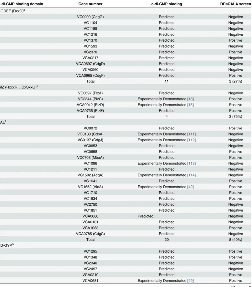
Table 2. Hit rate ofV.choleraeORFs encoding predicted and previously demonstrated cdiGMP-binding proteins.1
C-di-GMP binding domain Gene number c-di-GMP binding DRaCALA screen
GGDEF (RxxD)2
VC0900 (CdgG) Predicted Negative
VC1104 Predicted Negative
VC1185 Predicted Negative
VC1216 Predicted Negative
VC1370 Predicted Positive
VC1593 Predicted Negative
VC2370 Predicted Positive
VCA0217 Predicted Negative
VCA0697 (CdgD) Predicted Negative
VCA0960 Predicted Negative
VCA0965 (CdgF) Predicted Positive
Total 11 3 (27%)
PilZ (RxxxR. . .DxSxxG)
3
VC0697 (PlzA) Predicted Negative
VC2344 (PlzC) Experimentally Demonstrated [18] Positive
VCA0042 (PlzD) Experimentally Demonstrated [18] Positive
VCA0735 (PlzE) Predicted Positive
Total 4 3 (75%)
EAL4
VC0072 Predicted Positive
VC0130 (CdpA) Experimentally Demonstrated [111] Negative VC0137 (CdgJ) Experimentally Demonstrated [112] Negative
VC0653 Predicted Negative
VC0658 Predicted Positive
VC0703 (MbaA) Predicted Positive
VC1086 Experimentally Demonstrated [113] Negative
VC1211 Predicted Negative
VC1592 (AcgA) Experimentally Demonstrated [114] Negative
VC1641 Predicted Positive
VC1652 (VieA) Experimentally Demonstrated [42] Positive
VC1710 Predicted Positive
VC1934 Predicted Positive
VC2750 Predicted Negative
VC1851 Predicted Negative
VCA0080 Predicted Negative
VCA0101 Predicted Negative
VCA1083 Predicted Positive
VCA0785 (CdgC) Predicted Negative
Total 20 8 (40%)
HD-GYP5
VC1295 Predicted Positive
VC1348 Predicted Positive
VC2340 Predicted Negative
VC2497 Predicted Negative
VCA0210 Predicted Positive
VCA0681 Experimentally Demonstrated [49] Positive
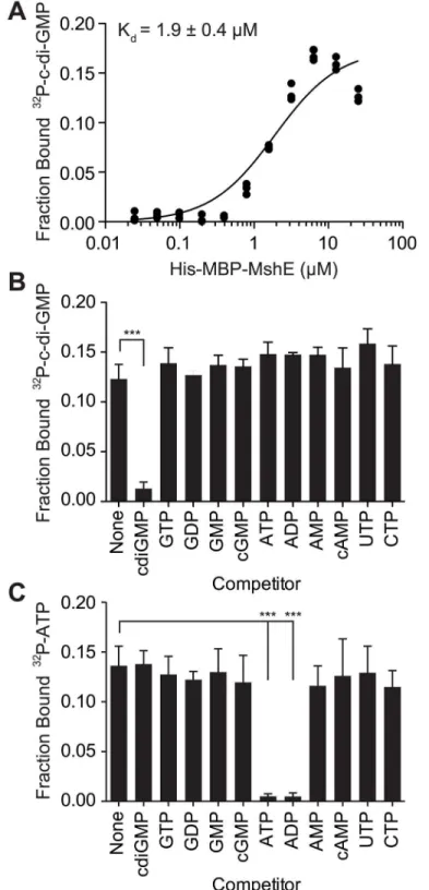
MshE specifically binds c-di-GMP with high affinity
To determine the affinity and specificity of c-di-GMP-binding to MshE, purified His-MBP-MshE was assayed for binding to32P-GMP by DRaCALA. The affinity of c-di-GMP-binding was determined by quantifying the fraction bound of32P-c-di-GMP in serial dilutions of His-MBP MshE (Fig 2A). Non-linear regression analysis of c-di-GMP-binding vs. protein concentration using a one site-binding model estimated the dissociation constant (Kd)
for c-di-GMP to be 1.9 ± 0.4μM. To determine the specificity of c-di-GMP-binding to MshE,
we measured the fraction bound of32P-c-di-GMP in the presence of unlabeled nucleotide com-petitors.32P-di-GMP-binding to His-MBP-MshE was significantly decreased by unlabeled c-di-GMP, but not by GTP, GDP, GMP cGMP, ATP, ADP, AMP, cAMP, CTP, or UTP (Fig 2B). MshE also binds ATP specifically since only ATP and ADP compete for32P-ATP binding (Fig 2C). These results indicate that MshE specifically binds c-di-GMP with micromolar affinity and at a site that is distinct from the ATP binding site.
A subset of ATPases that regulate type IV pili and type II secretion
systems bind c-di-GMP
MshE belongs to a family of ATPases associated with the biosynthesis and retraction of type IV pili and secretion by type II secretion systems. These phylogenetically related ATPases are called as T2SSE ATPases based on the type II secretion system (T2SS) nomenclature for Gen-eral secretion protein E (GspE) [54,55]. To determine if c-di-GMP-binding is a conserved fea-ture of T2SSE ATPases, we identified homologs of MshE and assayed them for c-di-GMP-binding. Protein-Blast search with the full-length MshE amino acid sequence against the com-plete protein sets ofV.choleraeO1 El Tor N16961 andP.aeruginosaPA14 identified five addi-tional ATPases inV.choleraeand nine inP.aeruginosawith an E value<1x10-15. These
ATPases include those required for type IV pili function: PilB (VC2424 and PA14_58750,) PilT (PA14_05180 and PA14_59340), PilU (PA14_05190), and TcpT (VC0835); and type II Table 2. (Continued)
C-di-GMP binding domain Gene number c-di-GMP binding DRaCALA screen
VCA0895 Predicted Positive
VCA0931 Predicted Positive
Total 8 6 (75%)
Transcription Factors
VC0065 (VpsR) Experimentally Demonstrated [20] Positive
VC2137 (FlrA) Experimentally Demonstrated [21] Negative
VCA0952 (VpsT) Experimentally Demonstrated [19] Positive
Total 3 2 (67%)
Total
Overall 46 22 (48%)
Experimental demonstrated 11 6 (55%)
1Only genes represented in theV.choleraeORF library are shown.
2GGDEF containing the RxxD I-site was determined by sequence alignment (S1 Fig) [27]. 3PilZ domains containing the RxxxR
. . .DxSxxG were determined by sequence alignment [16][38]. PlzB is omitted since it lacks the RxxxR. . .DxSxxG
motifs.
4EAL include all proteins containing the EAL domain (PF00563). 5HD-GYP is de
fined as described in [60].
Fig 2. C-di-GMP bind to MshE ATPase with high affinity, specificity and independently of ATP.(A) Fraction bound32P-c-di-GMP to decreasing concentrations of purified His-MBP-MshE. The dissociation constant (Kd) is indicated. Fraction bound of His-MBP-MshE to (B)32P-c-di-GMP and (C)32P-ATP in the presence of 100μM nucleotide competitors. P-value was determined by two-tailed t-test (***p0.001). All data are average of three independent assays and standard deviation is indicated by error bars.
secretion: GspE (VC2732), XcpR (PA14_23990), HxcR (PA14_55440), and HxrA
(PA14_29490) [56–63]. We constructed His-ORF fusions for eachV.choleraeandP. aerugi-nosaT2SSE ATPase and assayed c-di-GMP-binding by DRaCALA inE.coliwhole cell lysate
(Fig 3A). Expression ofV.choleraeMshE andP.aeruginosaPA14_29490, but not other T2SSE
ATPases, significantly increased the fraction bound of32P-c-di-GMP. These data identified MshE and PA14_29490 as c-di-GMP-binding receptor proteins, the only ones among the T2SSE family inV.choleraeandP.aeruginosa.
PA14_29490 is the reciprocal best BLAST hit for MshE [55]. Based on genomic context, MshE functions within the MSHA operon [64,65], while PA14_29490 is encoded within a putative T2SS operon [66,67]. Sequence comparison of T2SSE-type ATPases fromV.cholerae andP.aeruginosashowed that they differ substantially in protein length. MshE and
PA14_29490, both 575 aa long, were ~200 aa longer than PilT ATPases VC0462, VC0463, PA14_05180, PA14_05190, PA14_58760 and PA14_59340. Other T2SSE-type ATPases, anno-tated as GspE- and PilB-type, ranged in length from 469 to 562 aa, and only PA14_68820 was the same length as MshE. All these enzymes had very similar C-terminal ATPase domains and differed primarily in their N-terminal parts, with longer proteins containing an additional domain, referred to as T2SSE_N (PF05157) domain in the Pfam database [48]. A phylogenetic tree of the N-terminal fragments of T2SSE_N-containing ATPases showed that MshE is located in the branch related to PilB ATPases responsible for type IV pili, whereas
PA14_29490 is located in the branch related to GspE ATPases, which participate in type II secretion (Fig 3B). The canonical PilB and GspE ATPases that were tested (VC2424, VC2732 and their counterparts inP.aeruginosa) belong to branches of the tree that are distinct from those including MshE and PA14_29490. Expansion of the branches containing MshE (Fig 3C) and PA14_29490 (Fig 3D) reveal many homologous proteins that may also be receptors of c-di-GMP. Together, these results suggest that a subset of T2SSE ATPases represented by MshE and PA14_29490 are c-di-GMP-binding proteins.
PA14_29490 and MshE bind c-di-GMP specifically in the N-terminal
domain
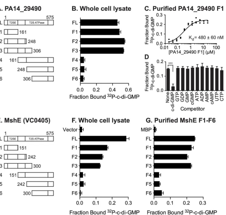
Both MshE and PA14_29490 lack known c-di-GMP binding protein sequence motifs. To locate the binding site(s) on these proteins, truncation analysis was performed on both pro-teins. The N-terminal T2SSE_N domain and C-terminal T2SSE ATPase domain of
PA14_29490 and MshE were separated at three points that were predicted to be at the ends of secondary structural elements (Fig 4A and 4E). These fragments were expressed inE.coliand the whole cell lysates were tested for c-di-GMP binding. The N-terminal fragments (F1-F3) of both PA14_29490 and MshE bound32P-c-di-GMP, while the C-terminal fragments (F4-F6) did not (Fig 4B and 4F). Purified fragment 1 of PA14_29490 binds c-di-GMP with a Kdof
480 ± 60 nM (Fig 4C) and this binding was competed away only with c-di-GMP (Fig 4D), indi-cating that binding is specific. Each of the fragments of MshE was purified and tested for bind-ing to32P-c-di-GMP. Only the purified N-terminal fragments (F1-F3) of MshE bound32 P-c-di-GMP, while the C-terminal fragments (F4-F6) did not (Fig 4G). These results indicate that the binding site for c-di-GMP is located in the N-terminal domain of the protein and is distinct from the ATPase domain in the C-terminus.
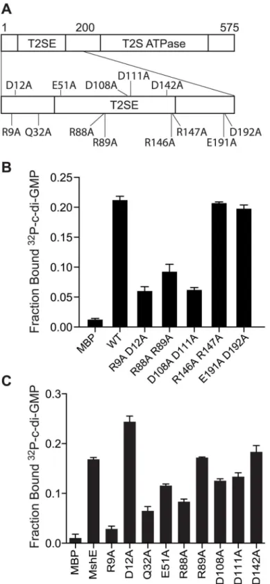
MshE binding to c-di-GMP requires a specific arginine residue
mshE(S3A Fig). ClustalW alignment revealed residues within the first 151 amino acids that are 100% identical among these proteins, including 4 conserved motifs: RLGDLLV, ARRxRAL, SDPADL, and DxxYRRT (S3B Fig). Since charged residues, in particular arginines, have been shown to participate in c-di-GMP binding in a variety of receptor proteins, mutant variants with alanine replacements within these 4 motifs were generated by site-directed mutagenesis including R9A/D12A, R88A/R89A, D108A/D111A, and R146A/R147A (Fig 5A). As a control, the conserved E191 and D192 residues, which are located outside of the first 151 amino acid fragment, were also changed to alanine. Purified R9A/D12A, R88A/R89A, and D108A/D111A variants were reduced for binding to c-di-GMP, whereas R146A/R147A and E191A/D192A did not affect binding to c-di-GMP (Fig 5A). These results indicate that motifs 1, 2 and 3 con-tribute to di-GMP binding, while motif 4 is dispensable. The E191A/D192A variant binds c-di-GMP similarly to the wild-type protein (Fig 5A), in agreement with the results from frag-ment analysis (Fig 4E). To determine the contribution of each amino acid residue within the Fig 3. C-di-GMP binds toV.choleraeandP.aeruginosahomologs of MshE.(A) Average fraction bound32P-c-di-GMP ofE.coliwhole cell lysate expressingVibrio choleraeandPseudomonas aeruginosahomologs of MshE. The dashed red line indicates background binding for a vector control strain. All data are average of three independent assays and standard deviation is indicated by error bars. (B) Unrooted phylogenetic tree of the T2SSE_N domain. Protein sequences present in the tree corresponding to proteins analyzed for c-di-GMP binding are highlighted in green (binds c-di-GMP), red (does not bind), or blue (candidate binding protein). The dark grey background corresponds to primarily type IV pili PilB sequences, and the light grey background corresponds to type II secretion protein E ATPase sequences. (C, D) Sub-trees containing VC0405 or PA14_29490 and closely related proteins.
first 3 motifs and the other conserved, charged amino acids, we generated and purified MshE variants with single alanine substitution in positions R9, D12, Q32, E51, R88, R89, D108, D111 and D142 (S4A Fig). The R9A variant had an 83% reduction in c-di-GMP binding, while Q32A and R88A variants had 61% and 50% reduction, respectively (Fig 5C). Interestingly, the R9/D12 residues represent an RxxD motif described in DGC I-site [26], PelD [31] and GIL domain of BcsE [40]. In those proteins, both residues are critical for c-di-GMP binding. In con-trast, the MshE D12A variant actually binds c-di-GMP better than wild-type MshE (Fig 5C), Fig 4. C-di-GMP binds MshE and PA14_29490 in the N-terminal T2SSE_N domain.Schematic of the truncations generated in (A) PA14_29490 and (E) MshE. C-di-GMP binding toE.coliwhole cell lysates expressing each fragment of (B) PA14_29490 and (F) MshE. (C) Fraction bound32P-c-di-GMP to decreasing concentrations of purified PA14_29490 fragment 1. (D) Fraction bound of purified PA14_29490 fragment 1 to32P-c-di-GMP in the presence of 100μM nucleotide competitors. (G) Fraction bound of32P-c-di-GMP to purified MshE and fragments 1
–6. Indicated p-value was determined by two-tailed t-test (***p0.001) by comparing the indicated samples. All data are average of three independent assays and standard deviation is indicated by error bars.
Fig 5. R9 is required for MshE to bind c-di-GMP.(A) Schematic of the charged and polar conserved residues in the first 200 amino acids of MshE targeted for site-directed alanine substitution. (B) Fraction bound of32P-c-di-GMP to purified MshE (WT) and indicated pairs of alanine substitutions. (C) Fraction bound of32P-c-di-GMP to purified MshE (WT) and indicated individual alanine substitution. All data are average of three independent assays and standard deviation is indicated by error bars.
indicating that MshE does not contain an I-site-like binding sequence. Each of these MshE var-iants was also tested for ATP binding. The R88A variant showed an 88% reduction in ATP binding and was the only variant that had a reduction by more than 50% (S4B Fig). Thus, the defect associated with c-di-GMP binding in R88A variant may be due to a general folding problem for this specific protein. Together, these results indicate MshE contains a novel c-di-GMP binding site that requires R9 residue with contribution from the Q32 residue.
MshE binding to c-di-GMP enhances ATPase activity
The effect of c-di-GMP on the ATPase activity of MshE was assessed by testing the WT MshE and the R9A proteins in the presence and absence of c-di-GMP. WT MshE produced 68μM of
phosphate from ATP without c-di-GMP and increased to 75 and 76μM with the addition of
10 and 33μM of c-di-GMP, respectively (S5 Fig). In contrast, R9A protein produced 60, 64,
and 58μM of phosphate with addition of 0, 10, and 33μM of c-di-GMP. These results indicate
that the ATPase activity is increased for WT MshE in response to c-di-GMP at concentrations above the dissociation constant, while c-di-GMP had no effect on the R9A protein. However, the magnitude of the enhanced ATPase activity is only about 10% suggesting that c-di-GMP may have additional effects on MshE interaction with the MshA substrate or other components of the MSHA export machinery.
C-di-GMP binding to MshE is required for MSHA function in attachment
and biofilm formation
MSHA is a responsible for initial attachment ofV.choleraeto surfaces and subsequent biofilm formation [68]. Recently, MSHA was demonstrated to reduce flagella mediated swimming motility [69]. We sought to determine whether MshE is the c-di-GMP receptor regulating MSHA activity by assessing the amount of MshA pili on the bacterial surface, the effect on swimming motility, and biofilm formation. The effect of MshE on the export of MshA to the surface ofV.choleraewas assessed by surface ELISA using antibodies specific to MshA (Fig 6A, WT andΔmshA). TheΔmshEmutant is defective for MshA export (Fig 6A). This defect was complemented by wild typemshEand theD12Aallele, but not theR9Aallele (Fig 6A). Com-plementation with theR88A/R89Aallele was able to restore the export of ~10% of MshA observed in wild type cells. A recent study revealed thatV.choleraeswimming motility is reduced by interaction of MSHA with surfaces [69].V.choleraewas assessed for motility through soft agar assay. Wild typeV.choleraehad reduced swimming motility and theΔmshE mutant has increased motility (Fig 6B, WT andΔmshEvector) recapitulating previous observa-tions [69]. The ability of either wild-typemshEormshEvariant under thetacpromoter in trans on a plasmid to complementΔmshEphenotypes were evaluated. Induction of either allele that binds c-di-GMP, wild-typemshEor theD12Avariant, reduced motility to wild type levels
(Fig 6B). Induction of the variant defective for c-di-GMP binding (R9A) failed to reduce
complemented withR9Ashowed small patches of biofilms similar toΔmshEwith the vector control (Fig 6D). Quantification of the biofilms revealed that both biomass (Fig 6E) and surface coverage (Fig 6F) are reduced forΔmshEcomplemented withR9A. Together, these results indi-cate that the ability of MshE to bind c-di-GMP via the R9 residue is required for MshA export to the cell surface and MSHA-mediated phenotypes.
Discussion
Evaluation of DRaCALA ORF screens for identification of ligand binding
proteins
DRaCALA screens are a relatively new method for identifying ligand binding proteins that combines arrayed protein libraries with a high-throughput biochemical assay of protein-ligand interactions. Two recent publications have successfully used DRaCALA screening to identify novel cyclic-dinucleotide protein receptors. A screen for c-di-AMP binding proteins in the Staphylococcus aureusstrain COL ORFeome identified PstA and KdpD [36]. The screen also identified KtrA, which was the only protein identified by affinity pull-down using c-di-AMP magnetic beads in the same study [36]. The crystal structures of PstA-c-di-AMP [70–72] and KtrA-c-di-AMP complexes [73] have been solved. KdpD binding motif has been recently iden-tified [74]. Since the DRaCALA screen, one other additional c-di-AMP receptor has been iden-tified in Gram-positive bacteria, pyruvate carboxylase ofListeria monocytogenes[38]. TheS. aureuspyruvate carboxylase has different residues within the binding motif than theL. mono-cytogenesand does not bind c-di-AMP [38]. Together these studies demonstrate that DRa-CALA was able to identify three novel bona-fide c-di-AMP receptors and correctly not detect a protein that does not bind c-di-AMP. Additionally, a DRaCALA screen for c-di-GMP binding proteins in theE.coliK12 ASKA overexpression gene library revealed three clones overexpres-sing putative c-di-GMP binding proteins: BcsE, IlvH and RimO [40]. Of these, BscE was fur-ther characterized and shown to contain a novel c-di-GMP binding domain, which the authors named GIL [40]. In conjunction with the results from these screens, our identification of MshE as a c-di-GMP receptor important for MSHA pilus function inV.choleraedemonstrates that DRaCALA is a powerful approach for finding new small-molecule receptors across different bacterial species.
The results of these three DRaCALA screening experiments allow us to further assess the limitations of the screen. Our screen of theV.choleraeORFeome identified many, but not all, of the expected binding proteins. These false negatives could be due to several factors. First, the enzymatic activity of these proteins may prevent detection. Phosphodiesterases active in the assay conditions could degrade the32P-c-di-GMP probe prior to application on nitrocellulose, while active diguanylate cyclases could produce excess c-di-GMP to compete for binding with the subsequently added32P-c-di-GMP probe. The failure to detect binding in many EAL and GGDEF domain proteins in our screen, as well as the lack of binding detected for all I-site-con-taining GGDEF proteins assayed in theE.coliDRaCALA screen [40], could be due to these fac-tors. Second, poor expression of the ORF can result in a false negative because DRaCALA relies on the expression of proteins above the Kd. In our assay of protein expression, we found that
only a subset of the ORFs tested had a protein band of the correct size on SDS-PAGE. Similarly, some false negatives of c-di-GMP binding proteins in theE.coliscreen were due to poor pro-tein expression as detected by propro-tein band intensity on SDS-PAGE [40]. Third, the DRaCALA with confocal microscopy 24 h post inoculation in a flow cell system. Scale bar represents 40μm. (E,F) COMSTAT quantitative analysis of biomass and surface coverage of biofilms from (D). Three images from each of two independent experiments were analyzed.
screen interrogates binding of individually expressed ORFs within a heterologous system. Thus proteins that require endogenous binding partners or activating factors to interact with ligand may not be active. Our screen also yielded false positives, which could be due to two factors. First, the expression of proteins that can activate the production of c-di-GMP binding proteins encoded in theE.coligenome can result in a positive signal. Second, the statistical method uti-lized to identify“positive”fraction bound may falsely identify proteins whose fraction bounds are near the cutoff, as was likely the case for Adk, a false positive result in theS.aurus c-di-AMP screen [36]. Both these categories of false positives can be detected after re-assaying bind-ing with purified protein.
We suggest several methods to increase the fidelity of DRaCALA-based screens: 1. Testing the ORFs with multiple fusion proteins can increase the likelihood of overexpressing recombi-nant proteins that retain ligand binding activity, 2. Express the ORFeome in a strain genetically modified to remove endogenous c-di-GMP signaling components and thus reduce the likeli-hood of false positives [75,76], and 3. Alter the buffer used to resuspend lysates—in the case of EAL domain PDE-As, resuspension in a buffer containing Ca2+rather than Mg2+can inhibit the activity of PDE-As [2] and increase the likelihood of detecting proteins that degrade c-di-GMP. Although DRaCALA did not identify all c-di-GMP proteins in theV.choleraegenome, DRaCALA is an unbiased approach that allows discovery of novel receptor protein such as MshE. We believe DRaCALA-based approach can be a powerful tool for the discovery of novel receptor proteins of other small nucleotide signaling molecules.
MshE is the founding member of a new class of c-di-GMP receptor
The MshE is a bona fide c-di-GMP binding protein was demonstrated by 1. High affinity bind-ing with the Kdof 1.9μM, 2. High specificity of binding based on competition assays, 3. Adefined binding site located in the N-terminal T2SSE_N domain and 4. The requirement of the conserved R9 residue for c-di-GMP binding in vitro and for MSHA function in vivo. MshE represents a new category of c-di-GMP binding proteins since it lacks any of the previously defined c-di-GMP binding domains (DGC I-sites, EAL, HD-GYP, PilZ or GIL) and is the first T2SSE ATPase demonstrated to bind c-di-GMP. The conserved R9 and D12 residues are remi-niscent of the RxxD c-di-GMP binding motif present in the I-site of DGCs or the RxGD bind-ing motif present in GIL domains. For both DGC I-sites and GIL domains, the R and D residues are required for c-di-GMP binding. In contrast, MshE requires only the R9 residue for binding to c-di-GMP, while the D12 residue of MshE is dispensable. In addition to MshE, only one other type II secretion/type IV pili ATPase, PA14_29490 fromP.aeruginosa, bound c-di-GMP. Other members of this subfamily likely will also have the ability to bind c-di-GMP, including XpsE fromX.campestris(Fig 3B, blue line). In contrast, related ATPases including PilT and PilU are unlikely to be regulated by c-di-GMP since they are shorter and lack the T2SSE_N domain. Although MshE and PA14_29490 both bind c-di-GMP in the N-terminal T2SSE_N domain, their sequence conservation within this domain is quite low. Nonetheless, proteins containing the T2SSE_N domain should be investigated for their ability to interact with c-di-GMP (Fig 3B, 3C and 3D).
Predicted structure of the c-di-GMP binding fragment of MshE
and Q32 residues. However, these residues were conserved in the GpsE/XpsEN domain from X.campestris(S6A Fig). Remarkably, theX.campestrisXpsEN was crystallized in two different forms that reflect two distinct conformational states that differ in the position of two N-termi-nal helices [78], which include the R9 and Q32 residues (S6B Fig). In one of the structures (PDB: 2D27), R9 and Q32 are positioned within a reasonable distance from each other and, upon the rotation of first helix, could form a potential c-di-GMP-binding site (S6B Fig). In the other structure (PDB: 2D28), the region around Q32 proved so flexible that the exact position of Q32 could not be resolved [78], which is consistent with the ability of this residue to move around and participate in ligand binding. Such flexibility has also been observed in the binding sites of PilZ-containing c-di-GMP receptors [18,51]. The conformational change of the two N-terminal helices of MshE upon binding c-di-GMP has the potential to alter its activity. Thus, type II secretion/type IV pili ATPases with the conserved arginine and glutamine residues cor-responding to R9 and Q32 of MshE represent candidate c-di-GMP receptors (S6B Fig).
Implications of MshE binding to c-di-GMP in the regulation MSHA during
the
V
.
cholerae
infection cycle
V.choleraecycles between environmental reservoirs and human infections in part using two type IV pili, MSHA and Tcp. MSHA contributes to theV.choleraeinfection cycle by maintain-ing an environmental reservoir through attachment to chitin surfaces [80,81] and tolerance to environmental hypotonic stress [82] through biofilm formation [83]. Upon entry into the next host, the preformed biofilms protectV.choleraefrom lower pH [84] and bile [85]. Subsequently, V.choleraeexpress toxin co-regulated pili (Tcp) to promote colonization [86,87] while concom-itantly turning off MSHA [88]. The repression of MSHA within the host is critical since a strain with themshapromoter replaced with the constitutive Placpromoter was outcompeted by the
wild type strain in an infant mouse infection model [88]. This defect can be attributed to binding of secretory IgA directly to MSHA since these strains do not show a competitive difference in mice lacking IgA [88] indicating that repression of MSHA during infection is a necessary for immune evasion. Based on recombination-based in vivo expression technology (RIVET) studies [46],tcpAis transcribed while themshoperon is repressed early in the infection; in contrast, the expression pattern is reversed at the late stage of infection. The decrease inmshtranscription early in the infection is likely regulated in part by the reduced levels of c-di-GMP since several DGCs are not expressed until late in the infection [46]. However, the precise mechanism for reg-ulation of the c-di-GMP signal is subject to strain specificity [49,89] and changes in the host microenvironment. Another mechanism to reciprocally regulate Tcp and MSHA is from the TcpJ pre-pilin peptidase. TcpJ has the unique property of processing both TcpA [90] and MshA [91], but cleavage of MshA by TcpJ leads to rapid degradation [91]. Our finding that MshE binding to c-di-GMP is required for MSHA production and function adds another mechanism to enhance the switch between Tcp and MSHA pili. Upon entering the host, the c-di-GMP levels are reduced, thus reducing both transcription of themshoperon and the function of existing export machinery to export MshA. In combination with the degradation of newly synthesized MshA by TcpJ,V.choleraecan quickly change from the immunogenic MSHA to the adhesive Tcp in the host. Late in the infection, this regulation is reversed allowing the bacteria exiting the host to express MSHA instead of Tcp to prepare for the environment.
Implications of MshE on c-di-GMP regulation of type IV pili and type II
secretion systems
surfaces and biofilm formation. These studies show that all of these effects are lost in themshE R9Amutant that is defective in binding to di-GMP. The implication of our results is that c-di-GMP binding to MshE is required for its activity in polymerizing MshA. This finding is intriguing since several type IV pili are regulated by c-di-GMP through different mechanisms. In bothXanthomonasandPseudomonasspp., type IV pili are regulated by PilZ and FimX pro-teins [92–95]. However, the precise mechanisms of regulation appear to be different. InX. axo-nopodis citriandX.campestris, PilZ binds FimX and the PilB ATPase, a homolog of MshE, to form a tripartite regulatory complex, but these interactions are not conserved inP.aeruginosa [94,96,97]. In addition to regulating the PilB ATPase, c-di-GMP interacts with a second PilZ-domain protein XC_2249 to regulate interactions the PilT and PilU retraction ATPases inX. campestris[98]. These studies and our MshE results indicate that c-di-GMP regulates type IV pili ATPases through a number of different mechanisms. The ability of PA14_29490 to bind di-GMP indicates that there may be also complex regulation of type II secretion systems by c-di-GMP.
Sustained sensing of c-di-GMP through multi-tiered regulation
C-di-GMP regulates major lifestyle changes in response to altered environmental cues. These changes incur a high cost in expenditure of cellular resources as well as an opportunity cost of committing to a sessile lifestyle. There is an emerging pattern in which c-di-GMP regulates the same phenotype at the transcriptional and post-translational levels. Examples of this include Pel polysaccharide synthesis inP.aeruginosa[31,99], and now MSHA inV.cholerae[100]. The idea of regulation by c-di-GMP first to activate gene expression and later to activate pro-tein function can be thought of as“sustained sensing”. Sustained sensing enables bacteria to repeatedly assess environmental and cellular conditions through multi-tiered regulation, and has been previously described for responses to iron availability, oxidative stress, and other sig-nals [101,102]. The concept of sustained signaling applied to c-di-GMP enables prediction of additional GMP receptors. In addition to known examples, there are additional c-di-GMP-regulated processes that could be regulated by sustained sensing. These include two clas-ses: 1. Operons that are transcriptionally regulated by c-di-GMP, but lack known c-di-GMP receptor proteins and 2. Operons that encode di-GMP receptor proteins, but lack known c-di-GMP transcriptional regulation. Examples of the first class include a number of biosynthetic operons of extracellular polysaccharides, such Vps and Eps inV.cholerae[19,100,103], Psl in P.aeruginosa[22],xagABCinX.campestris[32,104], and Bcam1330-Bcam1341 in Burkhol-deria cenocepacia[105]. These operons also likely encode a c-di-GMP binding protein to regu-late polysaccharide biosynthesis at the post-translational level. Examples of the second class include cellulose synthesis inE.coli[40], the alginate biosynthesis operon inP.aeruginosa[17] and the large adhesin protein (Lap) ofP.fluorescens[28]. Furthermore, c-di-GMP can also bind to riboswitches [106,107] to provide another regulatory tier in sustained sensing. There-fore, based on the emerging theme of sustained sensing of c-di-GMP through multi-tiered reg-ulation, we suggest that newly discovered processes regulated by c-di-GMP either
transcriptionally or post-translationally be investigated for additional levels of c-di-GMP control.
Materials and Methods
Gateway destination vector construction
fusions respectively. The gateway destination cassette was amplified from pRFA and cloned in frame with the N-terminal fusions to produce the gateway adapted vectors.
His-ORF and His-MBP-ORF expression library construction
TheV.choleraeN16961 ORF library was obtained from BEI. LR Clonase reactions were per-formed per NEB protocol using miniprepped pDONR vectors from theV.cholereaORFeome in combination with pVL791 Cb GW and pVL847 Gn GW destination vectors. Gateway reac-tions were transformed into anE.coli T7IQstrain (NEB) and recombinants were selected on LB agar plates containing either carbenecillin or gentamycin. Multiple colonies from individual transformations were inoculated in LB M9 rich media in 96-well plate format and grown over-night with shaking at 30°C. Overover-night cultures were subcultured 1:50 into fresh media and grown for 4 hours at 30°C with shaking. Protein expression was induced by addition of 1 mM IPTG and cultures were grown for an additional 4 hours after induction. 1.5 mL of induced cul-ture was centrifuged and cells were resuspended in 150μL of c-di-GMP-binding buffer
supple-mented with 10μg / mL DNAse, 250μg / mL Lysozyme and 1μM PMSF. 20μL aliquots were
transferred to 96-well microtiter plates and stored at -80°C.
DRaCALA
Whole cell lysates for DRaCALA screening were prepared by freeze-thawing resuspended cells in microtiter plates a total of 3 times. After the final thaw, 20μL of c-di-GMP-binding buffer
supplemented with 16 pM32P-c-di-GMP and 500 mM unlabeled GTP was added to whole cell lysate plates or purified proteins. 2μL of this mixture was then spotted in duplicate on
nitrocel-lulose using a 96-well pin tool. DRaCALA of purified proteins was performed with concentra-tions of protein and unlabeled competitor as indicated. Spots were allowed to dry completely (about 20 minutes) before exposing a phosphorimager screen and capturing with a Fujifilm FLA-7000. Photostimulated luminescence (PSL) from the inner spot and total PSL of the spot were quantitated with Fuji Image Gauge software. The fraction bound was calculated using measurements of the total area (Aouter), the area of the inner circle (Ainner), the total PSL
inten-sity (Itotal), and the inner intensity (Iinner) as described [39]. DRaCALA was also used to
deter-mine the ability of purified MshE and variants to bind c-di-GMP. Dissociation constants were estimated assuming a one-site binding model by a nonlinear regression of protein concentra-tion and fracconcentra-tion bound where, Fracconcentra-tion bound = (Maximum possible Fracconcentra-tion Bound)
[pro-tein concentration] / (Kd + [pro[pro-tein concentration]).
Identification of positive
V
.
cholerae
ORFs in primary and secondary
DRaCALA screens
In the primary screen of His-ORF and His-MBP-ORF libraries, mixtures of whole cell lysate and radiolabeled32P-c-di-GMP were spotted twice. To identify DRaCALA spots with signifi-cantly increased fraction bound32P-c-di-GMP, a positive cutoff three standard deviations above the mean fraction bound was created for each 96-well plate of whole cell lysates. Positive spots were iteratively removed from calculations of mean and standard deviation for individual plates, thereby decreasing the positive cutoff until no additional positives were identified. His-ORFs and His-MBP-His-ORFs for which both DRaCALA spots had positive binding were defined as positive and subjected to a secondary screen. Additionally, 8 His-ORF and 8 His-MBP-ORFs with only 1 DRaCALA spot with positive binding were also included in the secondary screen, but none of these ORFs were positive for c-di-GMP binding in the secondary screen.
His-ORF and His-MBP-ORF libraries. Each His-ORF and His-MBP-ORF lysate was spotted twice by DRaCALA and individual lysates were compared to a set of 8 lysates generated from vector controls. Positive fraction bound32P-c-di-GMP for DRaCALA spots was defined as those with at least 2 fold increase above the average fraction bound for the set of plate-matched vector controls. Additionally, each lysate was assayed by PCR to verify the size of the inserted ORF. Positive ORFs from this secondary screen displayed positive fraction bound32 P-c-di-GMP for both DRaCALA spots created from PCR positive lysates.
Generation of phylogenetic tree
Protein sequences corresponding to COG2840 were obtained from the EggNOG 4.1 database. Each sequence aligned to Pfam Family PF05157 (Type II Secretion System, protein E, N-termi-nal domain) using the version 10 HMM with HMMer 3.1 and the subsequences corresponding to the T2SSE N-terminal domain were extracted. The remaining 1437 domain sequences were aligned using the MAFFT 7.157b E-INS-i algorithm and trimmed using TrimAl 1.4 to elimi-nate columns with more than 90% gaps. An unrooted phylogenetic tree was constructed using FastTree 2.1.8.
Strains and plasmids
Strains used are listed inS2 Table. Plasmids are listed inS3 Table.
Generation of MshE fragments and site-directed alanine substituted
variants
Fragments of MshE and PA14_29490 were generated using the primers indicated inS4 Table. Site-directed alanine substitutions of MshE were generated by PCR amplification with the indi-cated primers, DpnI digest and transformation intoE.coliDH5α. pVL791-MshER88A, pVL791-MshER89A, pVL791-MshED111A were generated using the primers indicated inS3
Tableand the NEB Q5 Site-Directed Mutagenesis Kit. All constructs were verified by DNA
sequencing.
Protein expression and purification
V.choleraeMshE and variants were purified as previously described [39]. Briefly,E.coliT7Iq strains orE.coliBL21(DE3) containing expression plasmids were grown overnight, subcul-tured in fresh media and grown to OD600~1.0 when expression was induced with 1 mM IPTG.
Induced bacteria were pelleted and resuspended in 10 mM Tris pH 8, 100 mM NaCl, 25 mM imidazole and frozen at -80°C until purification. Proteins were purified over a Ni-NTA column and eluted with 10 mM Tris pH 8, 100 mM NaCl, 250 mM imidazole. Purified proteins were exchanged into 10 mM Tris pH 8, 100 mM NaCl using Sephadex G25. Proteins were aliquoted, and frozen at -80°C until use.
Plate motility assay
Motility plates consist of LB containing 0.3% agar supplemented with 20μg/mL ampicillin and
25 or 100μM IPTG where appropriate. Plates were poured and allowed to dry at room
Confocal laser scanning microscopy (CLSM) and flow cell biofilm studies
Inoculation of flow cells was done by diluting overnight-grown cultures to an OD600 of 0.04 and injecting into aμ-Slide VI0.4 (Ibidi, Martinsried, Germany). To inoculate the flow cellsur-face, bacteria were allowed to adhere at room temperature for 1 h. Flow of 2% v/v LB (0.02% tryptone, 0.01% yeast extract, 1% NaCl; pH 7.5) containing 20μg/mL ampicillin and 100μM
IPTG was initiated at a rate of 7.5 ml/h and continued for 24 h. Confocal images were obtained on a Zeiss LSM 5 PASCAL Laser Scanning Confocal microscope. Images were obtained with a 40X dry objective and were processed using Imaris (Bitplane, Zurich, Switzerland). Quantita-tive analyses were performed using the COMSTAT software package [108]. Statistical signifi-cance was determined using Oneway ANOVA with Dunnett’s Multiple Comparison test. Two biological replicates were performed in triplicate. Images presented are from one representative experiment.
Surface pilin ELISA
Surface pili composed of MshA were quantified using an ELISA based on a previously pub-lished protocol [92]. Briefly, overnight culture was diluted 1:100 in fresh LB medium and grown to OD6000.5 at 30°C. Cells (125μL) were added to a 96-well plate (Greiner Bio-One,
Monroe, NC) and incubated at 30°C for one hour. Cells were fixed with 100μL of methanol for
10 minutes at room temperature, then washed twice with PBS. Samples were blocked in 5% nonfat dry milk and immunoblotted with polyclonal rabbit anti-MshA (1:1000 dilution, gift of J. Zhu) and horseradish peroxidase (HRP)-conjugated secondary antibody (Santa Cruz Bio-technology, Santa Cruz, CA). After three washes in PBS, 100μL of TMB (eBioscience, San
Diego, CA) was added and incubated for 30 minutes at room temperature followed by the addi-tion of 100μL of 2N H2SO4. Absorbance was recorded at 490nm and the samples were
normal-ized to the change in WT. Two biological replicates were assayed in duplicate and statistical significance was determined with a Oneway ANOVA followed by a Dunnett’s Multiple Com-parison test.
Protein purification
E.coliBL21 harboring plasmids for gene expression were grown to an OD600of 0.4 at 30°C in
LB containing 100μg/mL ampicillin. Cultures were shifted to 18°C and IPTG was added to a
final concentration of 100μM. 16h post induction, cells were harvested by centrifugation at
10,000 x g for 15 minutes and stored at -80°C. Cell pellets were resuspended in GST Lysis Buffer (25mM Tris pH 8.0, 0.5M NaCl containing PI cocktail tablets (Roche Life Science, India-napolis, IN). Cells were lysed by sonication and cell lysate was cleared via centrifugation. Cleared lysate was loaded onto GS4B resin and washed with five column volumes of lysis buffer. Protein was eluted from the resin in 5mL elution buffer (25mM Tris pH8.0, 0.25M NaCl, 10mM glutathione). Samples were dialyzed against buffer (25mM Tris-HCl, 150mM NaCl, 250μM DTT, pH 7.5) overnight using 12 kDa cutoff dialysis tubing (Fisherbrand,
Pitts-burgh, PA) and concentrated to approximately 1mL using an Amicon 10KDa cutoff spin filter (EMD Millipore, Darmstadt, Germany). An aliquot of dialyzed protein was diluted in 6M gua-nidinium HCl and concentration determined via A280.
ATPase assay
Purified protein in buffer (25mM TrisHCl pH 7.5, 100mM NaCl) was added to the standard reaction mixture to a final concentration of 1μM with the indicated concentration of
c-di-GMP. After a 10 minute incubation at room temperature, ATP was added to a final concentra-tion of 10mM and reacconcentra-tions were incubated at 22°C for 30 minutes. Producconcentra-tion of inorganic phosphate was monitored by reading OD360and compared to a standard curve of solutions of
KH2PO4. The experiment was performed in triplicate. Significance was determined via
ANOVA and Bonferroni test.
Supporting Information
S1 Fig. Alignment ofV.choleraeGGDEF domains.V.choleraeproteins which encode a
GGDEF domain were aligned by Clustal-W. C-di-GMP binding was predicted by the presence of an RxxD motif (underlined in red) and DGC activity was predicted by the presence of a GGDEF motif (underlined in black). The presence of protein-encoding ORFs in theV.cholerae ORF library, potential c-di-GMP binding domains, prediction of activity, protein names,V. choleraeORF numbers, and residues comprising the GGDEF domain are indicated. VCA0560 encodes 2 GGDEF domains.
(PDF)
S2 Fig. Testing c-di-GMP binding to purified ORFs identified in the DRaCALA screen that lack known c-di-GMP binding domains.(A) Purified MBP-FliA (VC2066), MBP-RpoN (VC2529), MBP-TyrR (VC1308), and MBP-VCA0593 were separated on a 12% PAGE and stained with Coomassie Brilliant Blue. (B)32P-c-di-GMP binding to purified MBP-FliA (VC2066), MBP-RpoN (VC2529), MBP-TyrR (VC1308), and MBP-VCA0593. All data are average of three independent assays and standard deviation is indicated by error bars. (EPS)
S3 Fig. Conserved amino acids in MshE revealed by BLAST-P and ClustalW alignment.(A) BLAST-P scores for proteins most homologous to MshN, MshE and MshG. The genus and species and the scores for each gene are indicated. The maximum score is forV.cholerae. (B) ClustalW alignment of the first 237 amino acids of MshE homologs is shown. Amino acids shaded in gray are 100% identical in all homologs. Asterisks indicate charged and polar resi-dues targeted with site-directed mutagenesis. Position 151 indicates the C-terminus of Frag-ment 1 that retains c-di-GMP binding activity.
(EPS)
S4 Fig. Purification of MshE point mutants and their ability to bind32P-ATP.(A) Purified MshE and variants with indicated single alanine substitution were separated on a 12% PAGE and stained with Coomassie Brilliant Blue. (B)32P-ATP binding to purified MshE and variants with indicated single alanine substitution. All data are average of three independent assays and standard deviation is indicated by error bars.
(PDF)
S5 Fig. MshE binding to c-di-GMP enhances ATPase activity.ATPase activity of WT MshE and R9A proteins was assessed by detecting free phosphate released using the EnzCheck phos-phate assay in the presence of 0, 10 and 33μM c-di-GMP. Each condition was assayed with
three independent reactions. Statistical analyses were performed using ANOVA followed by Bonferroni Multiple Comparison test. (p<0.01).
(PDF)
PA14_29490 fromPseudomonas aeruginosa, GspE proteins fromXanthomonas campestris (UniProt: GPSE_XANCP, PDB: 2D27) andV.cholerae(GPSE_VIBCH, PDB: 2BH1). (B) Structure of the N-terminal domain of GSPE_XANCP (PDB: 2D27), Arg9 and Gln32 are shown in stick representation.
(PDF)
S1 Table. Fraction bound of32P-c-di-GMP from primary DRaCALA screen ofV.cholerae ORF library.
(PDF)
S2 Table. Strains used in this study.
(PDF)
S3 Table. Plasmids used in this study.
(PDF)
S4 Table. Primers used in this study.
(PDF)
Acknowledgments
We would like to that the members of the Lee lab and Dr. Wade Winkler for critical reading of the manuscript and Benjamin Abrams from UCSC Life Sciences Microscopy Center for his technical support.
Author Contributions
Conceived and designed the experiments: KGR SRH CJJ FHY VTL. Performed the experi-ments: KGR SRH CJJ XS. Analyzed the data: KGR SRH CJJ JRG MWO MYG FHY VTL. Con-tributed reagents/materials/analysis tools: KGR SRH CJJ. Wrote the paper: KGR SRH CJJ JRG MWO MYG FHY VTL.
References
1. Ross P, Weinhouse H, Aloni Y, Michaeli D, Ohana P, Mayer R, et al. Regulation of cellulose synthesis inAcetobacter xylinumby cyclic diguanylic acid. Nature. 1987; 325:279–81. PMID:18990795 2. Ross P, Mayer R, Benziman M. Cellulose biosynthesis and function in bacteria. Microbiol Rev. 1991;
55(1):35–58. PMID:2030672
3. Paul R, Weiser S, Amiot NC, Chan C, Schirmer T, Giese B, et al. Cell cycle-dependent dynamic locali-zation of a bacterial response regulator with a novel di-guanylate cyclase output domain. Genes Dev. 2004; 18(6):715–27. PMID:15075296
4. Chan C, Paul R, Samoray D, Amiot NC, Giese B, Jenal U, et al. Structural basis of activity and alloste-ric control of diguanylate cyclase. Proc Natl Acad Sci U S A. 2004; 101(49):17084–9. PMID:
15569936
5. Tal R, Wong HC, Calhoon R, Gelfand D, Fear AL, Volman G, et al. Threecdgoperons control cellular turnover of cyclic di-GMP inAcetobacter xylinum: genetic organization and occurrence of conserved domains in isoenzymes. J Bacteriol. 1998; 180(17):4416–25. PMID:9721278
6. Barends TR, Hartmann E, Griese JJ, Beitlich T, Kirienko NV, Ryjenkov DA, et al. Structure and mech-anism of a bacterial light-regulated cyclic nucleotide phosphodiesterase. Nature. 2009; 459
(7249):1015–8. doi:10.1038/nature07966PMID:19536266
7. Ryan RP, Fouhy Y, Lucey JF, Crossman LC, Spiro S, He YW, et al. Cell-cell signaling inXanthomonas campestrisinvolves an HD-GYP domain protein that functions in cyclic di-GMP turnover. Proc Natl Acad Sci U S A. 2006; 103(17):6712–7. PMID:16611728
9. Ross P, Aloni Y, Weinhouse C, Michaeli D, Weinberger-Ohana P, Meyer R, et al. An unusual guanyl oligonucleotide regulates cellulose synthesis inAcetobacter xylinum. FEBS Lett. 1985; 186(2):191–6. PMID:19160595
10. Ross P, Aloni Y, Weinhouse H, Michaeli D, Weinberger-Ohana P, Mayer R, et al. Control of cellulose synthesisAcetobacter xylinum. A unique guanyl oligonucleotide is the immediate activator of the cel-lulose synthase. Carbohydrate Research. 1986; 149(1):101–17.
11. Morgan JL, McNamara JT, Zimmer J. Mechanism of activation of bacterial cellulose synthase by cyclic di-GMP. Nat Struct Mol Biol. 2014; 21(5):489–96. doi:10.1038/nsmb.2803PMID:24704788 12. Galperin MY. A census of membrane-bound and intracellular signal transduction proteins in bacteria:
bacterial IQ, extroverts and introverts. BMC Microbiol. 2005; 5:35. PMID:15955239
13. Romling U, Galperin MY, Gomelsky M. Cyclic di-GMP: the first 25 years of a universal bacterial sec-ond messenger. Microbiol Mol Biol Rev. 2013; 77(1):1–52. doi:10.1128/MMBR.00043-12PMID: 23471616
14. Hengge R. Principles of c-di-GMP signalling in bacteria. Nat Rev Microbiol. 2009; 7(4):263–73. doi: 10.1038/nrmicro2109PMID:19287449
15. Schirmer T, Jenal U. Structural and mechanistic determinants of c-di-GMP signalling. Nat Rev Micro-biol. 2009; 7(10):724–35. doi:10.1038/nrmicro2203PMID:19756011
16. Amikam D, Galperin MY. PilZ domain is part of the bacterial c-di-GMP binding protein. Bioinformatics. 2006; 22(1):3–6. PMID:16249258
17. Merighi M, Lee VT, Hyodo M, Hayakawa Y, Lory S. The second messenger bis-(3'-5')-cyclic-GMP and its PilZ domain-containing receptor Alg44 are required for alginate biosynthesis inPseudomonas aeruginosa. Mol Microbiol. 2007; 65(4):876–95. PMID:17645452
18. Pratt JT, Tamayo R, Tischler AD, Camilli A. PilZ domain proteins bind cyclic diguanylate and regulate diverse processes inVibrio cholerae. J Biol Chem. 2007; 282(17):12860–70. PMID:17307739 19. Krasteva PV, Fong JC, Shikuma NJ, Beyhan S, Navarro MV, Yildiz FH, et al.Vibrio choleraeVpsT
regulates matrix production and motility by directly sensing cyclic di-GMP. Science. 2010; 327 (5967):866–8. doi:10.1126/science.1181185PMID:20150502
20. Srivastava D, Harris RC, Waters CM. Integration of cyclic di-GMP and quorum sensing in the control ofvpsTandaphAinVibrio cholerae. J Bacteriol. 2011; 193(22):6331–41. doi:10.1128/JB.05167-11 PMID:21926235
21. Srivastava D, Hsieh ML, Khataokar A, Neiditch MB, Waters CM. Cyclic di-GMP inhibitsVibrio cholerae
motility by repressing induction of transcription and inducing extracellular polysaccharide production. Mol Microbiol. 2013; 90(6):1262–76. doi:10.1111/mmi.12432PMID:24134710
22. Hickman JW, Harwood CS. Identification of FleQ fromPseudomonas aeruginosaas a c-di-GMP-responsive transcription factor. Mol Microbiol. 2008; 69(2):376–89. doi:10.1111/j.1365-2958.2008. 06281.xPMID:18485075
23. Leduc JL, Roberts GP. Cyclic di-GMP allosterically inhibits the CRP-like protein (Clp) ofXanthomonas axonopodispv. citri. J Bacteriol. 2009; 191(22):7121–2. doi:10.1128/JB.00845-09PMID:19633082 24. Tao F, He YW, Wu DH, Swarup S, Zhang LH. The cyclic nucleotide monophosphate domain of
Xanthomonas campestrisglobal regulator Clp defines a new class of cyclic di-GMP effectors. J Bac-teriol. 2010; 192(4):1020–9. doi:10.1128/JB.01253-09PMID:20008070
25. Tschowri N, Schumacher MA, Schlimpert S, Chinnam NB, Findlay KC, Brennan RG, et al. Tetrameric c-di-GMP mediates effective transcription factor dimerization to controlStreptomycesdevelopment. Cell. 2014; 158(5):1136–47. doi:10.1016/j.cell.2014.07.022PMID:25171413
26. Christen B, Christen M, Paul R, Schmid F, Folcher M, Jenoe P, et al. Allosteric control of cyclic di-GMP signaling. J Biol Chem. 2006; 281(42):32015–24. PMID:16923812
27. Galperin MY, Nikolskaya AN, Koonin EV. Novel domains of the prokaryotic two-component signal transduction systems. FEMS Microbiol Lett. 2001; 203(1):11–21. PMID:11557134
28. Newell PD, Monds RD, O'Toole GA. LapD is a bis-(3',5')-cyclic dimeric GMP-binding protein that regu-lates surface attachment byPseudomonas fluorescensPf0-1. Proc Natl Acad Sci U S A. 2009; 106 (9):3461–6. doi:10.1073/pnas.0808933106PMID:19218451
29. Navarro MV, De N, Bae N, Wang Q, Sondermann H. Structural analysis of the GGDEF-EAL domain-containing c-di-GMP receptor FimX. Structure. 2009; 17(8):1104–16. doi:10.1016/j.str.2009.06.010 PMID:19679088