ORIGINAL
ARTICLE
Validation and utilization of PCR for differential
diagnosis and prevalence determination of
Entamoeba
histolytica/Entamoeba dispar
in Salvador City, Brazil
Authors
Fred Luciano Neves Santos1
Marilda de Souza Gonçalves2
Neci Matos Soares3
1MSc, PhD Student, Health and Investigative Medicine Biotechnology (CPqGM-FIOCRUZ), Bahia, Brazil 2PhD; Professor, Clinical Hematology, Faculdade de Farmácia, Universidade Federal da Bahia; Researcher, Laboratory of Pathology and Molecular Biology, Centro de Pesquisa Gonçalo Moniz, Fundação Oswaldo Cruz, Bahia, Brazil
3PhD; Professor, Clinical Parasitology,Faculdade de Farmácia, Universidade Federal da Bahia, Bahia, Brazil
Submitted on: 7/3/2010 Approved on: 8/19/2010
Correspondence to: Fred Luciano Neves Santos
Departamento de Análises Clínicas e Toxicológicas, UFBA Rua Barão de Geremoabo S/N. Ondina,
40170115, Salvador, Bahia, Brazil Phone: 55 71 3283 6950 [email protected]
Financial Support: This study received financial support from FAPESB and CNPq grant 620219/2008-4.
We declare no conflict of interest.
ABSTRACT
Amoebiasis is an infection caused by Entamoeba histolytica and is a potential health risk in countries in which health barriers are inappropriate. Since the discovery of Entamoeba dispar, the prevalence of amoebiasis has been modified. Objective: This study has standardized the PCR technique applied for the diagnosis of different species of the E. histolytica/E. dispar complex and has evaluated the prevalence of infection among patients attending private and public clinical laboratories in Salva-dor City, Bahia State, Brazil. Results: Analysis of 52,704 stool samples by microscopic examination demonstrated that 1,788 (3.4%) were positive for the E. histolytica/E. dispar complex and infection occurred more often in samples originated from public clinical laboratories (5.0%) than those that came from private laboratories (3.2%). PCR performed in approximately 15% (262) E. histolytica/ E. dispar complex positive samples, randomly chosen, amplified 227 samples (86.6%), all of them positive for E. dispar. The non-amplified 35 samples (13.4%) were also negative for E. histolytica -spe-cific galactose adhesin. Moreover, to exclude a probable infection caused by E. hartmanni, morpho-metric analysis demonstrated that non-amplified samples had cyst sizes comparable to E. histolytica/ E. dispar (>10 µm). Conclusion: The absence of amplification of these samples indicates the presence of PCR inhibitors in the stool samples or the presence of DNA from Entamoeba species other than E. dispar,E. histolytica or E. hartmanni.
Keywords: amebiasis; Entamoeba histolytica; diagnosis; prevalence.
[Braz J Infect Dis 2011;15(2):119-125]©Elsevier Editora Ltda.
INTRODUCTION
Parasitic infections are endemic and represent a major public health problem in developing countries.1-6 In particular, Entamoeba
histo-lytica, the etiologic agent of amoebic colitis and liver abscess, causes human infections on a global scale, resulting in significant human suffering and death. Approximately 50 million people have this invasive disease annually, re-sulting in 100,000 deaths per year and making this the second most common cause of para-sitic death in humans.7 The high prevalence of
infection is due to fecal contamination of food and water supply; thus, the disease is predomi-nantly seen in developing countries.8
E. histolytica cysts measure 10-15 µm in diameter, possess a rigid cyst wall and can contain up to four nuclei. They are morpho-logically indistinguishable from cysts of the commensal Entamoeba dispar and share char-acteristics with cysts of Entamoeba hartmanni,
which are smaller than 10 µm in diameter. Op-tical microscopy is a desirable tool for the di-agnosis of amoebiasis:9 it is simple and cheap
to execute and does not require sophisticated technology. However, microscopy is unable to differentiate between species belonging to the
E. histolytica/E. dispar complex. Moreover, in-termittent releasing of cysts decreases the sen-sitivity of this method. Therefore, the posen-sitivity rate is enhanced by the use of concentration procedures, one of which is formalin-ether sed-imentation.10 Concentrated and purified cysts
of E. histolytica/E. dispar can improve diagnos-tic sensitivity to differentiate E. histolytica from
The prevalence of the E. histolytica/E. dispar complex differs among the five regions of Brazil with 2.5-11% in the South and Southeast, 19% in the North and the Ama-zon region and approximately 10% in the Northeast and Midwest.4,15 This variation in prevalence is associated with
regional differences in sanitation and socio-economic con-ditions, mainly related to housing, sewage facilities, water quality, and other as yet unknown factors.16
Given the medical importance of differentiating species that belong to the E. histolytica/E. dispar complex and the fact that the prevalence of each species is unknown in Salva-dor City, the purpose of this study was to perform a survey to determine the prevalence of E. histolytica and E. dispar
using a nested and multiplex PCR technique with genomic DNA extracted from stool specimens of individuals resi-dents in Salvador City.
MATERIALS AND METHODS
Sample details
A total of 52,704 stool samples were collected from pa-tients attending Datalab and NKB-Bahia groups (private clinical laboratories, n = 47,080) and the Clinical Labora-tory of Pharmacy Faculty and University Hospital, Univer-sidade Federal da Bahia (public clinical laboratories, n = 5,624), from February to August 2006. A single fresh stool specimen was collected from each patient, and diagnosis of the E. histolytica/E. dispar complex was performed by the spontaneous sedimentation technique.17 Daily, around
8-15 samples were positive for E. histolytica/E. dispar com-plex. From these, 262 samples were randomly selected for cysts concentration and extraction of genomic DNA. Ap-proximately 800 mg of each positive sample was preserved without fixative and stored at -20° C for immunologic di-agnosis.
This study was carried out in Salvador City (Bahia State, Northeast Brazil), which has a population of 2,892,625 in-habitants,18 and was approved by Committee of Ethics in
Re-search of the Gonçalo Moniz Institute number 100/2006. In-formed consent for participation was obtained from patients (or legal guardians in the case of minors) during collection of clinical specimens. A form for personal (age, sex) and epidemiologic data (e.g., race, signs and symptoms, drugs) was completed for all patients positive for E. histolytica/ E. dispar complex.
The sample was estimated for different scenarios based on the following parameters: error α = 0.05 and power of test (1–β) = 0.90, minimum detectable odds ratio (OR) = 2.0 and frequency of exposure 3.2%, according to Santos et al.19
Cyst concentration and morphometric analysis
To concentrate the cysts of these parasites, 25 E. histolytica/ E. dispar-positive samples were used to test the efficiency of
spontaneous sedimentation, formalin-ether and flotation by zinc sulfate or sucrose.20 The cysts were concentrated
from 5 g of fresh stools as described by Troll et al.21
Ap-proximately 25 µL of the concentrated sample and 50 µL of iodine were mixed, spotted on glass slides and covered by a coverslip (24 x 24 mm). The slides were analyzed by 40 x magnification, and the average of cysts was quantified in 20 microscopic fields.
Morphometric analysis of cysts was performed us-ing reticule calibration in a millimeter scale (mm). Each 0.01 mm corresponded to 10 µm in cyst size.
Extraction of genomic DNA
Cysts purified from 5 g of fresh stools by the formalin-ether technique were used for DNA extraction. A 50 µL pellet was washed four times with distilled water at 2,000 x g for 30s in an Eppendorf microfuge. The pellet was re-suspended in a small volume (50 - 100 µL) of a solution containing 100 mM Tris, pH 8.0, and 2.5 mM EDTA. The tubes were immersed in a mixture of dry ice and ethanol for 4 min and incubated at 50°C for 3 min. This process was repeated six times to rupture the cysts. The samples were then sonicated three times by being immersed in picked ice in an ultrasonic cleaner (model 250, Branson Sonifier, USA) for 30s at an amplitude of 35 without pulses. Then, 200 µL of buffer containing 100 mM Tris (pH 8.0), 1% so-dium dodecyl sulfate, 25 mM EDTA and 200 µg of pro-teinase K (Boehringer, Mannheim, Germany) was added to each tube, mixed, incubated at 50°C for 24h, boiled for 10 min and centrifuged at 12,000 x g for 5 min. The DNA in the supernatant was precipitated with 360 µL ice-cold isopropanol, resuspended with 20 µL of 10 mM Tris and 1 mM EDTA and frozen for analysis by PCR.
Multiplex PCR
Nested and multiplex PCR was carried out according to the protocol described by Evangelopoulos et al.,12 with
some modifications. A set of oligonucleotide primers based on small subunit rDNA (SSU-rDNA) sequences of E. histolytica and E. dispar were prepared. The outer primer set, E1 (5’-TGC TGT GAT TAA AAC GCT-3’) and E2 (5’-TTA ACT ATT TCA ATC TCG G-3’), which specifies a 1,076-bp fragment, is common to and specifi-cally designed for both species. The inner primer pair for pathogenic sequences, Eh-L (5’-ACA TTT TGA AGA CTT TAT GTA AGT A-3’) and Eh-R (5’-CAG ATC TAG AAA CAA TGC TTC TCT-3’), brackets a 427-bp region, whereas the inner primer pair Ed-L (5’-GTT AGT TAT CTA ATT TCG ATT AGA A-3’) and Ed-R (5’-ACA CCA CTT ACT ATA CCT ACC-3’) is specific for E. dispar, re-sulting in a 195-bp fragment.
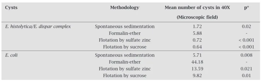
Table 1. Comparison of four techniques for concentration of amoeba cysts
Cysts Methodology Mean number of cysts in 40X p*
(Microscopic field)
E. histolytica/E. dispar complex Spontaneous sedimentation 1.72 0.02 Formalin-ether 5.88 - Flotation by sulfate zinc 0.72 < 0.001
Flotation by sucrose 0.64 < 0.001
E. coli Spontaneous sedimentation 5.71 0.008
Formalin-ether 44.18 - Flotation by sulfate zinc 13.59 0.021
Flotation by sucrose 9.82 0.01
The cysts were concentrated from 5 g of fresh stools with a positive diagnosis for E. histolytica/E. dispar or E. coli. The analysis of statistical significance between differences in numbers of cysts concentrated by formalin-ether or other methodologies was performed using the Wilcoxon Test.
10X PCR buffer (200 mM Tris-HCl, pH 8.4, 500 mM KCl), 0.2 mM of each dNTP, 1.5 mM MgCl2, 0.75U Taq
DNA polymerase (Gibco BRL, Rockville, MD) and 3 µL of DNA sample. An initial DNA amplification was per-formed using the E1, E2 primer set in a GenAmp PCR system 2400 (AB Applied Biosystems) thermal cycler. The first cycle of 5 min at 94°C was followed by 45 cy-cles of denaturation for 1 min at 94°C. Primers were an-nealed for 1.5 min at 47° C and elongated for 2.5 min at 72°C. As positive controls, 5 µL of DNA from cultured
E. histolytica strain HM-1:IMSS, grew in TYI-S-33 medium axenically, and 5 µL of DNA from cultured E. dispar strain MCR, grew in Pavlova medium polyxenically (kindly pro-vided by Dr. Maria Aparecida Gomes, Amoebiasis Labo-ratory, Universidade Federal de Minas Gerais, Brazil) were used. For subsequent amplifications, 5 µL of DNA from the first reaction and primer sets Eh-L, Eh-R and Ed-L, Ed-R were used under the conditions described above, except that the annealing temperature was 58°C. The analysis of PCR products was performed using gel electrophoresis. DNA fragments were separated on a 1.5% (w/v) agarose gel (Inv-itrogen Life Technologies, USA) containing 0.5 µg ethidium bromide/mL. Gels were photographed under ultraviolet il-lumination (Sigma Chem. Co., USA, model T1201).
Immunoenzymatic assay
The presence of E. histolytica-specific galactose adhesin was determined in 35 stool samples, without preservatives, that were negative by PCR and 60 randomly selected stool samples that were positive for E. dispar. The ELISA test was performed according to the manufacturer’s instructions (TechLab E. histolytica II ELISA, USA).
Statistical analysis
The statistics tests used in this study were performed using the SPSS program 15.0 for Windows. The Wilcoxon test was used to evaluate the statistical significance between analyzed variables; a two-tail p-value less than 0.05 was considered significant.
RESULTS
Cyst concentration
The effects of spontaneous sedimentation, formalin-ether and flotation by sulfate zinc or sucrose on the concentration of cysts of the E. histolytica/E. dispar complex were evalu-ated. The formalin-ether technique yielded more cysts than any other method analyzed with a mean of 5.88 cysts un-der 40 x magnification, varying from 0.1 to 8 cysts (Table 1). The mean number of E. histolytica/E. dispar complex cysts by formalin-ether was 3.42 (p = 0.02), 8.17 (p < 0.001) and 9.19 (p < 0.001) times more than that of the sponta-neous sedimentation, zinc sulfate and sucrose flotation procedures, respectively. Likewise, the formalin-ether tech-nique concentrated 7.73 (p = 0.008), 3.25 (p = 0.021) and 4.5 (p = 0.01) times more cysts of Entamoeba coli than the above mentioned techniques. Statistically significant dif-ferences between other concentration methods were not observed (p > 0.05).
Amplification of DNA extracted from stool samples
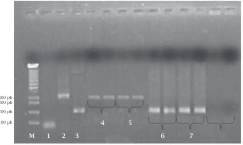
Figure 1. Limit detection of Multiplex-PCR in detection of DNA from E. dispar. Amplified PCR products using Eh-R/ Eh-L and Ed-L/Ed-R primers and a 1.5% electrophoresis gel stained by ethidium bromide. M, molecular weight lad-der (100-bp ladlad-der); Lane 1, negative control (presence of primer dimer); Lane 2, positive control (PCR products from 12 ng/mL DNA from E. histolytica – 427 bp); Lane 3, posi-tive control (PCR products from 18 ng/mL DNA from
E. dispar – 195 bp); Lane 4, PCR products from 9 ng/mL DNA from E. dispar; Lane 5, PCR products from 4.5 ng/mL DNA from E. dispar; Lane 6, PCR products from 2.25 ng/mL DNA from E. dispar ; Lane 7, PCR products from 1.12 ng/mL DNA from
E. dispar.
Figure 2. Evaluation of inter-testing reproducibility. Am-plified products in a 1.5% electrophoresis gel stained by ethidium bromide. PM, molecular weight ladder (100-bp ladder); Lane 1, negative control; Lane 2, positive control (DNA of E. histolytica – 427 bp); Lane 3, positive control (DNA of E. dispar – 195 bp); Lanes 4 to 9, clinical sample positive for E. dispar amplified monthly during a period of six months; Lane 10, clinical sample negative for the
E. histolytica/E. dispar complex (presence of primer dimer).
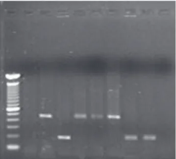
Figure 3. Evaluation of intra-testing reproducibility. Ampli-fied products in a 1.5% electrophoresis gel stained by ethid-ium bromide. M, molecular weight ladder (100-bp ladder); Lane 1, negative control (presence of primer dimer); Lane 2, positive control (DNA of E. histolytica – 427 bp); Lane 3, posi-tive control (DNA of E. dispar – 195 bp); Lanes 4 and 5, clinical samples spiked with 3.0 ng/mL of DNA from the HM-1 strain of E. histolytica; Lanes 6 and 7, clinical samples positive for
E. dispar; Lane 8, clinical sample negative for the E. histolytica/E. dispar complex.
Table 2. Prevalence of E. histolytica/E. dispar in laboratories from Salvador-BA
Samples
Laboratories Total (n) Positives (%)
Not public 47,080 1,507 (3.2%) Datalab 25,996 984 (3.8%) Dirceu Ferreira 18,501 473 (2.6%) Qualitech 2,583 50 (1.9%) Public 5,624 281 (5.0%) HUPES1 2,078 86 (4.1%)
Pharmacy School (UFBA)2 3,546 195 (5.5%)
Not public + public 52,704 1,788 (3.4%)
1Professor Edgard Santos University Hospital. 2Universidade Federal da Bahia.
PM 1 2 3 4 5 6 7 8 9 10
600 pb 300 pb 200 pb 100 pb
M 1 2 3 4 5 6 7
400 pb 300 pb 200 pb 100 pb
400 pb 300 pb 200 pb
100 pb 4 5
M 1 2 3 6 7
of parasites to either different concentrations of E. histol-ytica DNA (12 ng/mL, 6 ng/mL, 3 ng/mL, 1.5 ng/mL and 0.75 ng/mL; data not shown) or different concentrations of
E. dispar DNA (18 ng/mL, 9 ng/mL, 4.5 ng/mL, 2.25 ng/mL and 1.12 ng/mL; Figure 1). Multiplex PCR was capable of detecting the specific target sequence when a minimum of 1.5 ng/mL of DNA template was used for E. histolytica and when 2.25 ng/mL was used for E. dispar.
A positive stool sample for the E. histolytica/E. dispar
complex was fractionated into six aliquots and stored at -20°C. At monthly intervals, one aliquot was thawed, and its DNA was used to observe the inter-testing reproducibility of the PCR (Figure 2). Compatible profile bands are shown
with the species of amoeba tested. Similar results were ob-tained when the samples were amplified twice in the same reaction to demonstrate the reproducibility of intra-testing of PCR, as shown in Figure 3.
Analyses of stool samples
Figure 4. Analysis of 262 E. histolytica/E. dispar positive sam-ples by Multiplex-PCR. Amplified PCR products using Eh-R/ Eh-L and Ed-L/Ed-R primers and a 1.5% electrophoresis gel stained by ethidium bromide. M, molecular weight ladder (100-bp ladder); Lane 1, negative control (presence of primer dimer); Lane 2, positive control (DNA of E. histolytica – 427 bp); Lane 3, positive control (DNA of E. dispar – 195 bp); Lanes 4 to 6, clinical samples spiked with 3 ng/mL of DNA from the HM-1 strain of
E. histolytica; Lanes 7 and 8, clinical samples positive for
E. dispar; Lane 9, negative sample for the E. histolytica/E. dispar
complex.
Extraction of DNA from 262 samples positive for the
E. histolytica/E. dispar complex followed by PCR showed that 227 samples (86.6%) were positive for E. dispar, as dem-onstrated by amplification of the species-specific fragment (195 bp). No amplification was observed for pathogenic
E. histolytica. To evaluate the efficiency of detecting E. histolyt-ica fragments, we spiked negative samples with DNA template from E. histolytica strain HM-1:IMSS. Thereafter, the E. histo-lytica DNA fragment (495 bp) was amplified from all samples. This demonstrates that nested and multiplex PCR can be used for both E. histolytica and E. dispar species (Figure 4).
The mean number of cysts found in amplified samples was 116 cysts per gram of stool, while the mean number of cysts in the 35 non-amplified samples was 173 cysts. After successive dilution (1:20 to 1:160), these 35 samples remained negative. Even after having carried out the spike technique the samples did not amplify. In order to resolve this problem, non-amplified samples and 60 randomly cho-sen samples positive for E. dispar by PCR were submitted to an E. histolytica-specific galactose adhesin immunoenzy-matic assay. This technique demonstrated that all 35 non-amplified samples were negative for E. histolytica-specific galactose adhesin, suggesting a high PCR specificity in
E. histolytica diagnosis. Furthermore, to distinguish a prob-able infection caused by E. hartmanni, morphometric analy-sis demonstrated that non-amplified samples had cyst sizes comparable to E. dispar (> 10 µm).
Characteristics of E. dispar infection
The mean age of the 262 E. histolytica/E. dispar com-plex-positive individuals submitted to amplification re-actions was 35.7 years (with a range of 2 to 79 years), and the median was 35 (25 and 75% quartiles were 22.5 and 50, respectively). Sixty-four percent were male, and 65.5% were of African descent (p < 0.005), with a monthly income of less than 5 minimum wages (p < 0.005) and an educational level at the 2 grade (p < 0.005).
The study population was asymptomatic or pre-sented non-specific symptoms that could be attributed to amoebiasis. The primary reason for seeking medical attention was routine medical check-up, as reported by 67.2% of participants. Among the cases, 21.3% report-ed having gastrointestinal disturbances, flatulence and diarrhea, 11.5% reported using metronidazole, and no confirmation of E. histolytica infection. We identified 31 individuals with liquid stool samples (12% samples) and none with mucus or blood in their stools. A total of 45 individuals (17%) had loose stool samples, and 186 (71%) had formed stool samples.
DISCUSSION AND CONCLUSION
In this report, we described a PCR-based technique for selective identification of E. histolytica and E. dispar in stool samples. The technique consists of an initial am-plification of a 1,076-bp fragment of the SSU-rDNA se-quence of both species, followed by an additional ampli-fication for the two species differentiation. This multiplex PCR permits specific identification in a single reaction mixture and is therefore more cost-effective and less la-borious than other PCR-based methods.12 Some studies
have shown methods used to isolate DNA from in vitro amoeba cultures.5,27,28 However, the cultivation of
amoe-bae before concentrating cysts is more expensive and time-consuming.29 Here, we isolated DNA for PCR
am-plification of amoeba DNA directly from the stools, re-ducing time and the possibility of false negative results.
However, PCR presents some disadvantages. It can be difficult to purify DNA from stool samples due to hardness of the cyst wall and the presence of inhibiting substances, which can inhibit the Taq polymerase.13,30-32
According to Campos-Górgora et al.,33 the major
com-ponent of the Entamoeba cyst wall is chitin, a homopol-ymer of beta-(1,4)-linked N-acetyl-D-glucosamine that confers great rigidity and resistance. Therefore, the uti-lization of both chemistry and physical conditions was sufficient to yield a great quantity of genetic material to proceed with the PCR.
occurred in 13.3% of samples. It is likely that non-spe-cific substances inhibited amplification, since the num-ber of cysts found in these samples did not influence the reaction and the presence of non-specific substances in feces, indirectly evaluated by spike, showed that added DNA did not suffer similarly in amplification of succes-sive dilutions. Specific column chromatography could minimize the influence of enzymatic inhibitors present in feces. According to Verweij et al.,13 it is fast and
sim-ple to use. The authors observed that only 1.7% of 657 samples could not be amplified by PCR. We observed an inhibition in 13.3% of our samples, and the utilization of the columns might have lowered this figure. However, specific column chromatography use increases the cost of PCR, making this technique impracticable in labora-tory diagnosis.
In 1997, it was formally accepted that the species once called E. histolytica comprises two distinct species: the potentially invasive E. histolytica and the non-invasive, commensal E. dispar.7 The cysts and trophozoites of
these species cannot be distinguished microscopically. As no inexpensive or practical diagnostic procedures are currently available for the identification of E. histolytica
at health centers in countries with limited resources, we are left with the common practice of identifying the
E. histolytica/E. dispar complex as “E. histolytica.” The likely consequence of this is diagnosis and over-treatment, which could be the cause of anti-amoebic drug resistance.22 Therefore, a method capable of
differ-entiating both species is essential for appropriate treat-ment and follow-up of infected individuals.
As a result, there has been a lot of progress in the search for molecular methods to distinguish E. histolytica/E. dispar complex species in the last few decades. Coproantigen searching, for example, has in-numerable advantages relative to other methodologies. However, antigens can denature during the preservation process, yielding false negative results. Another alterna-tive to differentiating the species is PCR. This technique has been used frequently in epidemiological investiga-tions worldwide.5,23-26
In this report, the prevalence of E. histolytica/E. dispar based on fecal examination by optical micros-copy was 3.4%. This finding corroborates the results in the literature that state that the prevalence of in-fection varies a great deal throughout Brazil, reach-ing 19% in the Amazon and varyreach-ing from 2 to 29.5% in other regions.4,15,16,36 In the present study, the
analy-sis of 262 samples with E. histolytica/E. dispar using specific set of primers showed that 227 (86.6%) were positive for E. dispar. No amplification was observed in 35 samples (13.4%) that were negative for E. histo-lytica-specific galactose adhesin. Moreover, to exclude a
probable infection caused by E. hartmanni, morphometric analysis demonstrated that non-amplified samples had cyst sizes comparable to E. histolytica/E. dispar (> 10 µm). The absence of amplification of these samples indicates the presence of PCR inhibitors in the stool samples or the presence of DNA from Entamoeba species other than E. dis-par, E. histolytica or E. hartmanni.
In fact, Oliveira-Costa et al.37 in Belo Horizonte City,
Southeast Brazil, and Dourado et al.38 in Pernambuco
State, Northeast Brazil, described only the presence of E. dispar in their studies. Santos et al.5 found a prevalence
of 21% for four-nucleus amoebae in two slums in Rio de Janeiro State, Southeast Brazil, but only two samples were positive for E. histolytica. It appears that E. histol-ytica is more common in North and extreme Northeast Brazil and is rare in other regions.4,39
Our described protocol provides a method to sen- sitively and selectively detect and diferentiate
E. histolytica and E. dispar DNA directly from stool specimens without the need for prior cultivation. Apart from often unsuccessful and time-consuming cultiva-tion attempts, possible misdiagnoses by one strain dis-placing the other in mixed infections can be avoided. We believe that difficulties in the differential diagnosis of the E. histolytica/E. dispar complex need to be over-come for the adequate treatment of E. histolytica. PCR is expensive, and the majority of laboratories do not have adequate infrastructure for its use. The utilization of this method in Brazil is in its initial phase and restricted to research centers located in big cities. However, once the method is established and standardized, its cost will decrease. The main limitations to this technique are the difficulty in obtaining conserved DNA from cysts, and the presence of unspecific inhibitors. More inquiries about simpler, faster and cheaper methodologies should be encouraged and, after approval, should be established in laboratories that compose the public and private health network.
ACKNOWLEDGEMENTS
REFERENCES
1. Caballero-Salcedo A, Viveros-Rogel M, Salvatierra B et al.
Se-roepidemiology of amebiasis in Mexico. Am J Trop Med Hyg 1994; 50(4):412-9.
2. Kreidl P, Imnadze P, Baidoshvili L, Greco D. Investigation of an out-break of amoebiasis in Georgia. Euro Surveil 1999; 4(10):103-6.
3. Gatti S, Swierczynski G, Robinson F et al. Amebic infections
due to the Entamoeba histolytica-Entamoeba dispar complex: a
study of the incidence in a remote rural area of Ecuador. Am J Trop Med Hyg 2002; 67(1):123-7.
4. Benetton ML, Gonçalves AV, Meneghini ME et al. Risk factors
for infection by the Entamoeba histolytica/E. dispar complex:
an epidemiological study conducted in outpatient clinics in the city of Manaus, Amazon region, Brazil. Trans R Soc Trop Med Hyg 2005; 99(7):532-40.
5. Santos HL, Peralta RH, de Macedo HW, et al. Comparison of
mul-tiplex-PCR and antigen detection for differential diagnosis of
Enta-moeba histolytica. Braz J Infect Dis 2007; 11(3):365-70.
6. Mora L, García A, De Donato M, Urdaneta H. Epidemiologic
and molecular study of Entamoeba histolytica and Entamoeba
dispar strains in patients with diarrhea in Cumana, Sucre state, Venezuela. Invest Clin 2008; 49(2):225-37.
7. World Health Organization. Amoebiasis. Report on the WHO/Pan American Health Organization/UNESCO Expert Consultation, Mexico City. Geneva-WHO. W Epidemiol Rec 1997; 72:97-100. 8. Ashbolt NJ. Microbial contamination of drinking water and disease
outcomes in developing regions. Toxicology 2004; 198(1-3):229-38. 9. Chávez B, Martínez-Palomo A, De La Torre M.
Ultramicro-scopic structure of the cyst wall of Entamoeba invadens, E.
his-tolytica and E. coli. Arch Invest Med 1978; Suppl 1:113-6. 10. Ritchie LS. An ether sedimentation technique for routine
stool examinations. Bull U S Army Med Dep 1948; 8(4):326.
11. Acuna-Soto R, Samuelson J, De Girolami P et al. Application
of the polymerase chain reaction to the epidemiology of
path-ogenic and nonpathpath-ogenic Entamoeba histolytica. Am J Trop
Med Hyg 1993; 48(1):58-70.
12. Evangelopoulos A, Spanakos G, Patsoula E et al. A nested,
multiplex, PCR assay for the simultaneous detection and
dif-ferentiation of Entamoeba histolytica and Entamoeba dispar in
faeces. Ann Trop Med Parasitol 2000; 94(3):233-40.
13. Verweij JJ, Blotkamp J, Brienen EA et al. Differentiation of
Entamoeba histolytica and Entamoeba dispar cysts using poly-merase chain reaction on DNA isolated from faeces with spin columns. Eur J Clin Microbiol Infect Dis 2000; 19(5):358-61.
14. Zaman S, Khoo J, Ng SW et al. Direct amplification of
Enta-moeba histolytica DNA from amoebic liver abscess pus using polymerase chain reaction. Parasitol Res 2000; 86(9):724-8.
15. Silva EF, Gomes MA. Amebíase: Entamoeba histolytica/
En-tamoeba dispar. In: Neves DP, Melo AL, Genaro O et al. eds Parasitologia Humana. São Paulo: Atheneu Press, 2001.
16. Araújo R, Bichara CNC, Chaves LCL et al. Doenças infecciosas e
parasitárias – enfoque Amazônico. In: Leão RNQ. eds. CEJUP. Uni-versidade Federal do Pará, Instituto Evandro Chagas. Belém, 1997. 17. Hoffman WA, Pons JA, Janer JL. The sedimentation
concentra-tion method in schistosomiasis mansoni. Puerto Rico J Publ
Health Trop Med 1934; 9:283-298.
18. Brazilian Institute of Geography and Statistics. Contagem da População, 2007. http://www.ibge.gov.br/home/estatistica/ populacao/contagem2007/contagem_final/tabela1_1_16.pdf.
19. Santos FLN, Hiltner AN, Araújo FG et al. Prevalência e
per-fil epidemiológico das enteroparasitoses em Salvador (BA). In: 40º Congresso Brasileiro de Patologia Clínica e Medicina Laboratorial, Curitiba-PR, 2006.
20. Rey L. Parasitologia: parasitos e doenças parasitárias do homem nos trópicos ocidentais. Rio de Janeiro, BR: Guana-bara-Koogan Press, 2008.
21. Troll H, Marti H, Weiss N. Simple differential detection of
En-tamoeba histolytica and Entamoeba dispar in fresh stool speci-mens by sodium acetate-acetic acid-formalin concentration and PCR. J Clin Microbiol 1997; 35(7):1701-5.
22. Leiva B, Lebbad M, Winiecka-Krusnell J et al. Overdiagnosis
of Entamoeba histolytica and Entamoeba dispar in Nicaragua: a microscopic, triage parasite panel and PCR study. Arch Med Res 2006; 37(4):529-34.
23. Valle PR, Souza MB, Pires EM et al. Arbitrarily primed PCR
fingerprint of RNA and DNA in Entamoeba histolytica. Rev
Inst Med Trop São Paulo 2000; 42(5):249-53.
24. Evangelopoulos A, Legakis N, Vakalis N. Microscopy, PCR and ELISA applied to the epidemiology of amoebiasis in Greece. Parasitol Int 2001; 50(3):185-9.
25. Roy S, Kabir M, Mondal D et al. Real-time-PCR assay for
di-agnosis of Entamoeba histolytica infection. J Clin Microbiol
2005; 43(5):2168-72.
26. Vianna EN, Costa JO, Santos CK et al. An alternative
meth-od for DNA extraction and PCR identification of Entamoeba
histolytica and E. dispar in fecal samples. Parasitology 2009; 136(7):765-9.
27. Zindrou S, Orozco E, Linder E et al. Specific detection of
Enta-moeba histolytica DNA by hemolysin gene targeted PCR. Acta Trop 2001; 78(2):117-25.
28. Calderaro A, Gorrini C, Bommezzadri S et al. Entamoeba
his-tolytica and Entamoeba dispar: comparison of two PCR assays for diagnosis in a non-endemic setting. Trans R Soc Trop Med Hyg 2006; 100(5):450-7.
29. Tanyuksel M, Petri WA Jr. Laboratory diagnosis of amoebiasis. Clin Microbiol Rev 2003; 16(4):713-29.
30. Deuter R, Pietsch S, Hertel S, Müller O. A method for preparation of fecal DNA suitable for PCR. Nucleic Acids Res 1995; 23(18):3800-1. 31. Lantz PG, Tjerneld F, Hahn-Hägerdal B, Radström P. Use of
aqueous two-phase systems in sample preparation for poly-merase chain reaction-based detection of microorganisms. J Chromatogr B Biomed Appl 1996; 680(1-2):165-70.
32. Vandenberg N, van Oorschot RA. Extraction of human nu-clear DNA from feces samples using the QIAamp DNA Stool Mini Kit. J Forensic Sci 2002; 47(5):993-5.
33. Campos-Góngora E, Ebert F, Willhoeft U et al. Characterization
of chitin synthases from Entamoeba. Protist 2004; 155(3):323-30.
34. Monteiro L, Bonnemaison D, Vekris A et al. Complex
polysac-charides as PCR inhibitors in feces: Helicobacter pylori model.
J Clin Microbiol 1997; 35(4):995-8.
35. Wilson IG. Inhibition and facilitation of nucleic acid amplifi-cation. Appl Environ Microbiol 1997; 63(10):3741-51.
36. Cunha AS, Silva EF, Ferrari TCA et al. Amebíase em tópicos
e gastroenterologia. In: Castro L.P., Rocha P.R.S., Cunha A.S. eds. Amebiases. Rio de Janeiro: Medicis Press, 1991.
37. Oliveira-Costa J, Resende JA, Gomes MA. Prevalência de
En-tamoeba histolytica e Entamoeba dispar em amostras fecais de um laboratório privado na grande Belo Horizonte. In: Semana de Iniciação Científica da Universidade Federal de Minas Ger-ais, Belo Horizonte, MG, 2006.
38. Dourado A, Maciel A, Aca Ida S. Occurrence of Entamoeba
histolytica/Entamoeba dispar in ambulatory patients of Recife, PE. Rev Soc Bras Med Trop 2006; 39(4):388-9.
39. Braga LL, Gomes ML, Silva MW et al. Entamoeba histolytica