Electrical parameters and water
permeability properties of monolayers
formed by T84 cells cultured on
permeable supports
Laboratorio de Biomembranas, Departamento de Fisiología, Facultad de Medicina, Universidad de Buenos Aires, Buenos Aires, Argentina
M. Ozu, R. Toriano, C. Capurro and M. Parisi
Abstract
T84 is an established cell line expressing an enterocyte phenotype whose permeability properties have been widely explored. Osmotic permeability (POSM), hydraulic permeability (PHYDR) and transport-associated net water fluxes (JW-transp), as well as short-circuit current (ISC), transepithelial resistance (RT), and potential difference (∆VT) were measured in T84 monolayers with the following results: POSM 1.3 ± 0.1 cm.s-1 x 10⁻³; PHYDR 0.27 ± 0.02 cm.s-1; RT 2426 ± 109 Ω.cm2, and
∆VT 1.31 ± 0.38 mV. The effect of 50 µM 5,6-dichloro-1-ethyl-1,3-dihydro-2H-benzimidazol-2-one (DCEBIO), a “net Cl- secretory agent”, on T84 cells was also studied. We confirm the reported important increase in ISC induced by DCEBIO which was associated here with a modest secretory ∆JW-transp. The present results were compared with those reported using the same experimental approach applied to established cell lines originating from intestinal and renal epithelial cells (Caco-2, LLC-PK1 and RCCD-1). No clear association between PHYDR and RT could be demonstrated and high PHYDR values were observed in an electrically tight epithelium, supporting the view that a “water leaky” barrier is not necessarily an “electrically leaky” one. Furthermore, the modest secretory ∆JW-transp was not consistent with previous results obtained with RCCD-1 cells stimulated with vasopressin (absorptive fluxes) or with T84 cells secreting water under the action of Escherichia coli heat stable enterotoxin. We conclude that, while the presence of aquaporins is necessary to dissi-pate an external osmotic gradient, coupling between water and ion transport cannot be explained by a simple and common underlying mechanism.
Correspondence M. Parisi
Departamento de Fisiología, 7º piso Facultad de Medicina, UBA C1121ABG Buenos Aires Argentina
Fax: +54-11-4964-0503 E-mail: [email protected]
Received March 22, 2004 Accepted October 28, 2004
Key words
•Water absorption and
secretion
•Aquaporins
•Osmotic and hydrostatic
gradients
•T84 cells
Introduction
Epithelial barriers are classified as “tight” or “leaky” according to their electrical con-ductance (1), which in general decreases with barrier tightness. Two different
perme-ability coefficients of water net fluxes can be
measured: osmotic (POSM) and hydrostatic
(PHYDR) (2). Moreover, diffusional water
pressure gradients (JW-hydr) move across
the intercellular spaces, while the main route for those induced by an external osmotic gradient (JW-osm) is controversial.
Correlation between electric and water permeability properties is also especially im-portant when a so-called electrogenic ionic transport is observed, resulting in a net salt transfer across the barrier. A net fluid trans-fer (JW-transp) is frequently associated with
salt movement by a process that is not clearly understood (3,4). These water movements, driven by a salt transport-generated osmotic gradient, could also occur among or across epithelial cells.
The accepted water pathway for transcel-lular movements is the lipid bilayer itself (5), or specific water channels called aquaporins (6). Alternatively, solute-water cotransport, a controversial mechanism conceptually dif-ferent from those previously mentioned, has been proposed (7-9). In the present study, water fluxes and electrical parameters were measured in T84 monolayers, a cell line derived from a human colon carcinoma. The original colon epithelium expresses aqua-porins in different species, for example hu-mans (10) and rats (11). However, the T84 cell line does not express aquaporins (12) like other established cell lines (10,13). The Na+/H+ and Cl-/HCO
3- exchangers and the
Na+/HCO
3- cotransporter have been
de-scribed in these cells (14). In the present study, minute by minute recordings of the
transepithelial net water fluxes (JW-hydr;
JW-osm or JW-transp) were associated with
the measurement of the transepithelial po-tential difference (∆VT), transepithelial
re-sistance (RT) and short-circuit current (ISC)
under different experimental conditions. The results were compared with those reported for Caco-2, LLC-PK1 and RCCD-1 cells. Furthermore, the effects on water transfer of
5,6-dichloro-1-ethyl-1,3-dihydro-2H
-ben-zimidazol-2-one (DCEBIO), recently
re-ported as a “net Cl- secretory agent” in T84
cells (15), and of bumetanide were also tested
and compared with the reported effects of agents that increase active absorption or se-cretion (12,13,16,17).
Material and Methods
Cell culture
T84 cells, obtained from the American Type Culture Collection (Rockville, MD, USA), were grown to confluent monolayers in a 1:1 mixture of Dulbecco’s modified Eagle’s medium (DMEM) and Ham’s F-12
medium supplemented with 14 mM NaHCO3,
3.2 mM glutamine, 10 units/ml
penicillin-streptomycin, 15 mM HEPES/Na+, pH 7.4,
and 5% fetal bovine serum (Gibco BRL,
Grand Island, NY, USA) in a 5% CO2
atmos-phere at 37ºC. For these experiments, cells between passages 58 and 65 were seeded
onto Transwell holders (1.106
cells/Trans-well of 3-µm pore Nuclepore filters, 4.5 cm2
surface area; Corning-Costar Corp., Cam-bridge, MA, USA) and cultured for 10 to 12 days.
For the experiments, the T84 cells were bathed on either side with minimum medium containing 1:1 DMEM-Ham’s F-12 (Cata-log number: 12500-039; Gibco BRL-Life
Technologies) and 14 mM NaHCO3 plus 15
mM HEPES/Na+, pH 7.4, when bubbled with
5% CO2/95% O2.
Measurement of water fluxes
In order to perform water flux measure-ments across the T84 monolayers, the Trans-well holders with their bottom covered with the confluent cell layer were directly in-serted between two Lucite hemi-chambers so as to define two independent compart-ments, as previously described (12). One of them (serosal) was open to the atmosphere, while the other (mucosal) was hermetically sealed. A hydrostatic pressure difference (4.5
cm of H2O) was continuously applied;
to that on the serosal side. The closed cham-ber was connected with a small diameter polyethylene tube to the
net-water-measure-ment system where the net water flux (JW)
was recorded every minute, as described elsewhere (18). Briefly, the position of a liquid meniscus inside a capillary tube was photoelectrically detected. Displacements to the right or to the left were proportional to the amount of water moving across the tissue layer. The system sensitivity was 50 nl. The
data were computed in units of µl min-1
cm-2. Mucosa to serosa net movements
(ab-sorptive) were considered to be positive fluxes while serosa to mucosa net move-ments (secretory) were considered to be nega-tive fluxes. The serosal bath was
continu-ously bubbled with the appropriate CO2/O2
mixture to maintain pH at 7.4 ± 0.1 (37ºC). JW-hydr and JW-osm were the JW values
ob-served in the presence of a transepithelial hydrostatic pressure gradient or a
transepi-thelial osmotic gradient, respectively. JW
-transp is the remaining JW when the effects
of the external osmotic and hydrostatic gra-dients are subtracted.
Electrophysiological studies
Transepithelial voltage difference (∆VT)
and ISC were continuously recorded
employ-ing an automatic voltage clamp system (Physiological Instruments, San Diego, CA, USA) and Navycite (ME2AG4) electrodes. Before mounting the preparations in the
volt-age clamp system, RT was measured with a
Millicell-ERS electric resistance system (Mil-lipore, Bedford, MA, USA). During the
ex-periments, RT was measured every 90 s from
current deflections in response to a 1-mV/
second pulse. Regarding ∆VT, positive or
negative values indicate “serosal side posi-tive or negaposi-tive”. Electrophysiological and Jw measurements were carried out simulta-neously in the same epithelial layer.
DCEBIO was a generous gift from Prof. Robert J. Bridges, University of Pittsburgh,
USA. DMSO was used as solvent to test the effects of DCEBIO (50 µM on the mucosal side, and 50 and 75 µM on the serosal side) on water and ion transport.
Statistical analysis
Data are reported as means ± SEM. Sta-tistical significance was determined using
the paired or unpaired t-test, and a P
value <0.05 was considered to be statisti-cally significant.
Results
Osmotic and hydraulic permeability of T84 cells
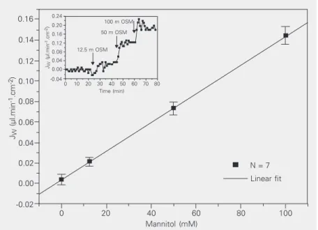
Figure 1 shows the JW values observed
when the osmotic gradient across T84 cell monolayers was increased. Gradients were generated by adding mannitol to the serosal bath (the spontaneous water movement, measured in the presence of a 4.5-cm
differ-Figure 1. Water flux as a function of osmotic gradients (JW-osm) applied from the serosal
side (12.5, 50 and 100 mM mannitol). Data are reported as means ± SEM (N = 7). The inset
shows a typical experiment in which water flux did not increase along the time course in the presence of an osmotic gradient. Osmotic permeability (POSM = 1.30 ± 0.09 x 10⁻³ cm
s-1) was calculated from the slope of the corresponding regression curve (R = 0.99 ± 0.04,
P < 0.001, t-test).
JW
(µl.min
-1.cm -2)
0.16
0.14
0.12
0.10
0.08
0.06
0.04
0.02
0.00
-0.02
0 20 40 60 80 100
Mannitol (mM)
N = 7 Linear fit
JW
(µl.min
-1.cm -2)
0.24 0.20 0.16 0.12 0.08 0.04 0.00 -0.04
0 10 20 30 40 50 60 70 80 Time (min)
12.5 m OSM 50 m OSM
Table 1). In this case, the spontaneous JW
was a secretory one and reverted to absorp-tive values under the action of the applied hydrostatic pressure.
Effects of DCEBIO on electrical parameters and water movements in T84 cells
It has been reported that DCEBIO
in-duces an important secretory Cl- response in
T84 cells, associated with a significant
in-crease in ISC as well (15). We explored the
possible effects of DCEBIO on water trans-fer and also tested the effects of bumetanide, an inhibitor of Cl- secretion, on this cell line
(17).
DMSO was employed as a solvent for both DCEBIO and bumetanide. It was added in a similar concentration to both sides of the cells and did not induce significant changes in JW, ISC, RT or ∆VT. The addition of
DCEBIO (50 µM in both mucosal and sero-sal baths) was followed by a secretory
re-sponse (∆JW-transp) together with the
ex-pected increase in both ISC and ∆VT (Table
2), effects that were reversed by 10 µM bumetanide. Figure 3 illustrates the results obtained when the concentration of DCEBIO increased on the serosal side after the initial stimulation of both sides. Finally, 10 µM bumetanide was added to the serosal bath.
Discussion
In the present study, we characterized the electrical parameters and water permeability properties of monolayers formed by T84 cells in culture. Low osmotic permeability was ob-served in this high resistance epithelium,
to-gether with rather high PHYDR values. An
im-portant difference in potential was associated with these properties, as previously reported (Table 1). We also report that the important increase in short circuit current induced by 50 µm DCEBIO was associated with
secre-tory JW while both responses were inhibited
by 10 µM bumetanide (Table 2).
Table 1. Osmotic permeability (POSM), hydraulic permeability (PHYDR), transepithelial
resistance (RT), and potential difference (∆VT) observed in different epithelial cell lines
cultured on permeable supports.
Caco-2a RCCD-1b T84c LLC-PK1d Transfected
LLC-PK1d
POSM (x10-3 cm.s-1) 35±6 5.0±0.4 1.3±0.1 7.4±4.8 49.9±8.2
PHYDR (cm.s-1) 2.7±0.3 1.0±0.1 0.27±0.02 0.20±0.03 0.25±0.04
RT (ohm.cm2) 160±15 2390±140 2426±109 323±13 244±20 ∆VT (mV) 0.28±0.03 10.5±1.2 1.31±0.38 ND ND
aParisi et al. (16); bChara O, Parisi M and Capurro C (unpublished results); cthe present
study; dToriano et al. (12). ND = not determined.
JW
-hydr (µl.min
-1.cm -2)
0.03
0.02
0.01
0.00
-0.01
-0.02
-0.03
∆Ph (atm)
Mean (N = 3) Linear fit
0.0040 0.0045 0.0050 0.0055 0.0060 0.0065 0.0070
JW
-hydr (µl.min
-1.cm -2)
0.06
0.04
0.02
0.00
-0.02
-0.04
0 10 20 30 40 50 60 70 Time (min)
∆h = 4.5 cm 5.3 6.2 7.1
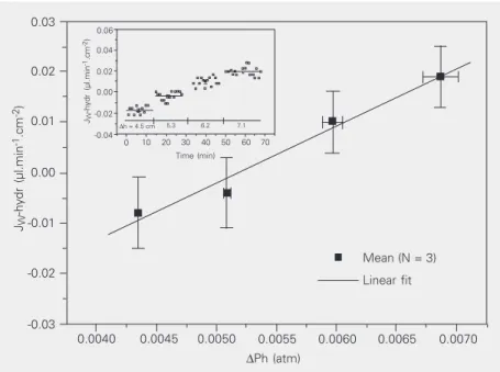
Figure 2. Hydraulic water flux (JW-hydr) across T84 monolayers as a function of different
hydrostatic gradients (∆Ph, atmospheres, N = 3). Gradients were applied from the mucosal side. The hydraulic permeability coefficient (PHYDR = 0.27 ± 0.02 cm.s-1) was calculated
from the slope of the corresponding regression curve (R = 0.99 ± 0.17, P < 0.02, t-test). The
inset shows that hydrostatic flux did not increase along the time course in the presence of
a hydrostatic gradient.
ence of H2O hydrostatic pressure, was not
significantly different from zero in this
ex-perimental series). POSM (Table 1) was
cal-culated from the slope of the regression curve
obtained from JW values and the applied
osmotic gradients (R = 0.99 ± 0.04, P < 0.001).
Mean JW values as a function of the
ap-plied hydrostatic gradient are presented in
Figure 2. PHYDR was calculated from the
Comparison of passive electrical and water permeability properties in different epithelial cell lines: the role of aquaporins
The results obtained with T84 cells cor-related with data previously reported for dif-ferent epithelial cell lines cultured on perme-able supports. All experiments were per-formed in our laboratory under similar ex-perimental conditions, permitting compari-son in a well-controlled manner. Figure 4 presents PHYDR values plotted against RT for
Caco-2, RCCD-1, T84, PK1, and LLC-PK1 cells transfected with AQP-2 (Table 1). It can be seen that high resistance values
(about 2380 Ω.cm2) were associated with
also rather high PHYDR values (about 1 cm
s-1) in RCCD-1 cells. On the other hand, in
LLC-PK1 cells much higher conductances
were associated with lower PHYDR values.
For both PHYDR and RT, the paracellular route
has been previously reported to be the most important one (2,19).
When POSM and RT were compared
(Fig-ure 5, Table 1), no clear correlation was observed. Values for LLC-PK1 cells trans-fected with AQP-2 clearly showed that an
important increase in POSM can be associated
with no significant changes in RT.
Finally, no statistically significant
corre-lation between PHYDR and POSM was observed
in non-transfected cells. Furthermore, it should be pointed out that after aquaporin transfection in LLC-PK1 cells, there was a
huge increase in POSM values with no changes
in PHYDR (Table 1). Previous studies have
demonstrated that T84, LLC-PK1 and RCCD-1 cell lines in culture lose aquaporin expres-sion (13,17,20). Several straightforward in-terpretations can be proposed on the basis of these observations and of previous and
pres-ent results: 1) PHYDR is a parameter that
represents water transfer across the paracel-lular pathway. Nevertheless, rather high
PHYDR values can be observed in electrically
tight epithelia (T84, RCCD-1), confirming that a “water leaky” barrier is not necessarily
Table 2. Effects of 5,6-dichloro-1-ethyl-1,3-dihydro-2H-benzimidazol-2-one (DCEBIO, 50 µM both in mucosal and serosal baths) and bumetanide (10 µM) on T84 cell monolayers.
Experimental - basal data DCEBIO + Bumetanide
∆JW-transp (x10-3 µl.min-1.cm-2) -13.1 ± 5.2* 18 ± 1.5+ ∆ISC (µA.cm2) 67.8 ± 22.2** -47.5 ± 22.5+
∆VT (mV) 12.0 ± 2.3* -7.0 ± 3.7+
Data are reported as means ± SEM (N = 4). ∆JW-transp = transport-associated net
water fluxes; ∆ISC = short-circuit current variation; ∆VT = transepithelial potential
difference.
*P < 0.001 for JW-transp and ∆VT (DCEBIO) against JW-transp and ∆VT (basal).
**P < 0.02 for ISC (DCEBIO) against ISC (basal). +P < 0.05 vs DCEBIO values (t-test).
∆
JW
-transp (µl.min
-1.cm -2) 0.008
0.006 0.004 0.002 0.000 -0.002 -0.004 -0.006
DCEBIO
(50 µM muc/ser) + DCEBIO
(25 µM ser)
+ Bumetanide (10 µm)
Figure 3. 5,6-Dichloro-1-ethyl-1,3-dihydro-2H -benzimidazol-2-one (DCEBIO)-induced variations in transport-associated fluxes (∆JW-transp) followed by an
ad-ditional DCEBIO stimulation (50 µM on the mucosal (muc) and serosal (ser) sides, followed by 25 µM on the serosal side). Fi-nally, bumetanide (10 µM, sero-sal side) reversed the secretory
effect of DCEBIO (mean ± SEM, N = 4). JW was measured until stable values were
achieved. Values were calculated (subtracting basal fluxes) in the absence of osmotic or chemical transepithelial gradients. *P < 0.02 compared to other groups (Student t-test).
Figure 4. Hydraulic permeability coefficients (PHYDR) and
trans-epithelial resistance (RT) for
dif-ferent cell lines. Caco-2 = Parisi et al. (16); RCCD-1 = Chara O, Parisi M and Capurro C (unpub-lished results); T84 = the pres-ent study; LLC-PK1 and trans-fected LLC-PK1 = Toriano et al. (12).
Figure 5. Osmotic permeability coefficients (POSM) and
trans-epithelial resistance (RT) for
dif-ferent cell lines. Caco-2 = Parisi et al. (16); RCCD-1 = Chara O, Parisi M and Capurro C (unpub-lished results); T84 = the pres-ent study; LLC-PK1 and trans-fected LLC-PK1 = Toriano et al. (12).
T84 Caco-2
LLC-PK1-AQP-2transf
LLC-PK1
RCCD-1 POSM
(cm.s
-1)
60
0 200 400 2200 2400 2600 RT (Ω.cm2)
50
40 30
20 10 0
PHYDR
(cm.s
-1)
3.0
T84 Caco-2
LLC-PK1-AQP-2transf
LLC-PK1 RCCD-1
0 200 400 2200 2400 2600 RT (Ω.cm2)
2.5 2.0 1.5 1.0 0.5 0.0
Water and ion coupling during absorption and secretion
The evaluation of the correlation of ISC, a
parameter frequently employed to evaluate ionic transport in epithelial barriers, and the
associated increases in JW-transp is more
complex. Three experimental situations were evaluated in previous work from our labora-tory and in the present study: the effects of
ADH on RCCD-1 (13), the effects of
Esche-richia coli heat stable enterotoxin on T84
(17), and the effects of DCEBIO on this last cell line reported in this paper (Table 3). Figure 6 shows the reported ∆JW-transp as a
function of the simultaneously measured
∆ISC. The observed ∆JW-transp values (open
columns) were compared with theoretical values (filled columns), which were calcu-lated assuming that: 1) the electrogenic trans-port, measured by the ISC, represents the total
ionic net transport across the barrier in a pure absorptive or secretory process, and 2) this ionic movement is coupled to an iso-tonic fluid transfer. In the case of RCCD-1 +
ADH, a modest increase in ISC was
accompa-nied by an important absorptive JW-transp,
higher than that predicted if the previously described conditions are accepted.
Further-more, when the effects of Escherichia coli
heat stable enterotoxin on T84 were
ana-lyzed, the observed increase in ISC was
coupled to a rather important secretory ∆JW
-transp. In fact, to accept isotonic movements, a non-electrogenic ionic transport must be postulated in both cases (17).
A completely different situation was ob-served in the DCEBIO-T84 experiments
re-ported here. An important increase in ISC
was associated with a modest ∆JW-transp. If
we assume that the fluid transfer is isotonic and that the ISC is the result of electrogenic
Cl- secretion, we can expect an induced ∆J
W-transp of 0.281 µl min-1 cm-2. The observed
∆JW was 20 times lower (Table 3, Figure 6).
These results can be explained if the
observed increments in ISC correspond to the
Table 3. ISC and JW-transp values under the effect of antidiuretic hormone (ADH), Escherichia coli heat stable enterotoxin (STa) and 5,6-dichloro-1-ethyl-1,3-dihydro-2H -benzimidazol-2-one (DCEBIO).
RCCD-1 stimulated T84 stimulated T84 stimulated with ADH (N = 6)a with STa (N = 8)b with DCEBIO (N = 4)c
Experimental ∆JW-transp 160 ± 20 -180 ± 40 -13.1 ± 5.2 (x10-3 µl.min-1.cm-2)
∆ISC (µA.cm-2) 2.24 ± 0.44 16.9 ± 4.32 67.8 ± 22.2 Theoretical ∆JW-transp 9.3 ± 1.8 -70.1 ± 17.9 -281.3 ± 109.3 (x10-3 µl.min-1.cm-2)
STa concentration: 0.25 µM; ADH concentration: 10 mM; DCEBIO: 50 µM. Theoretical values were calculated assuming isotonic flux derived from the electrogenic transport, measured by ISC (see the text). aCapurro et al. (13); bToriano et al. (17); cThe present study. For abbrevia-tions, see legend to Table 2.
∆
JW
-transp (x10
-3
µl.min
-1.cm -2)
200 150 100 50 0 -50 -100 -150 -200 -250 -300 -350 -400
RCCD-1 (ADH)
16.9 ± 4.32
2.24 ± 0.44
67.8 ± 22.2
∆Isc (µA.cm-2)
T84 (DCEBIO) T84 (STa)
Figure 6. Net water flux (∆JW-transp) and short-circuit current variation (∆ISC) for different
cell lines. The observed ∆JW-transp values (open columns) were compared with the
theoretical values (filled columns) assuming isotonic transport (see text). The data pre-sented correspond to the experimental ∆ISC values. Both parameters (∆JW-transp and ∆ISC)
were measured simultaneously in the same monolayer. RCCD-1 (ADH), Capurro et al. (13); T84 (STa), Toriano et al. (17); T84 (DCEBIO), the present study. ADH = antidiuretic hor-mone; Sta = Escherichia coli heat stable enterotoxin; DCEBIO = 5,6-dichloro-1-ethyl-1,3-dihydro-2H-benzimidazol-2-one.
an “electrically leaky” one (21); 2) in the tested native cells (not expressing aquaporins) no clear correlation was observed between POSM and RT. A relatively high POSM value
was observed in the absence of aquaporins in a very leaky epithelium (Caco-2), and 3)
after aquaporin transfection, POSM strongly
dissociated from PHYDR, probably because
combination of Na+ and Cl-
DCEBIO-in-duced currents. If the increases in Na+ and
Cl- currents were in opposite directions
(mu-cosa to serosa versus serosa to mu(mu-cosa fluxes), current values would add up but the associated water movements would be can-celed, at least partially. In this context, the
Jw-transp calculated from the ISC (accepting
isotonic movement) would be lower than that observed. On the other hand, if the increases in Na+ and Cl- currents were in the
same direction, the result would be that these ionic currents would subtract their values while water fluxes would be added.
Now the Jw-transp calculated from the
ISC (accepting isotonic movement) would be
higher than that observed. If pure Na+ or Cl
-increases were detected, appropriate counter-ions should assure electroneutrality under open circuit conditions. We conclude that the associated water fluxes cannot be
accu-rately determined from ISC measurements.
Water pathways in epithelial barriers
Net water and ionic fluxes can move across epithelial barriers through either trans-cellular or paratrans-cellular routes (2,22,23; for reviews, see Refs. 19,24). Our results indi-cate that in an epithelium showing important
PHYDR values (Caco-2), and in the absence of
aquaporins, the paracellular route could be a significant pathway for the osmotic flux. Conversely, in an epithelium presenting
lower PHYDR values (LLC-PK1), the
pres-ence of aquaporins was crucial to dissipate the external osmotic gradient (25). This last condition would be similar to that described for ADH target epithelia (12).
The route for the “transport-associated” water movement is a highly controversial topic (4), especially regarding secretory pro-cesses (26). The “standing gradient model” (27) postulates that the intercellular spaces play a central role in the so-called “isotonic transport-associated water movement”. Nev-ertheless, it has been reported that there is no
significant fluid flow through the tight junc-tion of MDCK cells, even when fluid ab-sorption is accelerated by the imposition of an external osmotic gradient (22). Discovery of aquaporin expression in different epithe-lial cells and particularly in leaky epithelia such as the proximal renal tubule has empha-sized the role of transcellular channel-medi-ated water transport. However, a report from our laboratory demonstrated that, in the case of RCCD-1 cells, an ADH-induced
absorp-tive JW-transp could develop in the absence
of aquaporins (13).
An additional pathway and mechanism
for water transport was proposed (i.e., Na+/
glucose cotransporter, SGLT), which would consist of a cotransport of water and solute
with a strict stoichiometry of two Na+, one
glucose and ~220-250 water molecules (9,28). This mechanism was also proposed to explain coupling of water and ions in secretory processes. Although this hypothesis has generated strong controversy (29), the authors consider that the local osmotic gra-dient generated by the solute transfer can explain water transfer (30). Alternatively, it has been recently proposed that electro-os-mosis can play a central role in water and ion coupling during active transport in a leaky epithelium (31). However, it should be pointed out that, although results obtained with epithelial cell monolayers represent use-ful experimental models, they cannot always be extrapolated to natural epithelia.
We conclude that JW-transp is not always
the result of a simple “secondary effect” due to the generation of an osmotic gradient anywhere inside the epithelial barrier. The role of water-solute cotransport in the phe-nomena described here remains an open ques-tion.
Acknowledgments
References
1. Frömter E & Diamond J (1972). Route of passive ion permeation in epithelia. Nature New Biology, 235: 9-13.
2. Parisi M, Amodeo G, Capurro C, Dorr R, Ford P & Toriano R (1997). Biophysical properties of epithelial water channels. Biophysical
Chemistry, 68: 255-263.
3. Whittembury G & Echevarria M (1994). Pathways for water absorp-tion in isosmotic transporting epithelia. Mount Sinai Journal of
Medicine, 61: 311-319.
4. Spring KR (1999). Epithelial fluid transport - A century of investiga-tion. News in Physiological Sciences, 14: 92-98.
5. Finkelstein A (1987). Water movements through lipid bilayers, pores and plasma membranes. In: Theory and Reality. Vol. 4. John Wiley and Sons, New York.
6. Agre P, Sasaki S & Chrispeels M (1993). Aquaporins: a family of water channel proteins. American Journal of Physiology, 265: F461-F468.
7. Meinild A, Loo D, Pajor A, Zeuthen T & Wright E (2000). Water transport by the renal Na+-dicarboxylate cotransporter. American
Journal of Physiology, 278: F777-F783.
8. Zeuthen T (1994). Cotransport of K+, Cl- and H
2O by membrane
proteins from choroid plexus epithelium of Necturus maculosus.
Journal of Physiology, 478: 203-219.
9. Zeuthen T, Meinild AK, Klaerke DA, Loo DD, Wright EM, Belhage B & Litman T (1997). Water transport by the Na+/glucose cotransporter
under isotonic conditions. Biologie Cellulaire, 89: 307-312. 10. Fischer H, Stenling R, Rubio C & Lindblom A (2001). Differential
expression of aquaporin 8 in human colonic epithelial cells and colorectal tumors. BMC Physiology, 1: 1.
11. Gallardo P, Olea N & Sepulveda FV (2002). Distribution of aquaporins in the colon of Octodon degus, a South American desert rodent.
American Journal of Physiology, 283: R779-R788.
12. Toriano R, Ford P, Rivarola V, Tamarappoo BK, Verkman AS & Parisi M (1998). Reconstitution of a regulated transepithelial water path-way in cells transfected with AQP2 and an AQP1/AQP2 hybrid containing AQP2-C terminus. Journal of Membrane Biology, 161: 141-149.
13. Capurro C, Rivarola V, Kierbel A, Escoudet B, Farman N, Blot-Chabaud M & Parisi M (2001). Vasopressin regulates water flow in a rat cortical collecting duct cell line not containing known aquaporins.
Journal of Membrane Biology, 179: 63-70.
14. Ramirez MA, Toriano R, Parisi M & Malnic G (2000). Control of cell pH in the T84 colon cell line. Journal of Membrane Biology, 177: 149-157.
15. Singh S, Syme C, Singh A, Devor D & Bridges R (2001). Benzimidazolone activators of chloride secretion: potential thera-peutics for cystic fibrosis and chronic obstructive pulmonary dis-ease. Journal of Pharmacology and Experimental Therapeutics, 296: 600-611.
16. Parisi M, Escobar E, Huet C, Ripoche P, Louvard D & Bourguet J (1993). Water handling in Caco-2 cells: effects of acidification of the medium. Pflügers Archiv, 423: 1-6.
17. Toriano R, Kierbel A, Ramirez MA, Malnic G & Parisi M (2001). Spontaneous water secretion in T84 cells: effects of STa entero-toxin, bumetanide, VIP, forskolin, and A-23187. American Journal of
Physiology, 281: G816-G822.
18. Dorr R, Kierbel A, Vera J & Parisi M (1997). A new data acquisition system for the measurement of the net water flux across epithelia.
Computer Methods and Programs in Biomedicine, 53: 9-14.
19. Whittembury G & Hill H (2000). Coupled transport of water and solutes across epithelia. In: Seldin D & Giebisch G (Editors), Kidney
Physiology and Pathophysiology. Lippincott Williams, New York,
341-362.
20. Katsura T, Ausiello DA & Brown D (1996). Direct demonstration of aquaporin-2 water channel recycling in stably transfected LLC-PK1 epithelial cells. American Journal of Physiology, 270 (Part 2): F548-F553.
21. Parisi M, Pisam M, Calamita G, Gobin R, Toriano R & Bourguet J (1995). Water pathways across a reconstituted epithelial barrier formed by Caco-2 cells: effects of medium hypertonicity. Journal of
Membrane Biology, 143: 237-245.
22. Kovbasnjuk O, Leader JP, Weinstein AM & Spring KR (1998). Water does not flow across the tight junctions of MDCK cell epithelium.
Proceedings of the National Academy of Sciences, USA, 95:
6526-6530.
23. Kierbel A, Capurro C, Pisam M, Gobin R, Christensen BM, Nielsen S & Parisi M (2000). Effects of medium hypertonicity on water perme-ability in the mammalian rectum: ultrastructural and molecular cor-relates. Pflügers Archiv, 440: 609-618.
24. Cereijido M, Shoshani L & Contreras RG (2000). Molecular physiolo-gy and pathophysiolophysiolo-gy of tight junctions. I. Biogenesis of tight junctions and epithelial polarity. American Journal of Physiology, 279: G477-G482.
25. Verkman A (2002). Physiological importance of aquaporin water channels. Annals of Medicine, 34: 192-200.
26. Masyuk AI, Marinelli RA & LaRusso NF (2002). Water transport by epithelia of the digestive tract. Gastroenterology, 122: 545-562. 27. Diamond JM & Bossert WH (1967). Standing-gradient osmotic flow.
A mechanism for coupling of water and solute transport in epithelia.
Journal of General Physiology, 50: 2061-2083.
28. Loo D, Zeuthen T, Chandy G & Wright E (1996). Cotransport of water by the Na+/glucose cotransporter. Proceedings of the
Na-tional Academy of Sciences,USA, 93: 13367-13370.
29. Lapointe JY, Gagnon MP, Gagnon DG & Bissonnette P (2002). Controversy regarding the secondary active water transport hypo-thesis. Biochemistry and Cell Biology, 80: 525-533.
30. Duquette P, Bissonnette P & Lapointe J (2001). Local osmotic gradients drive the water flux associated with Na(+)/glucose cotransport. Proceedings of the National Academy of Sciences, USA, 98: 3796-3801.
31. Sanchez JM, Li Y, Rubashkin A et al. (2002). Evidence for a central role for electro-osmosis in fluid transport by corneal endothelium.