Propolis and swimming in the prevention of atherogenesis and left
ventricular hypertrophy in hypercholesterolemic mice
Silva, DB.
a,b,c, Miranda, AP.
a, Silva, DB.
a,b,c, D`Angelo, LRB.
a, Rosa, BB.
a, Soares, EA.
d,
Ramalho, JGDC.
a, Boriollo, MFG.
cand Garcia, JAD.
b,c,d*
a Programa de mestrado em Ciência Animal, Universidade José do Rosário Vellano – UNIFENAS,
Campus Universitário, Rod. MG 179, Km zero, CEP 37130-000, Alfenas, MG, Brazil
bInstituto Federal Sul de Minas Gerais – IFSULDEMINAS, Campus Machado, Rodovia Machado,
Paraguaçú, Km 3, Santo Antônio, CEP 37750-000, Machado, MG, Brazil
cInstituto Federal Sul de Minas Gerais – IFSULDEMINAS,Campus Muzambinho, Estrada de Muzambinho,
Km 35, Morro Preto, CP 02, CEP 37890-000, Muzambinho, MG, Brazil
dFaculdade de Medicina e Medicina Veterinária, Universidade José do Rosário Vellano – UNIFENAS,
Rod. MG 179, Km zero, Campus Universitário, CEP 37130-000, Alfenas, MG, Brazil *e-mail: [email protected]
Received: August 30, 2013 – Accepted: January 2, 2014 – Distributed: May 31, 2015
(With 1 figure)
Abstract
Aims: The present study verified the effect of propolis alone and its association with swimming in dyslipidemia, left
ventricular hypertrophy and atherogenesis of hypercholesterolemic mice. Methods and Results: The experiments
were performed in LDLr–/– mice, fed with high fat diet for 75 days, and were divided into four experimental groups (n=10): HL, sedentary, subjected to aquatic stress (5 min per day, 5 times per week); NAT submitted to a swimming protocol (1 hour per day, 5 times per week) from the 16th day of the experiment; PRO, sedentary, submitted to aquatic stress and which received oral propolis extract (70 uL/animal/day) from the 16th day of the experiment; HL+NAT+PRO, submitted to swimming and which received propolis as described above. After 75 days, blood was collected for analysis of serum lipids. The ratio between the ventricular weight (mg) and the animal weight (g) was calculated. Histological sections of the heart and aorta were processed immunohistochemically with anti-CD40L antibodies to evaluate the inflammatory process; stained with hematoxylin/eosin and picrosirius red to assess morphological and morphometric alterations. The HL animals showed severe dyslipidemia, atherogenesis and left ventricular hypertrophy, associated with a decrease in serum HDLc levels and subsequent development of cardiovascular inflammatory process, characterized by increased expression of CD40L in the left ventricle and aorta. Swimming and propolis alone and\or associated prevented the LVH, atherogenesis and arterial and ventricular inflammation, decreasing the CD40L expression and increasing the HDLc plasmatic levels. Conclusion: Propolis alone or associated with a regular physical activity is beneficial in cardiovascular protection through anti-inflammatory action.
Keywords: dyslipidemia, left ventricular hypertrophy, propolis, LDLr–/– mice.
Própolis e natação na prevenção da aterogênese e hipertrofia ventricular
esquerda de camundongos hipercolesterolêmicos
Resumo
Objetivos: O presente estudo verificou o efeito do própolis associação ou não com a natação na dislipidemia, na hipertrofia ventricular esquerda e aterogênese de camundongos hipercolesterolêmicos. Métodos e Resultados: Os experimentos foram realizados em camundongos LDLr–/–, alimentados com dieta hiperlipídica por 75 dias, e divididos
1. Introduction
Inflammatory process (Kai et al., 2005), dysfunction of the endothelium (Aubin et al., 2006) and oxidative stress (Lang et al., 2000), in the cardiovascular environment resulting from dyslipidemia are the conditions that promote and sustain atherosclerosis and cardiac hypertrophy
Atherosclerosis is an inflammatory disease (Lusis, 2000) and the main hypotheses of atherogenesis described in the last decades are the reverse cholesterol transport (Zhang et al., 2003) and LDL oxidation (Navab et al., 2004). Both hypotheses pinpoint the pivotal role of
LDL oxidation as a starter, and HDL as an atherogenic
procrastinator (Navab et al., 2004). Evidences support a
central role in the interaction between ligand CD40 (CD40L) and CD40 (its membrane receptor) in the pathogenesis of
atherosclerosis (Lutgens and Daemen, 2002; André et al., 2002). The expression of CD40L correlates with the
severity of inflammation. Several studies have shown
that CD40L affects the endothelial function process. The CD40-CD40L interaction may trigger the production
of several proinflammatory cytokines and chemokines
(Thienel et al., 1999; Chakrabarti et al., 2007) and of matrix metalloproteinase expression and vascular endothelial
growth factor (Mach et al., 1999; Flaxenburg et al., 2004). The inhibition of CD40-CD40L signalizing can effectively reduce atherosclerosis in rats (Mach et al., 1998).
Studies by Garcia and Incerpi (2008) showed that the hypercholesterolemia induced left ventricular hypertrophy
through CD40L-CD40. The interaction between CD40 and CD40L activates Nfkappaβ pathway (Gelbmann et al., 2003) and promotes the phosphorylation of IKK (Kappaβ
kinase inhibitor), resulting in translocation of nuclear factor kappa β (NFkappa β) to the nucleus where it activates genes involved in inflammation and cell growth
(Vellaichamy et al., 2005). The NFkappa β activation participates in the development of cardiac hypertrophy in mice, characterized by increased collagen deposition (Vellaichamy et al., 2005). Studies have also shown that the prevention of left ventricular hypertrophy and resistance to neointimal lesion development in LDLr-/- mice fed
with standard diets can be related to lower activity of the CD40/CD40L pathway due to increased plasma levels of HDL (Garcia et al., 2011).
The regulation of lipid metabolism with drugs, natural
foods and physical activity is an important target to reduce
the risk of cardiovascular disease. These treatments have been used empirically, lacking in study methodology that allows more reliable conclusions. In the last decades, the association of physical activity with herbal medicines in
the prevention of cardiovascular disease has been growing.
The physical activity performed by individuals of varying
ages and levels of fitness was beneficial in the modification
of the levels and chemical composition of the fractions and
subfractions of HDLc and LDLc cholesterol, from small
and dense LDL-cholesterol processing, considered more
atherogenic, into large and less dense. Only a few did not find significant changes in HDL-cholesterol and LDL-cholesterol levels with aerobic exercise (Hurley, 1989).
Propolis is a bee product (Apis mellifera L.) which presents in its composition mainly flavonoids and other
compounds (Bankova et al., 2000; Marcucci et al., 2001),
and has been used in folk medicine in many countries since
ancient times, because it has antioxidant, antimicrobial
and anti-inflammatory properties (Wang, 1993; Marcucci, 1995; Banskota et al., 2002). In this manner, the present study aimed to verify the effect of propolis alone and its
association with swimming in dyslipidemia, left ventricular
hypertrophy and atherogenesis of hypercholesterolemic
mice that were receiving high-fat diet.
2. Material and Methods
2.1. Experimental protocol
The experiments were performed in male homozygous mice for the absence of the LDL receptor gene (LDLr–/–), generated in C57BL6 background, with three months of age and weight of 22 ± 3 g. The animals were bred in the
vivarium of José do Rosário Vellano University (Alfenas,
MG, Brazil) with controlled temperature and light / dark cycle (12 hours), were given throughout the experimental
period a hyperlipidemic diet containing 20% total fat, 1.25%
cholesterol and 0.5% cholic acid and were divided into four experimental groups: HL group (n = 10) – received a hyperlipidemic diet and were subjected to aquatic stress (5 min per day, 5 times per week, to mimic the aquatic stress associated with the experimental protocol)
(Evangelista et al., 2003), NAT group (n = 10) – received
a hyperlipidemic diet and were submitted to the swimming protocol five days a week, one hour a day for 60 days
(Evangelista et al., 2003); PRO group (n = 10) – received
a hyperlipidemic diet, were subjected to aquatic stress
(Evangelista et al., 2003) and were treated oral with 70 uL
of 85.7% propolis extract / animal / day, for 60 days; HL + NAT + PRO group (n = 10) - received a hyperlipidemic diet, were submitted to the swimming protocol 5 days per week, one hour a day for 60 days (Evangelista et al., 2003)
and were treated with 70 uL of 85.7% propolis extract / animal / day, orally, for 60 days. The swimming protocol and treatments were administered to mice after 15 days de HDLc e o desenvolvimento subsequente do processo inflamatório cardiovasculares, caracterizada pelo aumento da expressão do CD40L no ventrículo esquerdo e na aorta. Natação e a própolis isolado e \ ou associados preveniram a HVE, a aterogênese e a inflamação tanto na artéria quanto no ventrículo, diminuindo a expressão de CD40L, aumentando os níveis plasmáticos de HDLc. Conclusão: A Própolis isolada ou associada a uma atividade física regular é benéfica na proteção cardiovascular através da ação anti-inflamatória.
of the start of the high-fat diet. All animals received high
fat diet and water ad libitum.
After 75 days of experiment, the mice remained fasting
for 12 hours and right after were weighed and anesthetized
intraperitoneally using Xylazine / Ketamine (Bayer AS
and Parke-Davis) at dose of 6 and 40 mg / kg, respectively.
Blood was collected by puncturing the retro-orbital venous plexus, immediately centrifuged (3000 rpm per 10 min), and serum was obtained for biochemical analysis of triglycerides, total cholesterol and its HDLc, LDLc and
VLDLc fractions. Then, after thoracotomy, heart and
aorta were removed. The use of animals and experimental protocol were approved by the Ethics Committee on Animal Use (CEUA) of José do Rosário Vellano University (UNIFENAS), under protocol number 04A/2011.
2.2. Qualification of the constituent classes from the propolis extract
We analyzed the propolis sample provided by the Sul
de Minas Federal Institute’s apiary – Muzambinho
campus. It was determined the content of total phenols, total flavonoids and beeswax (Woisky and Salatino, 1998). High performance liquid chromatography techniques were employed coupled with mass spectrometry (HPLC/MS) and gas chromatography – mass spectrometry (CC/MS). Tests were performed in triplicate, which mean values,
expressed in percentage, relative to the constituent classes
from the sample were: total phenols (9,281±0,029%), total flavonoids (2.685±0.001%) and beeswax (14.514±0.144%).
2.3. Lipid serum analysis
Serum lipids (triglycerides, total cholesterol and HDLc) were measured by colorimetric enzymatic methods using protocols described in the commercial kits (In Vitro) by automation (Hedrick et al., 2001). VLDLc was calculated by dividing the plasma triglyceride levels by 5 (Tian et al., 2006). LDLc was determined by the formula: LDLc = total
cholesterol - (HDLc + VLDLc) (Friedewald et al., 1972).
2.4. Histological and immunohistochemical procedures
Immediately after removal, the heart was excised and the left ventricle was isolated, weighed and it was calculated the ratio of the left ventricular weight (mg) by the animal weight (g). Then, the ventricle was fixed for 24 hours in 10% formalin with the aorta. They were then embedded in paraffin for histological sections of four micrometers thick, according to Junqueira et al. (1979). In the immunohistochemical evaluation, histological
sections of the aorta and the left ventricles were treated with 3% hydrogen peroxide to block endogenous peroxidase activity. Nonspecific sites were blocked with 2% skimmed milk diluted in 10 mM PBS (phosphate-buffered saline) pH 7,4. The slides were incubated for 12 hours with rabbit polyclonal anti-CD40L antibody
(Santa Cruz 1:50) in a moist chamber. After incubation
with primary antibody, incubation was performed with biotinylated secondary antibody (Dako LSAB + kit) for one hour at 37 °C. To highlight immunoreactive areas, the
sections were incubated with the peroxidase conjugated
complex (Dako LSAB +) for 45 minutes at 37 °C and placed in a solution of chromogen (50 mg of DAB in 50
ml of PBS with 3 ml of 10% hydrogen peroxide) for three minutes. After counterstaining with Harris hematoxylin
(Sigma ) for 25 seconds, the slides were mounted and analyzed by light microscopy. Photomicrographs
were analyzed by LGMC-image software version 1.0
and the fractional percentages of the immunoreactive
area to aorta and myocardium CD40L were acquired
(Armstrong et al., 1998).
For analyzes of morphological and morphometric alterations, histological sections of the aorta and the left
ventricles were stained with hematoxylin / eosin and picrosirius red for qualitative analysis of collagen in the left ventricular wall. Four photomicrographs were obtained
from the same pre-established point of the cross sections of the aortas of each animal using the digital camera coupled
to the Leica IM50 program (version 1.20). Sections stained with picrosirius red were analyzed with non-polarized light. Each photomicrograph was analyzed and areas marked in red with picrosirius red were analyzed qualitatively. The LGMC-image vs 1.0 software, programmed to recognize colors and distinct shades, was used to highlight pixels of a particular color or specify shades within the field. The software highlighted a particular color within the field (based on the operator’s threshold settings) and calculated the occupied area. Nontissue spaces of the field were recognized (with operator-threshold settings) by the software and subtracted to provide the correct area of total tissue in the field. The ratio of collagen deposit area to total tissue area (×100%) was then calculated to provide a measure of percent area. Percent average was represented by the average of 11 fields from each histological section. In another analysis, the myocyte diameter within the field was measured using standard criteria. A point-to-point perpendicular line was placed across the longitudinal
axis of the myocyte at the level of the nucleus, and
this diameter was then measured by computer-imaging software. All longitudinally directed cardiomyocytes with a distinct cell border (at the level of the nucleus) within the sampling field were measured and averaged to provide a mean cardiomyocyte diameter. Transverse or oblique cut cardiomyocytes were excluded. We analyzed 5 different and complete cross-transverse cuts per ventricle, which
produced 15 to 20 measurements of cardiomyocyte per
histological section. The total measurements were 75 to
100 cardiomyocytes per animal.
2.5. Statistical methods
The data were expressed as mean ± SEM. The variance analysis (ANOVA) followed by the Tukey test were used
to compare the means of different groups. The value of
p < 0.05 indicated significant differences.
3. Results
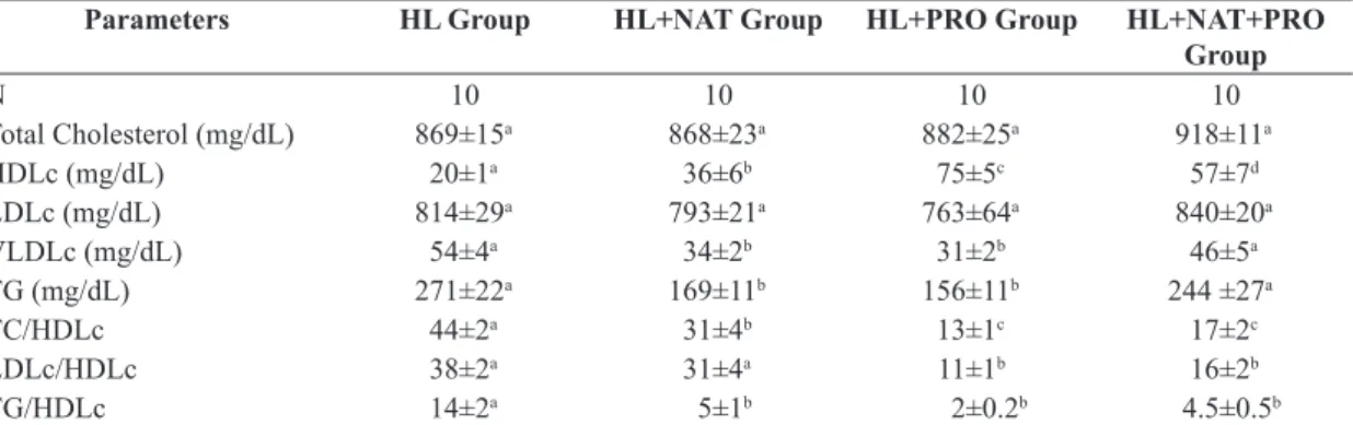
In the analysis of lipid profile, it was not observed significant differences in total cholesterol serum levels (TC)
and the LDLc fraction of animals in the four experimental groups (Table 1). However, it was observed an increase of
HDLc serum levels in mice of NAT, PRO and HL + NAT + PRO when compared with the ones from the HL group
(Table 1). Nevertheless, mice of the PRO group had higher
serum levels of this fraction compared with the mice of HL + NAT + PRO group. The mice of the latter showed greater HDLc serum levels than the mice of the NAT
group (Table 1). With respect to serum levels of VLDLc
fraction and TG, it was observed a decrease in mice from NAT and PRO groups compared to the HL and HL + NAT
+ PRO groups (Table 1). Yet, the TC / HDL, LDL / HDL
and TG / HDL relations were lower in the PRO and HL + NAT + PRO when compared to the HL group (Table 1).
In morphological and morphometric analysis of the
aorta, it was found that swimming and propolis alone in mice
of NAT and PRO respectively prevented the development of atherosclerotic lesions by preventing the increase of
the lesion area when compared to HL group (Table 2 and Figure 1). This was also followed by the prevention of the
inflammatory process in the aorta, in which the aorta of
the NAT and PRO mice exhibited a lesser immunoreactive
area for CD40L when compared to HL group (Table 2 and Figure 1). The mice that underwent swimming and were
treated with propolis, HL + NAT + PRO group, showed an area of injury similar to the one of HL and HL + NAT + PRO groups, but on the other hand, they showed a smaller immunoreactive area for CD 40L when compared with the
NAT and PRO groups (Table 2 and Figure 1).
In morphological and morphometric analysis of the left
ventricle, it was discovered that swimming and propolis
alone or in mice of NAT and PRO respectively prevented
Table 1. Comparison of total cholesterol (TC) serum levels and its HDLc, LDLc and VLDLc fractions, Triglycerides (TG)
and the TC / HDLc, LDLc / HDLc, TG / HDL relations among HL (hyperlipidemic), HL + NAT (hyperlipidemic submitted to swimming), HL + PRO (hyperlipidemic treated with propolis) and HL + NAT + PRO (hyperlipidemic submitted to swimming and treated with propolis) mice.
Parameters HL Group HL+NAT Group HL+PRO Group HL+NAT+PRO Group
N 10 10 10 10
Total Cholesterol (mg/dL) 869±15a 868±23a 882±25a 918±11a
HDLc (mg/dL) 20±1a 36±6b 75±5c 57±7d
LDLc (mg/dL) 814±29a 793±21a 763±64a 840±20a
VLDLc (mg/dL) 54±4a 34±2b 31±2b 46±5a
TG (mg/dL) 271±22a 169±11b 156±11b 244 ±27a
TC/HDLc 44±2a 31±4b 13±1c 17±2c
LDLc/HDLc 38±2a 31±4a 11±1b 16±2b
TG/HDLc 14±2a 5±1b 2±0.2b 4.5±0.5b
(ANOVA + Tukey test). Data were expressed as mean ± Standard Error of the Mean (SEM). Same superscript letters in rows do not differ by Tukey test (p <0.05).
Table 2. Comparison of aortic atherosclerotic lesion area, percentage of immunoreactive area for CD40L in the aorta
and myocardium / endocardium, the left ventricular weight (LVW) / Animals Weight (AW) ratio and the diameter of cardiomyocytes from HL (hyperlipidemic), HL + NAT (hyperlipidemic submitted to swimming), HL + PRO (hyperlipidemic treated with propolis) and HL + NAT + PRO (hyperlipidemic submitted to swimming and treated with propolis) mice.
Parameters HL
Group
HL+NAT Group
HL+PRO Group
HL+NAT+PRO Group
N 10 10 10 10
Atherosclerotic lesion area (μm2) 5552±1643a 484±398b 195±167b 109±109b Immunoreactive area % for CD40L in the
aorta
7.3±0.4a 4.3±0.8b 3.3±0.2b 1.8±0.4c
Immunoreactive area % for CD40L in the myocardial/endocardial
5.2±0.4a 1.8±0.2b 2.4±0.5b 0.5±0.1c
LVW/AW (mg/g) 4.5±0.1a 3±0.1b 2.9±0.1b 3.2±0.1b
Diameter of cardiomyocytes (μm) 25±0.6a 23±0.7a,b 20±0.8b 21±1b
the development of left ventricular hypertrophy, when compared with the HL group (Table 2 and Figure 1), preventing collagen deposition (Figure 1) without changing the cardiomyocytes diameter in the NAT group (Table 2).
However, the diameters of ventricular cardiomyocytes of mice of PRO and HL+ NAT + PRO (Table 2) did not increase. The mice in NAT and PRO groups exhibited a
lesser immunoreactive area for CD40L when compared to the HL group, showing a preventive effect in inflammatory
processes of the myocardium/ endocardium in isolated
conditions. However, when in associated conditions with the HL + NAT + PRO group, this effect was more
exacerbated, presenting a smaller immunoreactivity area
in these mice when compared to the NAT and PRO groups
(Table 2 and Figure 1).
4. Discussion
Studies in our laboratory showed that LDLr–/– mice with standard diet for rodents showed increased HDL serum levels, when compared with its C57Bl6 background, which prevented the inflammatory process and the insulin resistance and consequently showed resistance to the development of
atherosclerotic lesions (Garcia et al., 2011). In the present
study, the LDLr–/– mice fed with high-fat diet showed severe dyslipidemia associated with pronounced reduction Figure 1. Photomicrographs of histological sections of the aorta (panel a: stained with HE showing atherosclerotic
in serum levels of HDL and the subsequent development of cardiovascular inflammation process, characterized by the marked expression of CD40L both in the left ventricle and the aortic wall, formation of atheromatous plaque and left ventricular hypertrophy (LVH). The decrease in HDL serum levels associated with hypercholesterolemia, hypertriglyceridemia and the inflammatory process may
be the common denominator for the atherogenesis and
LVH in mice from the HL group in this study. Moreover, LDLr–/– mice fed with hyperlipidemic diets showed
insulin resistance (Garcia et al., 2011) and increased oxidative stress (Krieger et al., 2006), which sustain the
atherogenesis and LVH in this study.
With the appearance of oxidative hypothesis for atherosclerosis (Steinberg et al., 1989; Meilhac et al., 2001), experiments in vitro, in animal and human models,
have shown that oxidized lipids present pro-atherogenic
effects (Meilhac et al., 2001). In early events of the
atherogenic, oxidized low density lipoprotein (oxLDL) is
rapidly internalized and accumulated in macrophages, thus
forming the foam cells, which are found and deposited in
the subendothelial space (Lusis, 2000; Libby, 2002). The oxLDL is cytotoxic for endothelial cells, promoting the
expression of cytokines and cell proliferation, inhibiting vascular relaxation induced by nitric oxide (NO) and thereby triggering a cascade of inflammatory responses
(Smith, 2001). According to this hypothesis, the increase in
atheroma area of HL mice in the present study, associated with severe dyslipidemia was due to the inflammatory
process, determined by the increased expression of CD40L
with consequent generation of reactive oxygen species (ROS) and lower bioavailability of the NO, which induced
an increase of LDL oxidation and atherogenesis.
Imbalance between production and removal of reactive oxygen species (ROS) is called oxidative stress. Several
physiopathological and genetic situations can induce cardiac oxidative stress, such as hypercholesterolemia (Sato et al., 2004), mechanical stress in the myocardium (Aikawa et al., 2001) and inflammatory processes (Yao et al., 2006).
Therefore, the marked cardiac inflammatory process in HL mice associated with oxidative stress due to the increased expression of CD40L in cardiac tissue and subsequently reduced bioavailability of nitric oxide (NO) hold the pathway of left ventricular hypertrophy observed in the HL mice in this study, characterized by the increase in
collagen deposition and cardiomyocyte diameters.
In the present study, swimming and propolis alone and / or associated prevented LVH, atherogenesis, ventricular and arterial inflammation, decreasing the CD40L expression and increasing the plasma levels of HDLc.
Studies have shown that regular physical activity
promotes the reduction of the inactivation of nitric oxide (Fogarty et al., 2004; Traverse et al., 2000), increases the production of nitric oxide (Takamura et al., 2002), increases
the expression of endothelial nitric oxide synthase (eNOS)
(Griffin et al., 2001) and promotes angiogenesis induced
by vascular endothelial growth factor, and, with the net
effect of increasing the bioavailability of nitric oxide.
The increased bioavailability of nitric oxide in response to the decreased expression of CD40L in this study may be the factor that prevented ventricular hypertrophy and
atherogenesis in mice of NAT group. Therefore, LVH was attenuated by (1) an anti-inflammatory effect due to HDLc serum increase, detected as a decrease in CD40L overexpression, as well as fibrosis because decreasing
CD40-CD40L activity could lead to less severe vascular
inflammation, which in turn would lead to less fibrosis and cardiac derangements, (2) antioxidant activity and consequent decrease in oxidative stress, which resulted in anti-inflammatory effects and a return to homeostasis. Furthermore, the NO / cGMP pathway activates the
sarcolemmal KATP channels (Baker et al., 2001), with
probable inhibition of 70-kDa S6 kinase, attenuating
the hypertrophic response in hyperlipidemic models (Lee et al., 2004).
The prevention of atherogenesis by swimming in the present study was due to the lower oxidative stress and
increased bioavailability of the NO in response to the
increase in HDL serum concentration and increase of shear stress on the vascular wall caused by the increase in cardiac output while swimming in animals of NAT group. The HDL cholesterol is a lipoprotein that carries
out the reverse cholesterol transport, removing the excess of free cholesterol not only from cell membranes but also from the subendothelium and transporting it to the liver to be degraded (Shils et al., 2005), in addition to
its anti-inflammatory and antioxidant effect (Silva et al., 2006). The anti-inflammatory and antioxidant effect of
HDLc in NAT Group was visualized by the decrease
in the immunoreactive area to CD40L in the aorta and myocardium / endocardium of these animals.
In our studies, propolis [total phenols (9,281±0,029%), total flavonoids (2.685±0.001%) and beeswax (14.514±0.144%] alone or associated with swimming increased plasma levels of HDL and decreased expression of CD40L in both myocardium and aortic wall, preventing LVH and atherogenesis. Studies have shown that propolis is a natural antioxidant basically composed of flavonoids and phenolic compounds, which decrease the concentration and activity of superoxide dismutase (SOD), reducing
lipid peroxidation (Marcucci, 1995; Isla et al., 2001; Krol et al., 1990) in the initiation stage, because they act as antioxidants, eliminating superoxide and hydroxyl radicals.
It is proposed that flavonoids interrupt the chain reaction
of free radicals by donating hydrogen atoms to peroxyl
radical, forming a flavonoid radical. The flavonoid radical then reacts with the free radical ending in this manner the
propagation of the chain reaction (Bouziotas et al., 2004).
The increase of HDL, an antioxidant and anti-inflammatory
(Holvoet, 2008), in PRO and HL + PRO + NAT mice in this study explains the protective effect of propolis on the cardiovascular system, reducing oxidative stress and
lipid peroxidation, preventing atherogenesis and LVH in
these animals.
and ventricular inflammation, decreasing the CD40L expression and increasing the HDLc plasmatic levels. Propolis alone or associated with a regular physical activity is beneficial in cardiovascular protection through anti-inflammatory action.
Acknowledgements
The author wishes to thank FAPEMIG, CNPq and UNIFENAS for financial support; and Pharma Nectar for Qualification
of the Constituent Classes from the Propolis Extract.
REFERENCES
AIKAWA, R., NAGAI, T., TANAKA, M., ZOU, Y., ISHIHARA, T., TAKANO, H., HASEGAWA, H., AKAZAWA, H., MIZUKAMI, M., NAGAI, R. and KOMURO, I., 2001. Reactive oxygen species in mechanical stress-induced cardiac hypertrophy. Biochemical and Biophysical Research Communications, vol. 289, no. 4, p. 901-907. http://dx.doi.org/10.1006/bbrc.2001.6068. PMid:11735132 ANDRÉ, P., NANNIZZI-ALAIMO, L., PRASAD, SK. and
PHILLIPS, DR., 2002. Platelet-derived CD40L: the switch-hitting
player of cardiovascular disease. Circulation, vol. 106, no. 8, p. 896-899. http://dx.doi.org/10.1161/01.CIR.0000028962.04520.01. PMid:12186789
ARMSTRONG, AT., BINKLEY, PF., BAKER, PB., MYEROWITZ, PD. and LEIER, CV., 1998. Quantitative investigation of cardiomyocyte hypertrophy and myocardial fibrosis over 6 years after cardiac transplantation. Journal of the American College of Cardiology, vol. 32, no. 3, p. 704-710. http://dx.doi.org/10.1016/
S0735-1097(98)00296-4. PMid:9741515
AUBIN, MC., CARRIER, M., SHI, YF., TARDIF, JC. and PERRAULT, LP., 2006. Role of probucol on endothelial dysfunction
of epicardial coronary arteries associated with left ventricular
hypertrophy. Journal of Cardiovascular Pharmacology, vol. 47, no. 5, p. 702-710. http://dx.doi.org/10.1097/01.fjc.0000211763.13894.87. PMid:16775511
BAKER, JE., CONTNEY, SJ., SINGH, R., KALYANARAMAN, B., GROSS, GJ. and BOSNJAK, ZJ., 2001. Nitric oxide activates
the sarcolemmal K(ATP) channel in normoxic and chronically
hypoxic hearts by a cyclic GMP-dependent mechanism. Journal of Molecular and Cellular Cardiology, vol. 33, no. 2, p. 331-341. http://dx.doi.org/10.1006/jmcc.2000.1305. PMid:11162137 BANKOVA, VS., CASTRO, SL. and MARCUCCI, MC., 2000. Propolis: recent advances in chemistry and plant origin. Apidologie, vol. 31, no. 1, p. 3-15. http://dx.doi.org/10.1051/apido:2000102. BANSKOTA, AH., NAGAOKA, T., SUMIOKA, LY., TEZUKA,
Y., AWALE, S., MIDORIKAWA, K., MATSUSHIGE, K. and KADOTA, S., 2002. Antiproliferative activity of the Netherlands propolis and its active principles in cancer cell lines. Journal of Ethnopharmacology, vol. 80, no. 1, p. 67-73. http://dx.doi.
org/10.1016/S0378-8741(02)00022-3. PMid:11891088 BOUZIOTAS, C., KOUTEDAKIS, Y., NEVILL, A., AGELI, E., TSIGILIS, N., NIKOLAOU, A. and NAKOU, A., 2004. Greek
adolescents, fitness, fatness, fat intake, activity, and coronary heart disease risk. Archives of Disease in Childhood, vol. 89, no. 1, p. 41-44. PMid:14709501.
CHAKRABARTI, S., BLAIR, P. and FREEDMAN, JE., 2007. CD40-40L signaling in vascular inflammation. The Journal of
Biological Chemistry, vol. 282, no. 25, p. 18307-18317. http:// dx.doi.org/10.1074/jbc.M700211200. PMid:17459878 EVANGELISTA, FS., BRUM, PC. and KRIEGER, JE., 2003.
Duration-controlled swimming exercise training induces cardiac
hypertrophy in mice. Brazilian Journal of Medical and Biological Research = Revista Brasileira de Pesquisas Médicas e Biológicas, vol. 36, no. 12, p. 1751-1759. http://dx.doi.org/10.1590/S0100-879X2003001200018. PMid:14666261
FLAXENBURG, JA., MELTER, M., LAPCHAK, PH., BRISCOE, DM. and PAL, S., 2004. The CD40-induced signaling pathway in endothelial cells resulting in the overexpression of vascular
endothelial growth factor involves Ras and phosphatidylinositol 3-kinase. Journal of Immunology (Baltimore, MD.: 1950), vol. 172, no. 12, p. 7503-7509. http://dx.doi.org/10.4049/ jimmunol.172.12.7503. PMid:15187129
FOGARTY, JA., MULLER-DELP, JM., DELP, MD., MATTOX, ML., LAUGHLIN, MH. and PARKER, JL., 2004. Exercise training
enhances vasodilation responses to vascular endothelial growth
factor in porcine coronary arterioles exposed to chronic coronary occlusion. Circulation, vol. 109, no. 5, p. 664-670. http://dx.doi. org/10.1161/01.CIR.0000112580.31594.F9. PMid:14769688 FRIEDEWALD, WT., LEVY, RI. and FREDRICKSON, DS., 1972.
Estimation of the concentration of low-density lipoprotein cholesterol in plasma, without the use of preparatory ultracentrifugation.
Clinical Chemistry, vol. 18, p. 499-502. PMid:4337382. GARCIA, JAD. and INCERPI, EK., 2008. Fatores e mecanismos
envolvidos na hipertrofia ventricular esquerda e o papel anti-hipertrófico do óxido nítrico. Arquivos Brasileiros de Cardiologia, vol. 90, no. 6, p. 443-450. http://dx.doi.org/10.1590/S0066-782X2008000600010.
GARCIA, JAD., LIMA, CC., MESSORA, LB., CRUZ, AF.,
MARQUES, AP., SIMÃO, TP., SOARES, EA., RESCK, MCC., INCERPI, EK., OLIVEIRA, NM. and SANTOS, L., 2011. Efeito
anti-inflamatório da lipoproteína de alta densidade no sistema
cardiovascular de camundongos hiperlipidêmicos. Revista Portuguesa de Cardiologia, vol. 30, no. 10, p. 763-769. http://
dx.doi.org/10.1016/S0870-2551(11)70024-5. PMid:22118127 GELBMANN, CM., LEEB, SN., VOGL, D., MAENDEL, M.,
HERFARTH, H., SCHÖLMERICH, J., FALK, W. and ROGLER,
G., 2003. Inducible CD40 expression mediates NFkB activation and cytokine secretion in human colonic fibroblasts. Gut, vol. 52, no. 10, p. 1448-1456. http://dx.doi.org/10.1136/gut.52.10.1448. PMid:12970138
GRIFFIN, KL., WOODMAN, CR., PRICE, EM., LAUGHLIN,
MH. and PARKER, JL., 2001. Endothelium-mediated relaxation of porcine collateral-dependent arterioles is improved by exercise training. Circulation, vol. 104, no. 12, p. 1393-1398. http://dx.doi. org/10.1161/hc3601.094274. PMid:11560855
HEDRICK, CC., CASTELLANI, LW., WONG, H. and LUSIS, AJ., 2001. In vivo interactions of apoA-II, apoA-I, and hepatic lipase
contributing to HDL structure and antiatherogenic functions. Journal of Lipid Research, vol. 42, no. 4, p. 563-570. PMid:11290828.
HOLVOET, P., 2008. Relations between metabolic syndrome,
oxidative stress and inflammation and cardiovascular disease. Verhandelingen - Koninklijke Academie voor Geneeskunde van België, vol. 70, no. 3, p. 193-219. PMid:18669160.
ISLA, MI., NIEVA MORENO, MI., SAMPIETRO, AR. and VATTUONE, MA., 2001. Antioxidant activity of Argentine propolis extracts. Journal of Ethnopharmacology, vol. 76, no. 2, p. 165-170. http://dx.doi.org/10.1016/S0378-8741(01)00231-8. PMid:11390131
JUNQUEIRA, LC., BIGNOLAS, G. and BRENTANI, RR.,
1979. Picrosirius staining plus polarization microscopy, a specific method for collagen detection in tissue sections. The Histochemical Journal, vol. 11, no. 4, p. 447-455. http://dx.doi.org/10.1007/ BF01002772. PMid:91593
KAI, H., KUWAHARA, F., TOKUDA, K. and IMAIZUMI, T., 2005. Diastolic dysfunction in hypertensive hearts: roles of perivascular inflammation and reactive myocardial fibrosis. Hypertension research : official journal of the Japanese Society of Hypertension, vol. 28, no. 6, p. 483-490. http://dx.doi.org/10.1291/ hypres.28.483. PMid:16231753
KRIEGER, MH., SANTOS, KFR., SHISHIDO, SM., WANSCHEL, ACBA., ESTRELA, HFG., SANTOS, L., DE OLIVEIRA, MG.,
FRANCHINI, KG., SPADARI-BRATFISCH, RC. and LAURINDO, FRM., 2006. Antiatherogenic effects of S-nitroso-N-acetylcysteine
in hypercholesterolemic LDL receptor knockout mice. Nitric Oxide, vol. 14, no. 1, p. 12-20. http://dx.doi.org/10.1016/j. niox.2005.07.011. PMid:16198610
KROL, W., CZUBA, Z., SCHELLER, S., GABRYS, J., GRABIEC, S. and SHANI, J., 1990. Anti-oxidant property of ethanolic extract
of propolis (EEP) as evaluated by inhibiting the chemiluminescence
oxidation of luminol. Biochemistry International, vol. 21, no. 4, p. 593-597. PMid:2241984.
LANG, D., MOSFER, SI., SHAKESBY, A., DONALDSON, F. and LEWIS, MJ., 2000. Coronary microvascular endothelial cell redox state in left ventricular hypertrophy : the role of angiotensin II. Circulation Research, vol. 86, no. 4, p. 463-469. http://dx.doi. org/10.1161/01.RES.86.4.463. PMid:10700452
LEE, TM., LIN, MS., CHOU, TF., TSAI, CH. and CHANG, NC., 2004. Effect of pravastatin on left ventricular mass by activation of myocardial K ATP channels in hypercholesterolemic rabbits. Atherosclerosis, vol. 176, no. 2, p. 273-278. http://dx.doi. org/10.1016/j.atherosclerosis.2004.06.005. PMid:15380449
LIBBY, P., 2002. Atherosclerosis: the new view. Scientific American, vol. 286, no. 5, p. 46-55. http://dx.doi.org/10.1038/ scientificamerican0502-46. PMid:11951331
LUSIS, AJ., 2000. Atherosclerosis. Nature, vol. 407, no. 6801, p. 233-241. http://dx.doi.org/10.1038/35025203. PMid:11001066 LUTGENS, E. and DAEMEN, MJ., 2002. CD40-CD40L interactions in atherosclerosis. Trends in Cardiovascular Medicine, vol. 12, no. 1, p. 27-32. http://dx.doi.org/10.1016/S1050-1738(01)00142-6. PMid:11796241
MACH, F., SCHÖNBECK, U., FABUNMI, RP., MURPHY, C., ATKINSON, E., BONNEFOY, JY., GRABER, P. and LIBBY, P., 1999. T lymphocytes induce endothelial cell matrix metalloproteinase expression by a CD40L-dependent mechanism: implications for tubule formation. American Journal of Pathology, vol. 154, no. 1, p. 229-238. http://dx.doi.org/10.1016/S0002-9440(10)65269-8. PMid:9916937
MACH, F., SCHONBECK, U., SUKHOVA, GK., ATKINSON, E. and LIBBY, P., 1998. Reduction of atherosclerosis in mice by inhibition of CD40 signaling. Nature, vol. 394, no. 6689, p. 200-203. http://dx.doi.org/10.1038/28204. PMid:9671306
MARCUCCI, MC., FERRERES, F., GARCÍA-VIGUERA, C., BANKOVA, VS., DE CASTRO, SL., DANTAS, AP., VALENTE, PH. and PAULINO, N., 2001. Phenolic compounds
from Brazilian propolis with pharmacological activities. Journal of Ethnopharmacology, vol. 74, no. 2, p. 105-112. http://dx.doi.
org/10.1016/S0378-8741(00)00326-3. PMid:11167028 MARCUCCI, MC. and MARCUCCI, 1995. Propolis: chemical composition, biological properties and therapeutic activity. Apidologie, vol. 26, no. 2, p. 83-99. http://dx.doi.org/10.1051/ apido:19950202.
MEILHAC, O., RAMACHANDRAN, S., CHIANG, K.,
SANTANAM, N. and PARTHASARATHY, S., 2001. Role of
arterial wall antioxidant defense in beneficial effects of exercise
on atherosclerosis in mice. Arteriosclerosis, Thrombosis, and Vascular Biology, vol. 21, no. 10, p. 1681-1688. http://dx.doi.
org/10.1161/hq1001.097106. PMid:11597945
NAVAB, M., ANANTHRAMAIAH, GM., REDDY, ST., VAN LENTEN, BJ., ANSELL, BJ., FONAROW, GC., VAHABZADEH, K., HAMA, S., HOUGH, G., KAMRANPOUR, N., BERLINER, JA., LUSIS, AJ. and FOGELMAN, AM., 2004. Thematic review series: the pathogenesis of atherosclerosis the oxidation hypothesis
of atherogenesis: the role of oxidized phospholipids and HDL.
Journal of Lipid Research, vol. 45, no. 6, p. 993-1007. http:// dx.doi.org/10.1194/jlr.R400001-JLR200. PMid:15060092 SATO, K., KOMARU, T., SHIOIRI, H., TAKEDA, S., TAKAHASHI, K., KANATSUKA, H., NAKAYAMA, M. and SHIRATO, K., 2004.
Hypercholesterolemia impairs transduction of vasodilator signals
derived from ischemic myocardium: myocardium-microvessel
cross-talk. Arteriosclerosis, Thrombosis, and Vascular Biology, vol. 24, no. 11, p. 2034-2039. http://dx.doi.org/10.1161/01. ATV.0000143387.58166.c0. PMid:15331436
SHILS, ME., SHIKE, M. and OLSON, JA., 2005. Modern nutrition in health and disease. 10th ed. Baltimore: Lippincott
Williams & Wilkins. 2069 p.
SILVA, JFM., SOUZA, MC., MATTA, SR., ANDRADE, MR. and VIDAL, FVN., 2006. Correlation analysis between phenolic levels of Brazilian propolis extracts and their antimicrobial and antioxidant activities. Food Chemistry, vol. 99, no. 3, p. 431-435. http://dx.doi.org/10.1016/j.foodchem.2005.07.055.
SMITH, JK., 2001. Exercise and atherogenesis. Exercise and Sport Sciences Reviews, vol. 29, no. 2, p. 49-53. http://dx.doi. org/10.1097/00003677-200104000-00002. PMid:11337822 STEINBERG, D., PARTHASARATHY, S., CAREW, TE., KHOO, JC. and WITZTUM, JL., 1989. Beyond cholesterol. Modifications
of low-density lipoprotein that increase its atherogenicity. The New England Journal of Medicine, vol. 320, no. 14, p. 915-924. PMid:2648148.
TAKAMURA, M., PARENT, R. and LAVALLÉE, M., 2002. Enhanced contribution of NO to exercise-induced coronary
responses after α-adrenergic receptor blockade. American Journal of Physiology. Heart and Circulatory Physiology, vol. 282, no.
2, p. H508-H515. http://dx.doi.org/10.1152/ajpheart.00722.2001. PMid:11788398
THIENEL, U., LOIKE, J. and YELLIN, MJ., 1999. CD154
(CD40L) induces human endothelial cell chemokine production and migration of leukocyte subsets. Cellular Immunology, vol. 198, no. 2, p. 87-95. http://dx.doi.org/10.1006/cimm.1999.1583. PMid:10648122
Hyperlipidemia is a major determinant of neointimal formation
in LDL receptor-deficient mice. Biochemical and Biophysical Research Communications, vol. 345, no. 3, p. 1004-1009. http:// dx.doi.org/10.1016/j.bbrc.2006.04.180. PMid:16712797 TRAVERSE, JH., WANG, YL., DU, R., NELSON, D., LINDSTROM, P., ARCHER, SL., GONG, G. and BACHE, RJ., 2000. Coronary nitric oxide production in response to exercise and endothelium-dependent agonists. Circulation, vol. 101, no. 21, p. 2526-2531. http://dx.doi.org/10.1161/01.CIR.101.21.2526. PMid:10831528
VELLAICHAMY, E., SOMMANA, NK. and PANDEY, KN., 2005.
Reduced cGMP signaling activates NF-kappaB in hypertrophied hearts of mice lacking natriuretic peptide receptor-A. Biochemical and Biophysical Research Communications, vol. 327, no. 1, p. 106-111. http://dx.doi.org/10.1016/j.bbrc.2004.11.153. PMid:15629436
WANG, L., WANG, AN, MINESHITA, S., GA, I., 1993. Anti-inflammatory effects of propolis. Japanese Journal of Clinical Pharmacology and Therapeutics, vol. 24, p. 223-226.
WOISKY, RG. and SALATINO, A., 1998. Analysis of propolis:
some parameters and procedures for chemical quality control.
Journal of Apicultural Research, vol. 37, p. 99-105.
YAO, EH., YU, Y. and FUKUDA, N., 2006. Oxidative stress on progenitor and stem cells in cardiovascular diseases. Current Pharmaceutical Biotechnology, vol. 7, no. 2, p. 101-108. http:// dx.doi.org/10.2174/138920106776597685. PMid:16724944
ZHANG, Y., ZANOTTI, I., REILLY, MP., GLICK, JM., ROTHBLAT,