Pho to dynam ic the rapy: a ne w co nce pt
in m e dical tre atm e nt
Department of Radiation O ncology, Case Western Reserve University School of Medicine and University Hospitals of Cleveland, Cleveland, O H, USA
C.H. Sibata, V.C. Colussi, N.L. O leinick and T.J. Kinsella
Abstract
A new concept in the therapy of both neoplastic and non-neoplastic diseases is discussed in this article. Photodynamic therapy (PDT) involves light activation, in the presence of molecular oxygen, of certain dyes that are taken up by the target tissue. These dyes are termed photosensitizers. The mechanism of interaction of the photo-sensitizers and light is discussed, along with the effects produced in the target tissue. The present status of clinical PDT is discussed along with the newer photosensitizers being used and their clinical roles. Despite the promising results from earlier clinical trials of PDT, considerable additional work is needed to bring this new modality of treatment into modern clinical practice. Improvements in the area of light source delivery, light dosimetry and the computation of models of treatment are necessary to standardize treatments and ensure proper treatment delivery. Finally, quality assurance issues in the treatment process should be introduced.
Co rre spo nde nce
C.H. Sibata
Department of Radiation O ncology Case Western Reserve University 11100 Euclid Avenue Lerner Tower B-153A Cleveland, O H 44106-5000 USA
Fax: + 1-216-983-0335 E-mail: cxs81@ po.cwru.edu
Received O ctober 21, 1999 Accepted April 10, 2000
Ke y wo rds
·Photodynamic therapy (PDT)
·Light delivery
·Clinical PDT
·State of art of PDT
Intro ductio n
Photodynamic therapy (PDT) is a rela-tively new therapeutic modality for both neo-plastic and non-neoneo-plastic diseases, which involves light activation, in the presence of molecular oxygen, of certain dyes (photo-sensitizers) that have been somewhat selec-tively taken up by the target tissue. Photosen-sitizers are compounds that absorb energy from light of specific wavelengths and are capable of using that energy to induce reac-tions in other non-absorbing molecules. Therefore, all three components (photosen-sitizer, light and oxygen) have to be available for the treatment to occur.
The concept of PDT began with studies by Oscar Raab in 1900 on the effects of light and dyes on Paramecia (1). The modern era of PDT began with studies by Lipson and
Schwartz at the Mayo Clinic in 1960, who observed that injection of crude preparations of hematoporphyrin led to fluorescence of neoplastic lesions that could be visualized during surgery. Since then, considerable work has been done on how the process works, how to maximize efficacy using animal mod-els, and how to best treat human tumors (2). These pclinical and clinical studies re-cently resulted in the approval of the first
photosensitizing drug (Photofrin®
) for the treatment of selected tumors (3). Photofrin®
is a mixture of porphyrin oligomers (QLT Phototherapeutics Inc., Vancouver, BC,
Canada). Since Photofrin®
injection of Photofrin®
(4). Photofrin®
has been approved in Canada for the prophylac-tic treatment of bladder cancer, and The Netherlands, France, Germany, and Japan have approved it in patients with early and advanced stage cancer of the lung, digestive tract, and genitourinary tract (3). In the US, Photofrin®
is the only photosensitizer ap-proved by the FDA for clinical use for pal-liation of patients with completely obstruct-ing esophageal cancer, or of patients with partially obstructing esophageal cancer who, in the opinion of their physician, cannot be satisfactorily treated with Nd:YAG laser therapy, and for treatment of microinvasive endobronchial non-small cell lung cancer in patients for whom surgery and radiotherapy are not indicated (1,5). The FDA approval includes the uses of a sterile microlens and cylinder-diffusing fibers (1, 1.5, 2.0, 5.0 cm). Several second-generation photosensitiz-ers are being investigated for treatment of a variety of tumors (3,6), but also in non-cancer applications, including ophthalmol-ogy (7), dermatolophthalmol-ogy (6), cardiolophthalmol-ogy (8), virus inactivation (9), and blood purification
(10).These photosensitizers have been
de-signed to improve the selectivity of uptake and to take advantage of the greater depth of penetration of light of longer wavelengths than that used to activate Photofrin®
. Clini-cal trials are being conducted with these photosensitizers at many centers around the world in an effort to provide information for drug approval (3,6-8,10,11).
Ideally, the photosensitizer should be re-tained selectively in the tumor; in practice normal tissues, especially skin, also retain the photosensitizer, and the patients skin can become sensitive to light exposure (in-cluding sunlight) for a period of weeks. Due to the variable tumor selectivity of the photo-sensitizers, it is necessary to optimize the distribution of the delivered light to coincide with the geometric and optical characteris-tics of the targeted tumor tissue, thereby minimizing damage to surrounding normal
tissues. A variety of light sources can be used, provided that the intensity is high enough and the wavelength is within the appropriate range (12). Today, the use of lasers to activate photosensitizers has be-come synonymous with PDT. In fact, for treatment of many tumor sites, e.g., in the esophagus and the bronchus, only lasers pro-duce sufficient power to permit coupling to a fiber optic system and delivery of light to the appropriate site with minimal energy loss. The photosensitizer dose (mg/kg of body weight), time window for treatment (hours or days after injection of photosensitizer) and energy density (fluence) depend on the balance between concentration of the drug in the tumor (target) tissue (mg/g tissue) and in normal tissues at the time of light irradia-tion. The choices of these parameters have typically been based on drug pharmacoki-netics and pre-clinical trials done in experi-mental animals. This paper will present an overview of the therapeutic applications of PDT for neoplastic and non-neoplastic dis-eases.
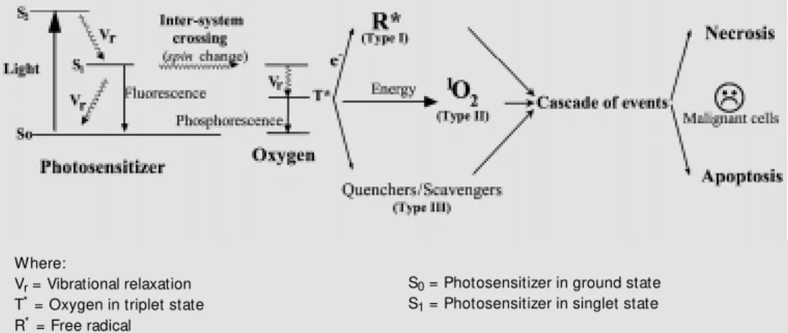
Me chanisms o f pho to dynamic the rapy
The photosensitizer in the triplet state can react with ground-state oxygen to produce a singlet-state oxygen (type II reaction), decay to the ground state by phosphorescence, or undergo type I and III reactions. Singlet oxy-gen (a non-radical but highly reactive form of oxygen) is generally accepted as the major damaging species in PDT (14). Other reac-tive oxygen species may also be involved in the tumor ablation caused by PDT (11).
The complex nature of the tumor response to PDT has yet to be fully elucidated. PDT depends on the photodynamic events in ma-lignant cells or within cells of the tumor vasculature. In the latter case, damage to the tumor vasculature can result in profound effects, including blood flow stasis, vascular collapse, and/or vascular leakage (15,16). Damage either to the malignant cells or to cells of the vasculature results in the death of tumor cells. PDT has been shown to induce apoptosis (a programmed process of cell death that is responsible for the orderly elimi-nation of cells during normal tissue develop-ment) in many cells in vitro,and in all animal tumors tested in vivo. Apoptosis in response to toxic agents proceeds from the signaling of cell stress and culminates in the activation of a cascade of cysteine proteases, termed caspases, that catalyze the final degradation of key cellular proteins, including the nuclear protein poly(ADP-ribose) polymerase (11,17,18). In animal systems, gross edema
and erythema are the first clinical signs of PDT response.
Light inte ractio n with tissue
In principle, the basic phenomena occur-ring when matter is exposed to light are the following:
- reflection andrefraction - absorption
- scattering
Reflection and refraction are closely
re-lated.Refraction is accounted for by a
dis-placement of the transmitted beam through
the medium. Refraction plays a role in the
optics of the instrumentation used to apply light for PDT.In opaque media, the effect of refraction is difficult to measure due to ab-sorption and scattering.In biological tissues, either water molecules or macromolecules such as proteins and pigments can absorb
light. Absorption occurs when an
electro-magnetic wave interacts with an elastically bound charged particle, which then vibrates at the frequency of the electromagnetic wave. Scattering, on the other hand, takes place at frequencies not corresponding to the natural frequencies of these particles.Scattering can be elastic or inelastic, depending on whether or not there is energy absorption during the scattering process.Both absorption and scat-tering processes will be present in most tis-sues, which are considered turbid media.
Figure 1 - Photodynamic therapy process.
Where:
Vr = Vibrational relaxation
T* = Oxygen in triplet state
R* = Free radical
S0 = Photosensitizer in ground state
The types of light interaction with tissue depend on the wavelength and on the proper-ties of the medium irradiated. There are five categories of interaction: photochemical, thermal, photoablation, plasma-induced
ab-lation, and photodisruption.These
interac-tions depend on the total power density
ap-plied to the tissue. For clinical PDT, only
photochemical interactions and possibly
ther-mal interactions are important.PDT has been
shown to be synergistic with sub-lethal hy-perthermia (19).In pre-clinical trials of PDT alone, the power density is maintained low enough to avoid thermal interactions.
Of the major proteins of blood that ab-sorb light, the most important quantitatively
is hemoglobin.Hemoglobin has significant
absorption near 425, 544, and 577 nm, ne-cessitating illumination of tissue at wave-lengths >600 nm to ensure significant
pen-etration. At wavelengths >1200 nm, light
absorption by water molecules becomes sub-stantial.For wavelengths >850-900 nm, the photons may not have sufficient energy to participate in a photochemical reaction. Therefore, the wavelength range between 600 and 800 nm has been determined as the practical therapeutic window for clinical PDT (Figure 2).
Photosensitization can also be affected within tissue by the presence of endogenous chromophores such as melanin, which com-petes with the photosensitizer for absorption of light.This is relevant when treating darker skinned patients or hyperpigmented lesions
of metastatic melanoma.The absorption of
light by the photosensitizer itself can limit light penetration in tissue, a phenomenon
called photosensitizer self-shielding (15). In addition, the excited-state photosensitizer molecule can undergo a side reaction lead-ing to loss of absorbance and photosensitiz-ing ability (a process called photobleachphotosensitiz-ing) (15). This process can modify the reciproc-ity between photosensitizer level and light, since with irradiation there will be a
progres-sive loss of sensitizer.Photobleaching can
be an advantage because, as the photosensi-tizer near the surface bleaches, light can
penetrate deeper into the tissue. After the
completion of treatment, photobleaching can be used to accelerate the clearance of drug from the body (20).
Light so urce s
The light source for PDT can be an ordi-nary light bulb, a diode array emitting a broad band incoherent spectrum, or a laser. Initially, PDT was performed with broad-spectrum light sources such as xenon arc lamps or slide projectors equipped with red
filters to eliminate short wavelengths.
How-ever, the light intensities are low with these devices. Furthermore, PDT using these sources is limited to directly accessible sites such as the skin.Such sources are still used for in vitro and pre-clinical invivo studies of
tumors implanted in or under the skin.The
advantages of lamps as light sources for PDT are their relatively low cost, simplicity and
reliability (21). However, they cannot be
used for optical fiber delivery because of the poor coupling efficiency into single fibers.
Lasers have become the standard light sources for PDT applications, due to their
monochromatic character, high power out-put, and ease of coupling to fiber optics for endoscopic light delivery within a body cav-ity or for interstitial implants.Lasers can be used for both therapeutic and diagnostic
ap-plications in PDT.The most commonly used
lasers for PDT are the tunable dye lasers, due to their versatility with respect to wavelength
selection. The US FDA approved Photofrin®
in conjunction with a specific device, either
a laser system produced by LaserScope®
(http://www.laserscope.com) or a system
from Coherent® (http://www.cohr.com).The
main disadvantage of pumped dye lasers is the high capital and running costs and poor
reliability in the clinical environment. One
of the more practical recent advances in PDT is the availability of diode lasers at wave-lengths compatible with currently used
pho-tosensitizers (12).These systems have
mini-mum electrical power requirement and are cooled thermoelectrically to provide a
com-pact laser system.The potential advantages
of the diode lasers for PDT are the low capital cost, negligible running costs, high reliability, small size and portability.These systems are very attractive for both clinical and pre-clinical investigations.
A laser delivery system for PDT consists of light source entrance optics, a beam guide, and target optics. Currently, coherent mono-chromatic lasers and single-strand optical fibers provide the flexibility needed to de-liver light to subsurface lesions through sur-face, interstitial, intracavitary or endoscopic techniques. The choice of the delivery sys-tem will be governed by the characteristics of the laser system on the one hand and the
tissue application on the other. The spatial
distribution of a beam coming directly from the laser is usually of a gaussian shape (monomode) or a summation of gaussian
profiles (multimode). The end of the fiber
optic cable can be modified to shape the light
to match the tissue to be treated. For
ex-ample, introducing a lens at the end of the fiber optic spreads the beam uniformly over
a specified area and can make the beam more
uniform in intensity. These properties are
important for the design and computation of target optics or modifications of the fiber tip. For a therapeutic system, the light on the target tissue can be seen and verified through the endoscope, either by eye or using a video
camera.The tip of the fiberscope is usually
maintained 1-2 cm from the target. An excel-lent overview of the evolution of endoscopic light delivery systems for PDT has recently been published (22).
Light do sim e try
The most important requirements for the diagnostic and therapeutic photoirradiation systems employed in PDT are that sufficient light must reach the target sites and its inten-sity must be properly verified at the
treat-ment site by light detectors.There are four
kinds of light detectors generally used in the
optics field. The thermopile relies on the
measurement of a temperature increase re-sulting from light absorbed in a material. Since the measurement is dependent upon significant thermal change, the use of a ther-mopile is limited to high power applications and is therefore well suited for laser opera-tions.The photodiodedirectly converts light into an electrical current or voltage.Diodes show a large spectral sensitivity variation and must be calibrated at the wavelength of
interest for quantitative measurements.Use
use-ful in the detection of low light levels.These detectors have been used alone or in combi-nation to verify light delivery before treating
a tumor.The most common device for power
measurement is the integrating sphere. The integrating sphere is a hollow sphere coated inside with barium sulfate, a diffuse white reflectance coating that offers greater than 97% reflectance between 450 and 900 nm. Integrating spheres are used as sources of uniform radiance and as input optics for measuring total power.
The success of PDT is dependent among other factors on the total light dose delivered
into the target tissue (Table 1). The unit
giving the total energy delivered is the joule (J) and is determined by watt (W) multiplied
by time (s).The number of photons (N) in a
joule depends upon the wavelength (l) of
the light. If two different wavelengths of
light are used, the number of photons per joule varies as the inverse ratio of the wave-length (hc/l, where h = 6.623 x 10⁻³⁴
J is Plancks constantand c = 2.998 x 10⁸
ms-1
is the speed of light).
Another factor that must be considered in PDT is the delivered rate of the light (fluence rate = W/area).Fluence rate, and thus treat-ment time, depends on the light source used. If the light is delivered at a high rate, signifi-cant heating of a molecule and its
surround-ing may take place. Usually, fluence rates
higher than 200 mW/cm2
(for microlens) or 400 mW/cm (for cylindrical diffusers) are not used due to thermal effects that can damage the normal tissues.
Applying a given light intensity in W/ area to the surface of a tissue does not imply that we know precisely what happens to the light intensity as the photons penetrate into the underlying tissue.It is necessary to meas-ure light fluence within the tissue during the PDT session and the distribution of the pho-tosensitizer to better quantify the effects of
PDT.The ideal PDT irradiation would
con-sist of optimizing the distribution of the de-livered light to match the geometric
distribu-tion of the photosensitizer and the optical properties of the target tissue in order to minimize the damage to the surrounding
nor-mal tissues. Ascertaining the geometrical
and optical properties of the target tissue and surrounding normal tissues for the individual patient is one of the more difficult aspects of
PDT dosimetry.This requires both
theoreti-cal and experimental techniques for
plan-ning and monitoring the irradiation.
Treat-ment planning is generally the physicists responsibility, and it consists of optimizing the light delivery (balance between geom-etry, power density and treatment time) con-sidering the laser system available.
It is not always possible to rely upon the reading of the laser display for accurate
do-simetry during PDT.Although most lasers
have a power calibration port on the system, the measurement cannot always be performed under conditions that are comparable with the clinical application.Since the concentra-tion of the photosensitizer in tissue will af-fect the biological response, an acceptable means of measuring the photosensitizer in the target tissue and adjacent normal tissues is also desirable.The goal of light dosimetry is to optimize the distribution of the light dose in the treatment volume by selecting the
best irradiation geometry.A recent
publica-tion (23) presented the concepts of light distribution in biological tissues in terms of simple expressions for spherical, cylindrical and planar geometry resulting from point sources, line sources (cylindrical diffusers), and planar sources (microlenses) which can be used as a guideline for clinical applica-tions.
Central to light therapy planning is com-putation of the spatial distribution of the light dose in the target and surrounding
tis-sues.The computational models are usually
Techniques to measure photosensitizer con-centration non-invasively in vivo are being developed (24) using elastic scattering
meth-ods.The development of suitable
computa-tional models involves testing the dose pre-dictions against experimental measurements using both in vivo and tissue-simulating phan-toms (25).
O xyge n and o xyge natio n strate gie s fo r PD T
The efficacy of PDT with photosensitiz-ers which localize in tumor tissue is most likely related to the yield of singlet oxygen (1
O2) in the tumor (14). The yield of 1O2, in
turn, depends on the concentration of oxy-gen in the tissue (26). Some tumors may contain regions with oxygen concentrations too low for PDT to be optimally efficient (27). Both blood supply and oxygen con-sumption determine the amount of free oxy-gen available in a tissue. Truly hypoxic cells are thus very resistant to PDT. Tumor oxy-genation may be improved by breathing a perfluorochemical emulsion or carbogen
(95% O2, 5% CO2), which may modify the
effect of PDT under certain conditions. The PDT reaction mechanism itself may con-sume oxygen at a rate sufficient to inhibit further PDT effects (28,29). It has been sug-gested that hyperbaric oxygen could enhance the PDT effect (30). In a recent study using the Walker 256 tumor model in Wistar rats, the enhancement of PDT effects under hy-perbaric hyperoxia was demonstrated (31). The increased depth of tumor damage was evaluated by measuring histological sections following PDT treatment of tumors at 3
at-mospheres (atm) and controls at 1 atm. Ex
vivo morphometric analysis showed a total
loss of cell viability in treated over control tissue. More experimental studies in this direction are warranted.
Another simpler approach for overcom-ing limitations of oxygen diffusion is to frac-tionate light delivery (e.g., 30 s on, 30 s off)
or to reduce the fluence rate. Such protocols allow oxygen diffusion to compete with oxy-gen consumption and can provide improved tumor response (32,33).
Pho to se nsitize rs and clinical trials
Numerous clinical trials of PDT have been carried out (phase I, II, III and IV) for
treatment of malignant lesions.PDT has been
shown to be most efficacious for small tu-mors because of the light depth limitation.
Photofrin®
(labs @ 630 nm) is the first
photo-sensitizer to be approved for clinical PDT, and additional trials are ongoing. Photofrin®
-PDT is considered easier to perform than Nd-YAG ablation, and is especially advan-tageous in situations where Nd-YAG laser irradiation is difficult to carry out due to tumor location or tumor size such as found in
advanced stage esophageal tumors (34).For
lung cancer, Photofrin®
-PDT has been ap-proved for early and advanced non-small cell lung cancer.In these cases, it was con-cluded that PDT is superior to Nd-YAG irradiation for relief of dyspnea, cough, and
hemoptysis. Photofrin®
-PDT has been ap-proved in Canada since 1993 for prophylac-tic treatment of papillary bladder tumors in patients at a high risk for recurrence.
Photofrin®
-PDT is being used in trials of high grade dysplasia as found in Barretts
esophagus.Biel (35) has reported excellent
results in treatment of early stage head and neck cancer. Photofrin®
-PDT has been tested also for adjuvant therapy such as combining PDT with resection of brain tumors (36,37), surgery for pleural cancers, especially ma-lignant mesothelioma (38) and debulking surgery for intraperitoneal tumors (39).
In addition, several second-generation photosensitizers are undergoing clinical test-ing. These second-generation compounds are generally pure, can be activated by light in the range of 630-800 nm, and share in com-mon a lower incidence of prolonged cutane-ous photosensitivity than Photofrin®
Table 1 - The basic units used in photodynamic therapy.
Name (definition) Concept (symbol)
Photosensitizer Drug dose mg/kg body w eight or mg/m2
Drug concentration mg drug/g tissue
Wavelength of light absorption labs (nm = 10-9 m)
Light delivery Number of photons N
Energy of photons Joule (J)
Energy density, fluence, intensity J/cm2 (microlens) or J/cm (cylinder diffuser)
Pow er, radiant pow er, radiant energy flux Watt (W) = J/s
Pow er density, fluence rate, irradiance W/cm2 (microlens) or W/cm
(cylinder diffuser)
Optical penetration d (cm)
Oxygen Partial pressure atm = kgf/cm2, mmHg, torr
Concentration mM (10-6 M )
Results Treatment penetration depth z (cm)
these is d-aminolevulic acid (ALA or
Levu-lan®
; DUSA Pharmaceuticals, Toronto, Ontario, Canada; labs @ 630 nm), a precursor
to the photosensitive protoporphyrin IX in the heme biosynthetic pathway. A limitation of Photofrin®
and ALA is their low extinc-tion at their absorpextinc-tion peak furthest into the red region (630 nm). Promising clinical re-sults have been obtained using ALA in a variety of superficial malignant and non-malignant lesions such as squamous cell car-cinoma of the skin, Bowens disease, myco-sis fungoides, psoriamyco-sis (40,41) and solar keratosis (21).
Benzoporphyrin derivative monoacid ring
A (BPD-MA or Verteporfin®
; QLT Photo-therapeutics Inc.; labs @ 692 nm) is a liposomal
formulation. It appears to be useful for the treatment of age-related macular generation
and choroidal melanoma (42).BPD-PDT also
has been tested for treatment of atherosclerotic plaques (43) and psoriasis (44,45).
Lutetium texaphyrin/Lutex (LutrinTM
;
Pharmacyclics, Sunnyvale, CA, USA; labs @
732 nm) is a water-soluble photosensitizer that recently entered phase I clinical trials. It accumulates preferentially in malignant tis-sue (via an increased lipoprotein receptor
mechanism).Lutex has been tested for
pho-todynamic therapy of cardiovascular disease
and for treatment of certain skin lesions (46).
Tin ethyl etiopurpurin(SnET2 or
Purly-tin®
; Miravant, Santa Barbara, CA, USA; labs @ 664 nm) is being used in a phase II/III
open-label, randomized study for women with advanced breast cancer and Kaposis sarcoma in patients with AIDS, and phase I/ II clinical testing for age-related macular
degeneration.FDA has given its approval to
begin a clinical study of Purlytin®
for pros-tate cancer as well (47).
Tetra(m-hydroxyphenyl)chlorin (mTHPC
or Foscan®
; Scotia Pharmaceuticals, Kent-ville, Nova Scotia, Canada; labs@ 652 nm) is
undergoing clinical testing in recurrent head and neck cancers in Europe and the US (48),
and mono-L-aspartyl chlorin e6 (NPe6; labs
@ 664 nm) has recently entered clinical trials for superficial malignancies of the skin and nasopharynx (49).
An advantage of using ALA, BPD-MA, and Lutex for PDT treatment is the ability to complete both drug administration and light exposure on the same day as a routine office procedure (41).
The silicon phthalocyanine (Pc 4; labs@
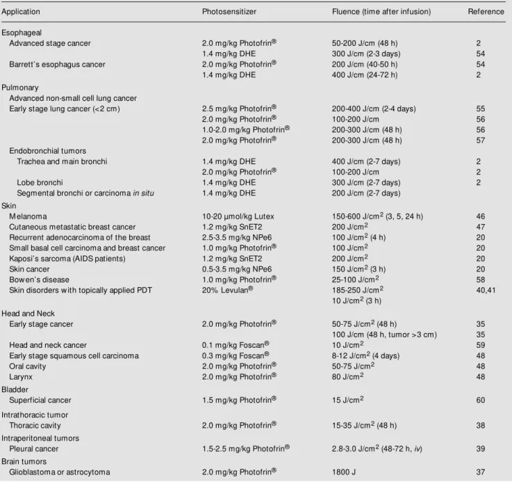
Table 2 - PDT clinical applications.
Every effort w as made to compile all the published protocols.
Application Photosensitizer Fluence (time after infusion) Reference
Esophageal
Advanced stage cancer 2.0 mg/kg Photofrin® 50-200 J/cm (48 h) 2
1.4 mg/kg DHE 300 J/cm (2-3 days) 54
Barrett’s esophagus cancer 2.0 mg/kg Photofrin® 200 J/cm (40-50 h) 54
1.4 mg/kg DHE 400 J/cm (24-72 h) 2
Pulmonary
Advanced non-small cell lung cancer
Early stage lung cancer (<2 cm) 2.5 mg/kg Photofrin® 200-400 J/cm (2-4 days) 55
2.0 mg/kg Photofrin® 100-200 J/cm 56
1.0-2.0 mg/kg Photofrin® 200-300 J/cm (48 h) 56
2.0 mg/kg Photofrin® 200-300 J/cm (48 h) 57
Endobronchial tumors
Trachea and main bronchi 1.4 mg/kg DHE 400 J/cm (2-7 days) 2
2.0 mg/kg Photofrin® 100-200 J/cm 2
Lobe bronchi 1.4 mg/kg DHE 300 J/cm (2-7 days) 2
Segmental bronchi or carcinoma in situ 1.4 mg/kg DHE 200 J/cm (2-7 days) Skin
M elanoma 10-20 µmol/kg Lutex 150-600 J/cm2 (3, 5, 24 h) 46
Cutaneous metastatic breast cancer 1.2 mg/kg SnET2 200 J/cm2 47
Recurrent adenocarcinoma of the breast 2.5-3.5 mg/kg NPe6 100 J/cm2 (4 h) 20
Small basal cell carcinoma and breast cancer 1.0 mg/kg Photofrin® 100 J/cm2 20
Kaposi’s sarcoma (AIDS patients) 1.2 mg/kg SnET2 200 J/cm2 20
Skin cancer 0.5-3.5 mg/kg NPe6 150 J/cm2 (3 h) 20
Bow en’s disease 1.0 mg/kg Photofrin® 25-100 J/cm2 58
Skin disorders w ith topically applied PDT 20% Levulan® 185-250 J/cm2 40,41
10 J/cm2 (3 h)
Head and Neck
Early stage cancer 2.0 mg/kg Photofrin® 50-75 J/cm2 (48 h) 35
100 J/cm (48 h, tumor >3 cm) 35
Head and neck cancer 0.1 mg/kg Foscan® 10 J/cm2 59
Early stage squamous cell carcinoma 0.3 mg/kg Foscan® 8-12 J/cm2 (4 days) 48
Oral cavity 2.0 mg/kg Photofrin® 50-75 J/cm2 48
Larynx 2.0 mg/kg Photofrin® 80 J/cm2 48
Bladder
Superficial cancer 1.5 mg/kg Photofrin® 15 J/cm2 60
Intrathoracic tumor
Thoracic cavity 2.0 mg/kg Photofrin® 15-35 J/cm2 (48 h) 38
Intraperitoneal tumors
Pleural cancer 1.5-2.5 mg/kg Photofrin® 2.8-3.0 J/cm2 (48-72 h, iv) 39
Brain tumors
Glioblastoma or astrocytoma 2.0 mg/kg Photofrin® 1800 J 37
of the Drug Decision Network of the US National Cancer Institute, Pc 4 has com-pleted evaluation of pre-clinical pharmaco-kinetics, efficacy and toxicology and has been recommended for clinical testing. A-mong the desirable features of Pc 4 are its chemical purity, its high extinction coeffi-cient (e >2 x 10⁵
at 672 nm) affording deep
tissue penetration of light, and its rapid clear-ance from skin, limiting the extent and
dura-tion of cutaneous photosensitivity.
Consid-erable biological data regarding the efficacy of Pc 4-PDT are available in human tumor cells in vitro and in xerografts systems as
well as in animal tumors (10,11,51,52). A
target-ing of the tumor parenchyma (52).
Table 2 shows the current status of PDT treatment and the time after drug injection generally used for the clinically approved and experimental photosensitizers.
Co nclusio n
Photodynamic therapy is a potentially effective and safe treatment approach for superficial human cancers and selected
be-nign conditions.The technique can be used
as an adjuvant therapy with surgery,
radia-tion or chemotherapy.Major late effects are
limited to skin photosensitization for up to 6 weeks after Photofrin®
injection. Newer gen-eration photosensitizers are being tested which may produce less photosensitivity.
PDT is an exciting multi-disciplinary area involving research and direct clinical
appli-cation of the research. A cancer center is
multi-disciplinary in nature making it a natu-ral environment for a PDT center. It provides alternative therapy for many patients who cannot have any other type of treatment, and
it also benefits medical education.
Refine-ment of this technique will require collabo-rative research efforts in several fields, in-cluding chemical synthesis, pharmacokinet-ics of the photosensitizers to establish opti-mum treatment times, physics to develop better light sources and delivery methods to ensure proper light delivery, and to improve
treatment through visualization of the target. One of the most pressing issues in deal-ing with the several clinical trials bedeal-ing imple-mented is the issue of light dosimetry and the proper delivery of light dose. Usually, the light dose is given in terms of external power density delivered by the light system. The light dose actually received by the photosen-sitizer and tissue may be higher or lower depending on the geometry of irradiation. A proper definition of light dose is also needed. An attempt to define light dose similar to what is used in radiation oncology was
at-tempted by Profio and Doiron (53).The use
of this definition calls for knowledge of the photosensitizer concentration in tissue, which
is not yet known. Another issue is quality
assurance of the treatment. Presently, most treatments are done without proper verifica-tion of the equipment, whether or not the light distribution is uniform, the light deliv-ery system is properly calibrated, and if the
patient has been properly immobilized. To
ensure optimum treatment and comparison between results of different clinical trials, a quality assurance program of treatment method and light delivery should be estab-lished.Finally, the future progress of photo-dynamic therapy will require input from phys-ics, engineering, and computer science to develop models for light dosimetry and treat-ment planning.
Re fe re nce s
1. Laser M edical Research Foundat ion. [http://209.41.253.5:80/pdt@lmrf]. Date accessed: 10 September 1999.
2. Colussi VC, Nicola EM D & Nicola JH (1996). Phot ot herapy, phot ochem o-therapy and some photosensitizers (in Port uguese). Revist a da Associação M édica Brasileira, 42: 229-236.
3. Dougherty TJ, Gomer CJ, Henderson BW, Jori G, Kessel D, Korbelik M , M oan J & Peng Q (1998). Photodynamic therapy (Review ). Journal of the National Cancer Institute,90: 889-905.
4. M cCaughan Jr JS & Williams TE (1997). Photodynamic therapy for endobronchial malignant disease: A prospective four-teen-year study. Journal of Thoracic and Cardiovascular Surgery,114: 940-947. 5. Nat ional Cancer Inst it ut e. [ht t p://
cancernet.nci.nih.gov]. Date accessed: 10 September 1999.
6. Colussi VC, Feyes DK & M ukhtar H (1998). Perspectives of photodynamic therapy for skin diseases. Skin Pharmacology and Ap-plied Skin Physiology,11: 336-346. 7. Husain D, M iller JW, Kenney AG, M ichaud
N, Flotte TJ & Gragoudas ES (1997). Pho-todynamic therapy and digital angiogra-phy of experimental iris neovasculariza-tion using liposomal benzoporphyrin de-rivative. Ophthalmology, 104: 1242-1250. 8. Woodburn KW, Fan Q, Kessel D, Wright M , M ody TD, Hemmi G, M agda D, Sessler JL, Dow WC, M iller RA & Young SW (1996). Phot ot herapy of cancer and atheromatous plaque w ith texaphyrins.
Journal of Clinical Laser M edicine and Sur-gery, 14: 343-348.
Salzberg S, Wagner P & M alik Z (1998). Herpes simplex virus proteins are dam-aged follow ing photodynamic inactivation w ith phthalocyanines. Journal of Photo-chemistry and Photobiology. B, Biology, 44: 77-83.
10. Ben-Hur E, Oetjen J & Horow itz B (1997). Silicon phthalocyanine Pc 4 and red light cause apoptosis in HIV-infected cells. Pho-tochemistry and Photobiology, 65: 456-460.
11. Colussi VC, Feyes DK, M ulvihill JW, Li Y-S, Kenney M E, Elmets CA, Oleinick NL & M ukhtar H (1999). Phthalocyanine (Pc 4) photodynamic therapy of human OVCAR-3 tumors. Photochemistry and Photobiol-ogy, 69: 236-241.
12. Wilson BC (1998). Light sources for pho-todynamic therapy. Photodynamic Ther-apy New s, 1: 6-8.
13. Laustriat G (1986). M olecular mechanisms of photosensitization. Biochimie, 68: 771-778.
14. Weishaupt KR, Gomer CJ & Dougherty TJ (1976). Identification of singlet oxygen as the cytotoxic agent in cancer photoinacti-vation of a murine tumor. Cancer Re-search, 36: 2326-2329.
15. Henderson BW & Dougherty TJ (1992). How does photodynamic therapy w ork?
Photochemistry and Photobiology, 55: 145-157.
16. Peng Q, M oan J & Nesland JM (1996). Correlation of subcellular and intratumoral photosensitizer localization w ith ultra-structural features after photodynamic therapy. Ultrastructural Pathology, 20: 109-129.
17. Zaidi SIA, Oleinick NL, Zaim M T & M ukhtar H (1993). Apoptosis during pho-todynamic therapy-induced ablation of RIF-1 tumors in C3H mice: Electron mi-croscopic, histopathologic and biochemi-cal evidence. Photochemistry and Photo-biology, 58: 771-776.
18. Kessel D, Luo Y, Deng Y & Cang CK (1997). The role of subcellular localization in initiation of apoptosis by photodynamic therapy. Photochemistry and Photobiol-ogy,65: 422-426.
19. Chen Q, Chen H & Hetzel FW (1996). Tumor oxygenation changes post-photo-dynamic therapy. Photochemistry and Photobiology, 63: 128-131.
20. Taber SW, Fingar VH & Wieman TJ (1998). Photodynamic therapy for palliation of chest w all recurrence in patients w ith breast cancer. Journal of Surgical Oncolo-gy,68: 209-214.
21. Fritsch C, Stege H, Saalmann G, Goerz G, Ruzicka T & Krutmann J (1997). Green
light is effective and less painful than red light in photodynamic therapy of facial so-lar keratoses. Photodermatology, Photo-immunology and Photomedicine, 13: 181-185.
22. van den Bergh H (1998). On the evolution of some endoscopic light delivery sys-tems for photodynamic therapy. Endos-copy,30: 392-407.
23. Jacques SL (1998). Light distributions from point, line and plane sources for pho-tochemical reactions and fluorescence in turbid biological tissues. Photochemistry and Photobiology, 67: 23-32.
24. M ourant JR, Johnson TM , Los G & Bigio IJ (1999). Non-invasive measurement of chemotherapy drug concentrations in tis-sue: preliminary demonstrations of in vivo
measurements. Physics in M edicine and Biology,44: 1397-1417.
25. Wagnieres G, Cheng S, Zellw eger M , Utke N, Braichotte D, Ballini JP & van den Bergh H (1997). An optical phantom w ith tissue-like properties in the visible for use in PDT and fluorescence spectroscopy.
Physics in M edicine and Biology, 42: 1415-1426.
26. M oan J & Sommer S (1985). Oxygen de-pendence of the photosensitizing effect of hematoporphyrin derivative in NHIK 3025 cells. Cancer Research, 45: 1608-1610.
27. Vaupel P & Thew s G (1974). PO2
distribu-tion in tumour tissue of DS-carcinoma.
Oncology,30: 475-484.
28. Trom berg BJ, Orenstein A, Kim el S, Barker SJ, Hyatt J, Nelson JS & Berns M W (1990). In vivo tumor oxygen tension measurements for the evaluation of the efficiency of photodynamic therapy. Pho-tochemistry and Photobiology, 52: 375-385.
29. Chen Q, Chen H, Shapiro H & Hetzel FW (1996). Sequencing of combined hyper-thermia and photodynamic therapy. Ra-diation Research, 146: 293-297. 30. Jirsa Jr M , Poucková P, Dolezal J, Pospisil
J & Jirsa M (1991). Hyperbaric oxygen and photodynamic therapy in tumor-bear-ing nude mice. European Journal of Can-cer, 27: 109.
31. Colussi VC (1997). Int ensif icação da terapia fotodinâmica do câncer pela varia-ção da pressão parcial do oxigênio no tecido: Efeitos físicos e biológicos. Doc-t oral Doc-t hesis, UniversiDoc-t y of Cam pinas/ UNICAM P, São Paulo, Brazil.
32. Foster TH, Hartley DF, Nichols M G & Hilf R (1993). Fluence rate effects in photody-namic therapy of multicell tumor sphe-roids. Cancer Research, 53: 1249-1254.
33. Sitnik TM , Hampton JA & Henderson BW (1998). Reduction of tumour oxygenation during and after photodynamic therapy in vivo: effects of fluence rate. British Jour-nal of Cancer, 77: 1386-1394.
34. Light dale CJ, Heier SK, M arcon NE, M cCaughan Jr JS, Gerdes H, Overholt BF, Sivak Jr M V, Stiegmann GV & Nava HR (1995). Photodynamic therapy w ith porfimer sodium versus thermal ablation therapy w ith Nd:YAG laser for palliation of esophageal cancer: a multicenter random-ized trial. Gastrointestinal Endoscopy, 42: 507-512.
35. Biel M A (1998). Photodynamic therapy and the treatment of head and neck neo-plasia. Laryngoscope, 108: 1259-1268. 36. M uller PJ & Wilson BC (1995).
Photody-namic therapy for recurrent supratentorial gliomas. Seminars in Surgical Oncology, 11: 346-354.
37. Popovic E, Kaye A & Hill J (1996). Photo-dynamic therapy of brain tumors. Journal of Clinical Laser M edicine and Surgery, 14: 251-256.
38. Takita H & Dougherty TJ (1995). Intracavi-tary photodynamic therapy for malignant pleural mesothelioma. Seminars in Surgi-cal Oncology, 11: 368-371.
39. DeLaney TF, Sindelar WF, Tochner Z, Sm it h PD, Friauf W S, Thom as G, Dachow ski L, Cole JW, Steinberg SM & Glatstein E (1993). Phase I study of debulking surgery and phot odynam ic therapy for disseminated intraperitoneal tumors. International Journal of Radiation Oncology, Biology, Physics, 25: 445-457. 40. Kennedy JC, M arcus SL & Pottier RH (1996). Photodynamic therapy (PDT) and photodiagnosis (PD) using endogenous photosensitization induced by 5-aminolev-ulinic acid (ALA): mechanisms and clinical results. Journal of Clinical Laser M edicine and Surgery, 14: 289-304.
41. Peng Q, W arloe T, Berg K, M oan J, Kongshaug M , Giercksky KE & Nesland JM (1997). 5-Aminolevulinic acid-based photodynamic therapy. Clinical research and future challenges. Cancer, 79: 2282-2308.
42. Husain D, M iller JW, M ichaud N, Connolly E, Flotte TJ & Gragoudas ES (1996). Intra-venous infusion of liposomal benzopor-phyrin derivative for photodynamic ther-apy of experimental choroidal neovascu-larization. Archives of Ophthalmology, 114: 978-985.
Watanabe heritable hyperlipidemic rabbits and balloon-injured New Zealand rabbits.
Photochemistry and Photobiology, 65: 877-883.
44. Taylor CR, Kw angsukstith C, Wimberly J, Kollias N & Anderson RR (1999). Turbo-PUVA: dihydroxyacetone-enhanced pho-tochem otherapy for psoriasis: a pilot study. Archives of Dermatology, 135: 540-544.
45. Tanew A, Radakovic-Fijan S, Schemper M & Honigsmann H (1999). Narrow band UV-B phototherapy vs photochemotherapy in the treatment of chronic plaque-type pso-riasis: a paired comparison study. Ar-chives of Dermatology,135: 519-524. 46. Woodburn KW, Fan Q, Kessel D, Luo Y &
Young SW (1998). Photodynamic therapy of B16F10 murine melanoma w ith lute-tium texaphyrin. Journal of Investigative Dermatology,110: 746-751.
47. Razum N, Snyder A & Doiron D (1996). SnEt2: Clinical update. Proceedings of SPIE (Society for Photo-optical Interna-tional Engineering), 2675: 43-46. 48. Grosjean PSJ & Wagnieres G (1996). Tetra
(m-hydroxyphenyl) chlorin clinical photo-dynamic therapy of early bronchial and oesophageal cancer. Laser in M edicine Science, 11: 227-235.
49. Taber SW , Fingar VH, Coot s CT &
Wieman TJ (1998). Photodynamic therapy using mono-L-aspartyl chlorin e6 (Npe6) for the treatment of cutaneous disease: a phase I clinical study. Clinical Cancer Re-search,4: 2741-2746.
50. Oleinick NL, Antunez AR, Clay M E, Rihter BD & Kenney M E (1993). New phthalo-cyanine photosensitizers for photody-namic therapy. Photochemistry and Pho-tobiology, 57: 243-247.
51. Separovic D, He J & Oleinick NL (1997). Ceramide generation in response to pho-todynamic treatment of L5178Y mouse lymphoma cells. Cancer Research, 57: 1717-1721.
52. Anderson CY, Freye K, Tubesing KA, Li Y-S, Kenney M E, M ukhtar H & Elmets CA (1998). A comparative analysis of silicon phthalocyanine photosensitizers for in vivo photodynamic therapy of RIF-1 tu-mors in C3H mice. Photochemistry and Photobiology, 67: 332-336.
53. Profio AE & Doiron DR (1981). Dosimetry considerations in phototherapy. M edical Physics, 8: 190-196.
54. Overholt BF, Panjehpour M & Haydek JM (1999). Phot odynam ic t herapy f or Barrett’s esophagus: follow -up in 100 pa-tients. Gastrointestinal Endoscopy,49: 1-7.
55. Edell ES & Cortese DA (1992).
Photody-namic therapy in the management of early superficial squamous cell carcinoma as an alternative to surgical resection. Chest, 102: 1319-1322.
56. Kato H, Okunaka T & Shimatani H (1996). Photodynamic therapy for early stage bronchogenic carcinoma. Journal of Clini-cal Laser M edicine and Surgery, 14: 235-238.
57. Cortese DA, Edell ES & Kinsey JH (1997). Photodynamic therapy for early stage squamous cell carcinoma of the lung.
M ayo Clinic Proceedings,72: 595-602. 58. Jones CM , M ang T, Cooper M , Wilson
BD & Stoll Jr HL (1992). Photodynamic therapy in the treatment of Bow en’s dis-ease. Journal of the American Academy of Dermatology, 27: 979-982.
59. Berenbaum M , Bonnet R & Cheorettan E (1993). Selectivity of meso-tetra-(hydroxy-phenyl) porphyrins and chlorins and photofrin in causing photodamage in tu-mor, skin, muscle and bladder. Lasers in M edical Science, 8: 235-243.