ISSN 0100-879X
BIOMEDICAL SCIENCES
AND
CLINICAL INVESTIGATION
www.bjournal.com.br
www.bjournal.com.br
Volume 43 (10) 914-1009 October 2010
Faculdade de Medicina de Ribeirão Preto Campus
Ribeirão Preto
Institutional Sponsors
The Brazilian Journal of Medical and Biological Research is partially financed by
analiticaweb.com.br S C I E N T I F I C Hotsite of proteomics metabolomics
developped by:
Braz J Med Biol Res, October 2010, Volume 43(10) 942-949
doi: 10.1590/S0100-879X2010007500092
Modulation of ROS production in human leukocytes by ganglioside
micelles
Modulation of ROS production in human
leukocytes by ganglioside micelles
M. Gavella
1, M. Kveder
2and V. Lipovac
11Laboratory of Cell Biochemistry, Vuk Vrhovac University Clinic for Diabetes, Endocrinology and Metabolic Diseases, Zagreb, Croatia 2Division of Physical Chemistry, Laboratory for Magnetic Resonances,
Ruđer Bošković Institute, Zagreb, Croatia
Abstract
Recent studies have reported that exogenous gangliosides, the sialic acid-containing glycosphingolipids, are able to modulate many cellular functions. We examined the effect of micelles of mono- and trisialoganglioside GM1 and GT1b on the production of reactive oxygen species by stimulated human polymorphonuclear neutrophils using different spectroscopic methods. The
results indicated that exogenous gangliosides did not influence extracellular superoxide anion (O2.-) generation by
polymorpho-nuclear neutrophils activated by receptor-dependent formyl-methionyl-leucyl-phenylalanine. However, when neutrophils were stimulated by receptor-bypassing phorbol 12-myristate 13-acetate (PMA), gangliosides above their critical micellar concentra-tions prolonged the lag time preceding the production in a concentration-dependent way, without affecting total extracellular O2.- generation detected by superoxide dismutase-inhibitable cytochrome c reduction. The effect of ganglioside GT1b (100
µM) on the increase in lag time was shown to be significant by means of both superoxide dismutase-inhibitable cytochrome c
reduction assay and electron paramagnetic resonance spectroscopy (P < 0.0001 and P < 0.005, respectively). The observed phenomena can be attributed to the ability of ganglioside micelles attached to the cell surface to slow down PMA uptake, thus increasing the diffusion barrier and consequently delaying membrane events responsible for PMA-stimulated O2.- production.
Key words: Ganglioside micelles; Reactive oxygen species; Human polymorphonuclear neutrophils
Introduction
Correspondence: M. Gavella, Laboratory of Cell Biochemistry, Vuk Vrhovac University Clinic for Diabetes, Endocrinology and Metabolic Diseases, 4a Dugi Dol, 10000 Zagreb, Croatia. Fax: +385-123-1515. E-mail: [email protected]
Received February 21, 2010. Accepted September 2, 2010. Available online September 17, 2010. Published October 18, 2010.
Gangliosides, the sialic acid-containing glycosphin-golipids that constitute the plasma membranes of vari-ous cells, regulate many different cellular functions (1). A number of studies have suggested that they appear mainly in specialized membrane microdomains known as the lipid rafts, which are envisaged as lateral assemblies
of sphingolipids, cholesterol and a specific set of proteins,
proposed to function in many biological processes (2,3). Recent studies have shown that NADPH oxidase activity in neutrophils is dependent on the presence of lipid rafts (4-6). Studies of lipid raft properties in the presence of exogenously supplied gangliosides have demonstrated that ganglioside monomers can be incorporated into the membrane, where they behave as endogenous gangliosides in lipid raft subdomains and possibly disturb multiple raft-dependent signal transduction pathways (7,8). The ability of exogenous ganglioside monomers to enhance oxygen radi-cal production by different cell types and by neuronal cells in particular, has also been reported (9,10). In this context,
Avrova et al. (11,12) have shown that monosialoganglioside GM1, supplied in picomolar concentrations, increased the phorbol 12-myristate 13-acetate (PMA)-induced generation of superoxide anion (O2.-) by human neutrophils.
In contrast to ganglioside monomers, which are inserted into cell membrane rafts, the exogenous gangliosides at concentrations above their respective critical micellar concentrations (cmc) in aqueous solution aggregate into micelles of large molecular mass (13). When added to a cell suspension, they either loosely associate with the cell surface, tightly attach to the cell membrane, or fuse with it in such a way that ganglioside monomers are inserted into the cell membrane (14,15).
Modulation of ROS in leukocytes by ganglioside micelles 943
www.bjournal.com.br Braz J Med Biol Res 43(10) 2010
to examine the effect of ganglioside micelles on neutrophil production of superoxide anions. Therefore, the present study was designed to investigate how ganglioside micelles
influence the kinetics of O2.- production by human
polymor-phonuclear neutrophils (PMN) activated by N-formyl-me-thionyl-leucyl-phenylalanine (fMLP), a receptor-dependent chemoattractant, and PMA, a receptor-bypassing agonist, a protein kinase C (PKC) activator (18,19).In general, these agonists promote NADPH oxidase assembly at the plasma membrane, leading to superoxide release primarily in the extracellular milieu. In this study, we used superoxide dis-mutase (SOD)-inhibitable ferricytochrome c reduction and electron paramagnetic resonance (EPR) spectroscopy to measure extracellular superoxide production.
Since micellar properties depend on the structural char-acteristics of gangliosides (20), we compared the effects of monosialoganglioside GM1 and trisialoganglioside GT1b on extracellular superoxide generation by PMN.
Material and Methods
Chemicals
Monosialoganglioside GM1, trisialoganglioside GT1b, fMLP, PMA, cytochrome c and dextran (MW = 400,000-500,000) were obtained from Sigma (USA). The spin trap, 5-diethoxyphosphoryl-5-methyl-1-pyrroline-N-oxide (DEPMPO), was purchased from Calbiochem/Merck (Germany). Ficoll-Plaque was purchased from Pharmacia Biotech (Sweden). All other reagents used were laboratory-grade chemicals from Kemika (Croatia).
Isolation and activation of PMN
Leukocytes were isolated from freshly collected EDTA-anticoagulated peripheral blood of 11 healthy volunteers (6 men and 5 women). The study was approved by the Vuk Vrhovac University Clinic’s Ethics Committee and written informed consent was obtained from the participating sub-jects. The cells were separated by sedimenting erythrocytes with 3% dextran in saline at room temperature for 45 min. The leukocyte-rich plasma was collected and centrifuged for 5 min at 300 g, and sedimented red cells were removed by hypotonic lysis. PMN were isolated on Ficoll-Paque accord-ing to Boyum (21), washed twice with Hank’s balanced salt solution (HBSS, pH 7.4) and the pellet was resuspended in
HBSS, modified by the deletion of calcium and magnesium
ions. Ca-Mg-free buffer provided the best preparation since sample loss due to cell aggregation was markedly reduced (22). Moreover, as Ca2+ ions form a complex with the sialic
acid from the terminal part of gangliosides (23), they had to be eliminated from this study.
For EPR measurements PMN were resuspended in chelex-pretreated phosphate buffer (PB, 0.1 M, pH 7.4) containing deferroxamine (2 mM/L) (17).
PMA was dissolved in dimethyl sulfoxide (stock solu-tion), fMLP in HBSS (stock solusolu-tion), and further dilutions to
achieve experimental concentrations were prepared in either HBSS or phosphate-buffered saline (PBS) as indicated.
The amount of exogenous gangliosides bound by biological membranes depends on various parameters such as ganglioside concentration, temperature, cell type, divalent cations in the incubation medium, and duration of incubation, as indicated previously (14,15).
In our experiments gangliosides were dissolved in H2O,
and appropriate amounts of stock solution were added to the PMN suspension immediately preceding the addition of respiratory burst stimulators in cytochrome c and EPR measurements.
Measurement of osmolarity denoted that the isotonicity of the cell suspension was not changed by the addition of gangliosides. To establish whether they had an impact on superoxide formation, they were also added to PMN in the absence of the two stimulators, revealing no production of O2.-. Spectroscopic measurements were performed at
room temperature.
Cell viability was assessed using a lactate dehydroge-nase (LDH) release assay. Treatment of cells was equal to that in the cytochrome c and EPR measurements. Disrup-tion of the cell membrane with Triton X-100 was used as a positive control. No difference in LDH release between the control untreated cells and ganglioside-treated cells after stimulation with PMA was observed.
Assessment of superoxide radical formation by cytochrome c reduction
Extracellular production of superoxide anion was mea-sured by SOD-inhibitable reduction of ferricytochrome c
(18,24). Briefly, human peripheral neutrophils (1 x 10⁶/mL)
were suspended in HBSS containing 80 µM ferricytochrome c, to which the activators fMLP and PMA were added at concentrations of 100 and 162 nM, respectively, to obtain a maximal response of neutrophils (18). The assay was performed in the presence and absence of SOD (90 U/mL). Gangliosides were added to the neutrophil suspension prior to the addition of the activators. The rate of SOD-inhibitable reduction of cytochrome c was measured continuously by recording the increase in absorption at 550 nm using a Pye Unicam SP-8 (UK) spectrophotometer. The amount of superoxide O2.- was calculated using the molar extinction
coefficient of 2.1 x 104 cm-2 mM-1. Results are reported as
nM O2.- per 106 cells.
Assessment of superoxide radical formation by EPR spectroscopy
PMN (5 x 10⁶ cells/mL) containing the DEPMPO spin trap
settings: microwave power, 20 mW, modulation amplitude, 0.1 mT, modulation frequency, 100 kHz. The concentration of superoxide spin adduct formed upon PMN stimulation was estimated from the comparison of spectral intensity with Fremy’s salt solution as a standard (27).
Statistical analysis
Data are reported as means ± SEM. The effects of different ganglioside concentrations were analyzed by the Student t-test. Multiple between-group comparisons were carried out by means of ANOVA, whereas post hoc analysis of differences was performed using the Scheffé test. P
val-ues <0.05 were considered to be statistically significant.
Results
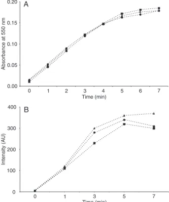
Extracellular O2.- production was investigated by two
dif-ferent experimental methods: SOD-inhibitable cytochrome c reduction and EPR spectroscopy. The time course of extracellular superoxide generation by fMLP-stimulated PMN measured by SOD-inhibitable cytochrome c reduc-tion is shown in Figure 1A. The chemotactic peptide fMLP induced a rapid increase in the production of O2.-, which
reached its maximum after several minutes. The presence
of GM1 or GT1b gangliosides (100 µM) had no influence
on any aspect of extracellular O2.- production. Similarly,
in EPR spectroscopy with the DEPMPO spin trap, no
sig-nificant difference in time course of DEPMPO-OOH signal
detection was observed in the presence of GM1 and GT1b gangliosides (Figure 1B).
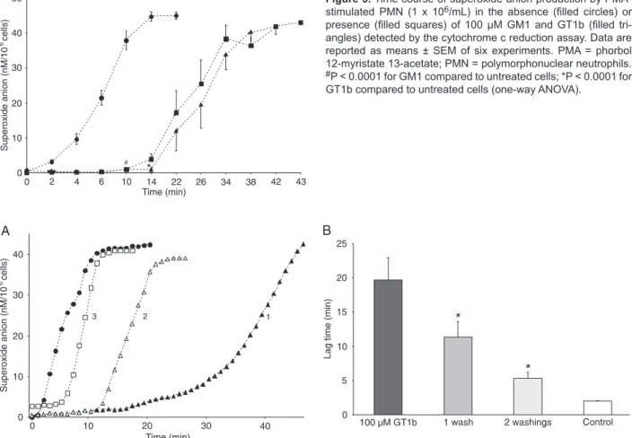
On the other hand, a short lag time of 0.3-3 min preced-ing the increase in absorbance, indicative of an extracel-lular O2.- production measured by cytochrome c reduction,
was observed in PMA-activated PMN, in agreement with literature data (18,24). The concentration-dependent experi-ments were conducted to investigate the effect of GM1 and GT1b at concentrations below and above their cmc [(2 ± 1) x 10-8 M and (1 ± 0.5) x 10-5 M, respectively, at pH 7.4 and
20°C] (28) on the lag time of superoxide anion generation measured by cytochrome c reduction. No difference in the onset of superoxide production in the presence of either GM1 or GT1b could be observed compared with the control samples containing no gangliosides at low concentrations (0.01-0.1 and 0.01-5 µM for GM1 and GT1b, respectively).
A significant increase in lag time, however, occurred in the
presence of ganglioside concentrations higher than 0.5 µM GM1 (P < 0.01) and 5 µM GT1b (P < 0.001) compared to neutrophils without gangliosides (Figure 2A and B). Further experiments were carried out using the concentration of 100 µM GM1 and GT1b.
Time-dependent superoxide generation by PMA-stimulated PMN (1 x 10⁶/mL) in the presence of 100 µM
GM1 and GT1b detected by cytochrome c reduction is shown in Figure 3. In the presence of both gangliosides, the onset of O2.- generation compared to untreated cells
was significantly delayed (12.6 ± 0.9 and 18.2 ± 1.2 min for
GM1 and GT1b, respectively, compared to 1.6 ± 0.2 min
in the untreated cells; P < 0.0001); moreover, a significant
difference in the onset of O2.- production between GM1
and GT1b was observed (P < 0.01). However, maximum absorbance, indicating maximal PMA-induced superoxide anion release, was the same in the presence and in the absence of gangliosides.
The data showed that exogenously added gangliosides
influenced the first phase of the PMA-induced response,
whereas the total extracellular O2.- production was not
affected.
The effect of the washing of PMN after exposure to gangliosides on the onset of PMA-activated PMN response was tested in separate experiments. After washings and adjustment of cells to their original count, a gradual decline of lag time was observed compared to the unwashed cells. The lag time was reduced by 43 ± 3.3 and 73 ± 1.2% (N = 3) after one and two washings, respectively, indicat-ing that loosely adherindicat-ing ganglioside micelles could be removed (Figure 4).
Figure 1. Time course of extracellular superoxide anion
genera-tion by PMN stimulated with fMLP in the absence (filled circles) and presence of 100 µM GM1 (filled squares) and GT1b (filled
Modulation of ROS in leukocytes by ganglioside micelles 945
www.bjournal.com.br Braz J Med Biol Res 43(10) 2010
Figure 3. Time course of superoxide anion production by PMA-stimulated PMN (1 x 10⁶/mL) in the absence (filled circles) or presence (filled squares) of 100 µM GM1 and GT1b (filled tri -angles) detected by the cytochrome c reduction assay. Data are reported as means ± SEM of six experiments. PMA = phorbol 12-myristate 13-acetate; PMN = polymorphonuclear neutrophils. #P < 0.0001 for GM1 compared to untreated cells; *P < 0.0001 for GT1b compared to untreated cells (one-way ANOVA).
Figure 2. Lag time of the onset of superoxide anion generation by PMN (1 x 10⁶/mL) stimulated by PMA in the absence and presence of different concentrations of GM1 (A, open columns) and GT1b (B, filled columns) as detected by the cytochrome c reduction assay. Data are reported as means ± SEM. A significant increase in lag time (min) occurs at the point where gangliosides reach concentra -tions higher than 0.5 µM GM1 (P < 0.01) and 5 µM GT1b (P < 0.001) with respect to neutrophils without gangliosides. PMN = polymor-phonuclear neutrophils; PMA = phorbol 12-myristate 13-acetate.
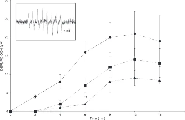
EPR spectroscopy
In order to demonstrate the presence of extracellular superoxide radicals in stimulated neutrophils, EPR spec-troscopy with the DEPMPO spin trap was applied. The experimental spectra of PMN stimulated by PMA are typical of DEPMPO-OOH adduct formation (25) (Figure 5, inset). Since in the presence of SOD, which cannot penetrate the cell membrane (29), no EPR spectra could be measured, it can be assumed that the observed DEPMPO-OOH was due to the trapping of extracellular superoxide anion (25). The time course of superoxide spin adduct formation indicated again a short delay in the onset of superoxide generation when PMN were activated by PMA. However, when PMN were exposed to 100 µM GM1 or GT1b the formation of DEPMPO-OOH was additionally delayed with respect to the samples without gangliosides (Figure 5). This phenomenon
was statistically significant in the presence of 100 and 200
µM GT1b (5.4 ± 1.2 and 11.5 ± 1.7 min, respectively; P < 0.005), while in the presence of 200 µM GM1 a tendency towards an increase in lag time (7.7 ± 1.9 min; P < 0.52) was observed in comparison with 0.7 ± 0.4 min in the untreated cells. In contrast to data obtained by cytochrome c reduc-tion, the effect of gangliosides on the total ROS generation
could not be quantitatively evaluated in EPR experiments due to the complex decay of spin adduct within the time course of the measurements (25).Therefore, only the delay in the onset of superoxide production in the presence of gangliosides was considered to be relevant.
Discussion
Exogenously added gangliosides have been involved in a variety of cell properties and biological events. In this study, we investigated the impact of micelles of two different types of gangliosides on extracellular superoxide produc-tion by activated human PMN detected by cytochrome c reduction assay and EPR spectroscopy. To activate PMN we used fMLP and PMA, each with different pathways but both resulting in the activation of NADPH oxidase. The peptide fMLP exerts its effect via receptor-dependent multiple signal-ing pathways, includsignal-ing lipid kinases, production of second messengers by various phospholipases, and the activation of PKC. This activation usually results in rapid O2.-
produc-tion, whereas activation by PMA, a receptor-independent activator, causes prolonged generation of O2.- until
neces-sary substrates and cofactors are depleted (30).
Figure 5. Influence of GT1b and GM1 micelles on the onset of superoxide production by PMA-activated PMN (5
x 106/mL) detected by EPR spectroscopy in the presence of the DEPMPO spin trap. The time-dependent super-oxide anion generation by PMA-stimulated PMN in the absence (filled circles) or presence (filled squares) of 100 µM GM1 and GT1b (filled triangles). Data represent means ± SEM of five experiments. The typical experimental
Modulation of ROS in leukocytes by ganglioside micelles 947
www.bjournal.com.br Braz J Med Biol Res 43(10) 2010
The delay in the generation of O2.-that we observed
only in PMA-activated PMN in the presence of gangliosides, as opposed to the unchanged O2.-generation using fMLP,
was probably due to different mechanisms of the activator used. It may be presumed that ganglioside micelles do not interfere with fMLP binding to its receptors. This indicates the importance of membrane involvement in the PMN activation by PMA, as the activator bypassing receptor-mediated signal pathways diffuses through the cell membrane to activate a PKC-mediated cascade of events leading to the produc-tion of superoxide. Therefore, it cannot be excluded that ganglioside micelles sterically interfere with PMA stimulation across the cell membrane. In this context, by adhering to the cell surface, gangliosides might effectively increase the diffusion barrier for a stimulant responsible for triggering membrane events, and thus induce a delay in superoxide production. It should be emphasized that, despite this delay, the total ROS generating capacity of PMA-activated PMN
was not influenced by the presence of ganglioside micelles
as measured by cytochrome c reduction.
Exogenously supplied ganglioside micelles have mainly been described to bind loosely to the cultured cell surface (13,14,31), in contrast to ganglioside monomers that fuse with the membrane (8,10). Our experimental data also point to a loose association, as we were able to almost completely remove exogenously added ganglioside micelles by repeated washings of PMN.
In the presence of higher concentrations of ganglioside micelles at the cell surface the delay of superoxide genera-tion was found to be more pronounced, with
trisialoganglio-side GT1b at a concentration of 100 μM exhibiting a more significant effect than GM1.
The structural properties of ganglioside micelles, which are mainly governed by the respective hydrophilic parts of these amphiphilic compounds, could provide an explanation
for the observed phenomena. GM1 has five sugar rings and
only one carboxylic group, whereas GT1b has seven sugar rings and three sialic acid residues. As a consequence, GT1b micelles exhibit a higher negative charge due to a higher sialic acid content compared to GM1. Because of a greater hydrophilic repulsive contribution and a larger number of hydrogen-bonding groups, GT1b molecules are prone to form smaller and more spherical aggregates that exhibit a two times lower aggregation number (176) than GM1 (301) (3). The difference in their respective surface curvatures has been evidenced in their mean hydrodynamic diameters (13), which we have found to be lower in GT1b micelles (9 nm) than in GM1 micelles (11 nm) (17). Based on the reports from the literature pointing to the importance of GM1 and GT1b headgroups in conformational properties of aggregate surfaces (32), the more pronounced
modula-tion of superoxide producmodula-tion observed in the presence of GT1b micelles compared to those of GM1 can be ascribed to the better steric shielding from PMA uptake by PMN when smaller GT1b aggregates adhere to the cell surface.
The idea that ganglioside micelles could be responsible for the increased diffusion barrier across the cell membrane
is in agreement with our previous findings that exogenous
GT1b acts as an inhibitor of hydrogen peroxide diffusion across the sperm membrane (33).
Gangliosides are known to be present in increased con-centrations in different diseases, especially neoplasms, due to their overexpression and shedding from the cell surface (34-36). Evidence suggests that tumor-derived gangliosides
influence specific aspects of the immune response (34).In
comparison with the physiological levels of gangliosides in healthy individuals (10 nM), the levels of serum-shed gangliosides, such as those in neuroblastomas, have been found to be 10-50 times higher (37,38). In our study, the effect of exogenous gangliosides on PMN production of oxygen radicals occurred at ganglioside concentrations higher than the cmc. Tumor-derived gangliosides form micelles depending on their structure; the greater the hy-drophobicity, the more shed gangliosides in micellar form (38). Although gangliosides in their natural states are known to mainly exist in the form of micelles (31,38), some studies have reported on their association with serum lipoproteins (39,40). Our results on the effect of ganglioside micelles on the in vitro oxygen radical production by PMN point to their possible relevance in in vivo conditions. Further studies are required to determine whether gangliosides, either in their free micellar form or bound to lipoproteins, are able to provide micellar coverage of leukocytes, thus modulating their superoxide anion production.
The attachment of ganglioside micelles to the surface of the cell increases the membrane diffusion barrier for the signal translocation to the interior of the cell. This study shows that exogenous GM1 and GT1b micelles, depend-ing on their concentrations and structure, delay the onset but not the total extracellular production of superoxide radicals in PMA-activated PMN detected by the cytochrome c reduction method.
Acknowledgments
We wish to express our thanks to Mariastefania Antica
for her efforts and helpful discussions, Ljiljana Paović for her excellent and reliable assistance and Lovorka Perković
References
1. Cantu L, Corti M, Brocca P, Del Favero E. Structural aspects of ganglioside-containing membranes. Biochim Biophys Acta 2009; 1788: 202-208.
2. Simons K, Ikonen E. Functional rafts in cell membranes. Nature 1997; 387: 569-572.
3. Sonnino S, Mauri L, Chigorno V, Prinetti A. Gangliosides as components of lipid membrane domains. Glycobiology 2007; 17: 1R-13R.
4. Shao D, Segal AW, Dekker LV. Lipid rafts determine
ef-ficiency of NADPH oxidase activation in neutrophils. FEBS Lett 2003; 550: 101-106.
5. Vilhardt F, van Deurs B. The phagocyte NADPH oxidase depends on cholesterol-enriched membrane microdomains for assembly. EMBO J 2004; 23: 739-748.
6. Peshavariya H, Dusting GJ, Di Bartolo B, Rye KA, Barter PJ, Jiang F. Reconstituted high-density lipoprotein suppresses leukocyte NADPH oxidase activation by disrupting lipid rafts. Free Radic Res 2009; 43: 772-782.
7. Simons M, Friedrichson T, Schulz JB, Pitto M, Masserini M, Kurzchalia TV. Exogenous administration of gangliosides displaces GPI-anchored proteins from lipid microdomains in living cells. Mol Biol Cell 1999; 10: 3187-3196.
8. Pitto M, Palestini P, Ferraretto A, Flati S, Pavan A, Ravasi D, et al. Dynamics of glycolipid domains in the plasma mem-brane of living cultured neurons, following protein kinase C activation: a study performed by excimer-formation imaging. Biochem J 1999; 344 (Part 1): 177-184.
9. Tyurina YY, Tyurin VA, Avrova NF. Ganglioside GM1 protects cAMP 3’5’:phosphodiesterase from inactivation caused by lipid peroxidation in brain synaptosomes of rats. Mol Chem Neuropathol 1993; 19: 205-217.
10. Min KJ, Pyo HK, Yang MS, Ji KA, Jou I, Joe EH. Ganglio- Ganglio-sides activate microglia via protein kinase C and NADPH oxidase. Glia 2004; 48: 197-206.
11. Avrova NF, Ivanova VP, Tyurin VA, Gamalei IA, Kliubin IV, Schepetkin IA, et al. [Modulation by super-low concentra-[Modulation by super-low concentra-tions of ganglioside GM1 of oxidative burst in murine mac-rophages and human neutrophils]. Bull Exp Biol Med 1994; 117: 44-46.
12. Avrova NF, Zakharova IO, Tyurin VA, Tyurina YY, Gamaley IA, Schepetkin IA. Different metabolic effects of ganglioside GM1 in brain synaptosomes and phagocytic cells. Neuro-chem Res 2002; 27: 751-759.
13. Sonnino S, Cantu L, Corti M, Acquotti D, Venerando B. Ag-gregative properties of gangliosides in solution. Chem Phys Lipids 1994; 71: 21-45.
14. Saqr HE, Pearl DK, Yates AJ. A review and predictive models of ganglioside uptake by biological membranes. J Neuro-chem 1993; 61: 395-411.
15. Schwarzmann G. Uptake and metabolism of exogenous glycosphingolipids by cultured cells. Semin Cell Dev Biol 2001; 12: 163-171.
16. Sokolova TV, Furaev VV, Victorov IV, Andreeva NA, Av-rova NF. Stimulation by gangliosides of viability of rat brain neurons and of neuronal PC12 cell line under conditions of oxidative stress. J Evol Biochem Phys 2005; 41: 415-423. 17. Gavella M, Kveder M, Lipovac V, Jurasin D,
Filipovic-Vincek-ovic N. Antioxidant properties of ganglioside micelles. Free Radic Res 2007; 41: 1143-1150.
18. Watson F, Robinson J, Edwards SW. Protein kinase C-de-pendent and -indeC-de-pendent activation of the NADPH oxidase of human neutrophils. J Biol Chem 1991; 266: 7432-7439. 19. DeCoursey TE, Ligeti E. Regulation and termination of
NADPH oxidase activity. Cell Mol Life Sci 2005; 62: 2173-2193.
20. Brocca P, Cantu L, Corti M, Del Favero E, Raudino A.
Col-lective phenomena in confined micellar systems of ganglio -sides. Physica A 2002; 304: 177-190.
21. Boyum A. Isolation of mononuclear cells and granulocytes from human blood. Isolation of monuclear cells by one cen-trifugation, and of granulocytes by combining centrifugation and sedimentation at 1 g. Scand J Clin Lab Invest Suppl 1968; 97: 77-89.
22. Seeds MC, Parce JW, Szejda P, Bass DA. Independent stimulation of membrane potential changes and the oxida-tive metabolic burst in polymorphonuclear leukocytes. Blood 1985; 65: 233-240.
23. Leskawa KC, Rosenberg A. The organization of ganglio-sides and other lipid components in synaptosomal plasma membranes and modifying effects of calcium ion. Cell Mol Neurobiol 1981; 1: 373-388.
24. Babior BM, Kipnes RS, Curnutte JT. Biological defense mechanisms. The production by leukocytes of superoxide, a potential bactericidal agent. J Clin Invest 1973; 52: 741-744.
25. Roubaud V, Sankarapandi S, Kuppusamy P, Tordo P, Zweier JL. Quantitative measurement of superoxide generation and oxygen consumption from leukocytes using electron paramagnetic resonance spectroscopy. Anal Biochem 1998; 257: 210-217.
26. Morse PD. Data acquisition and manipulation on the IBM PC for ESR spectroscopy. Biophys J 1987; 51: 440a.
27. Wertz JE, Bolton J. Electron spin resonance: elementary theory and practical applications. New York: McGraw-Hill; 1972.
28. Ulrich-Bott B, Wiegandt H. Micellar properties of glycosphin-golipids in aqueous media. J Lipid Res 1984; 25: 1233-1245.
29. Barbacanne MA, Souchard JP, Darblade B, Iliou JP, Nepveu F, Pipy B, et al. Detection of superoxide anion released extracellularly by endothelial cells using cytochrome c
re-duction, ESR, fluorescence and lucigenin-enhanced chemi -luminescence techniques. Free Radic Biol Med 2000; 29: 388-396.
30. Sheppard FR, Kelher MR, Moore EE, McLaughlin NJ, Baner-jee A, Silliman CC. Structural organization of the neutrophil NADPH oxidase: phosphorylation and translocation during priming and activation. J Leukoc Biol 2005; 78: 1025-1042. 31. Sharom FJ, Ross TE. Association of gangliosides with the
lymphocyte plasma membrane studied using radiolabels and spin labels. Biochim Biophys Acta 1986; 854: 287-297. 32. Brocca P, Cantu L, Corti M, Del Favero E, Raudino A. Inter-micellar interactions may induce anomalous size behavior in micelles carrying out bulky heads with multiple spatial arrangements. Langmuir 2007; 23: 3067-3074.
Modulation of ROS in leukocytes by ganglioside micelles 949
www.bjournal.com.br Braz J Med Biol Res 43(10) 2010
Int J Androl 2010; 33: 536-544.
34. Olshefski R, Ladisch S. Intercellular transfer of shed tumor cell gangliosides. FEBS Lett 1996; 386: 11-14.
35. Gavella M, Lipovac V, Mrzljak V. Lipid-bound sialic acid in diabetes. Horm Metab Res 1989; 21: 280-281.
36. Sverko V, Hadzija M, Gavella M, Lipovac V, Slijepcevic M, Radacic M. Lipid bound sialic acid concentration in mice with myeloid leukemia and alloxan diabetes. Horm Metab Res 1993; 25: 446-448.
37. Ladisch S, Wu ZL, Feig S, Ulsh L, Schwartz E, Floutsis G, et al. Shedding of GD2 ganglioside by human neuroblastoma.
Int J Cancer 1987; 39: 73-76.
38. Kong Y, Li R, Ladisch S. Natural forms of shed tumor gan-gliosides. Biochim Biophys Acta 1998; 1394: 43-56. 39. Valentino LA, Ladisch S. Localization of shed human tumor
gangliosides: association with serum lipoproteins. Cancer Res 1992; 52: 810-814.
40. Rebbaa A, Portoukalian J. Distribution of exogenously added gangliosides in serum proteins depends on the