UNIVERSIDADE FEDERAL DO CEARÁ
FACULDADE DE FARMÁCIA, ODONTOLOGIA E ENFERMAGEM PROGRAMA DE PÓS-GRADUAÇÃO EM ODONTOLOGIA
MARY ANNE SAMPAIO DE MELO
DETERMINAÇÃO DE PARÂMETROS EFETIVOS E SEGUROS PARA O USO DA TERAPIA FOTODINÂMICA ANTIMICROBIANA EM DENTINA CARIADA:
ESTUDO IN VITRO
MARY ANNE SAMPAIO DE MELO
DETERMINAÇÃO DE PARÂMETROS EFETIVOS E SEGUROS PARA O USO DA TERAPIA FOTODINÂMICA ANTIMICROBIANA EM DENTINA CARIADA: ESTUDO
IN VITRO
Dissertação apresentada ao Programa de Pós-Graduação em Odontologia da Faculdade de Farmácia, Odontologia e Enfermagem da Universidade Federal do Ceará, como requisito parcial para obtenção do Título de Mestre em Odontologia.
Área de Concentração Clínica Odontológica.
Orientadora: Profa Dra Lidiany Karla Azevedo Rodrigues
Co-Orientadora: Dra Iriana Carla Junqueira Zanin
M486e Melo, Mary Anne Sampaio de
Determinação de parâmetros efetivos e seguros para o uso da terapia fotodinamica antimicrobiana em dentina cariada: estudo in vitro
/ Mary Anne Sampaio de Melo. – Fortaleza, 2009. 42 f.
Orientadora: Profa Dra Lidiany Karla Azevedo Rodrigues Dissertação (Mestrado) – Universidade Federal do Ceará. Faculdade de Farmácia, Odontologia e Enfermagem. Programa de Pós-Graduação em Odontologia.
1. Cárie dentária 2. LED 3. Streptococcus mutans 4. Fotossensilização 5. Terapia antimicrobiana. I. ) Rodrigues, Lidiany Karla Azevedo (orient.). II. Título.
MARY ANNE SAMPAIO DE MELO
DETERMINAÇÃO DE PARÂMETROS EFETIVOS E SEGUROS PARA O USO DA TERAPIA FOTODINÂMICA ANTIMICROBIANA EM DENTINA CARIADA: ESTUDO
IN VITRO
Dissertação apresentada à Faculdade de Farmácia, Odontologia e Enfermagem da Universidade Federal do Ceará como requisito parcial para obtenção do Título de Mestre em Odontologia.
Aprovada em: ____/____/______
BANCA EXAMINADORA
_______________________________________________ Profa. Dra. Lidiany Karla Azevedo Rodrigues (Orientadora)
Universidade Federal do Ceará - UFC
_______________________________________________ Prof. Dr Eduardo Bedê Barros
Universidade Federal do Ceará - UFC
_________________________________________________ Prof. Dr. Jaime Aparecido Cury
Dedico esta dissertação aos meus pais, MARY SAMPAIO e ANÍSIO MELO.
Cada palavra dessa dissertação é um agradecimento pelo amor, carinho, dedicação, preocupação, confiança, educação e tantos outros gestos realizados por vocês durante todos os dias de minha vida.
AGRADECIMENTOS ESPECIAIS
À DEUS, por ter me dado a vida e por tê-la mantido sempre com tantas alegrias e repleta de
pessoas especiais.
Aos meus professores orientadores,
Profa. Dra. LIDIANY KARLA AZEVEDO RODRIGUES, por ter me recebido como
orientanda. Sua amizade, calma, otimismo e imensurável conhecimento guiaram meus passos nos últimos dois anos. A integralidade de suas atitudes como professora é um exemplo a ser seguido. Sua capacidade de estimular a “realização de sonhos” emana garra, vontade e entusiasmo, despertando pessoas muito melhores na essência de cada um de seus alunos.
Profa Dra. IRIANA CARLA JUNQUEIRA ZANIN, pela co-orientação, pelo tempo dispensado para meu aprendizado nesta área que me era nova. Sua acolhida, a amizade, o constante incentivo e a confiança depositada me fizeram evoluir pessoalmente e cientificamente.
....obrigada pelo estímulo ao estudo, ao trabalho, ao amor pela profissão e à valorização
da amizade e do respeito. Com certeza os ensinamentos provindos do convívio dos últimos anos vão muito além do âmbito da Odontologia.
Aos Profs. SÉRGIO LIMA SANTIAGO, HAROLDO CÉSARPINHEIRO BELTRÃO, RODRIGO OTÁVIO CITÓ CÉSAR REGO, JOSÉ JEOVÁ SIEBRA MOREIRA NETO e demais professores pela convivência e ensinamentos durante o curso de Mestrado.
Aos colegas e colaboradores do Laboratório de Microbiologia, Parasitologia e Imunologia do Curso de Odontologia da Universidade Federal do Ceará – Campus Sobral, EMANUELA DE LIMA REBOUÇAS, ELIANE DOAS SANTOS PEREIRA, FRANSCISCO RULIGLÉSIO ROCHA pelo apoio e ajuda nos trabalhos laboratoriais.
Aos funcionários da Pós-Graduação em Odontologia da UFC, GERMANO MAHLMANN MUNIZ FILHO e LÚCIA RIBEIRO MARQUES LUSTOSA, pelo auxílio e disponibilidade.
À JULIANA LIMA MARQUES PAIVA, amiga e companheira de todos os momentos no laboratório. Obrigada pelo carinho, pelo companheirismo, pelo apoio e compreensão nos momentos estressantes. Serei sempre grata por tudo que aprendi com você pessoalmente e profissionalmente.
À FÁTIMA MARIA CAVALCANTE BORGES, com quem tive o prazer de trabalhar
durante o curso. Sua coragem, sabedoria e tranqüilidade transmitida me serviram de
exemplo.
Às amigas SUYANE MARIA LUNA CRUZ DE VASCONCELOS, ROSANE PONTES
DE SOUZA, MARIA DENISE RODRIGUES DE MORAIS E DANIELLA DA SILVA
BEZERRA por toda a ajuda nos experimentos laboratoriais. Obrigada por terem me dado
apoio.
Aos alunos de Iniciação Científica DIEGO MARTINS DE PAULA e JOÃO PAULO SARAIVA WENCESLAU pelo empenho, bom humor e auxílio na execução do nosso trabalho.
Aos colegas do curso de Mestrado, ALRIETA HENRIQUE TEIXEIRA, VANARA
Ao amigo do curso de Doutorado em Química Inorgânica/UFC FERNANDO BARROSO DE ALBUQUERQUE FILHOpor toda a colaboração dada à realização das análises espectroscópicas deste trabalho.
Aos amigos do curso de Mestrado/Doutorado em Física/UFC JOHNNY PETER MACEDO FEITOSA, ALEXANDRE ROCHA PASCHOAL, LUCIANA MAGALHÃES REBELO, ERIVELTON FAÇANHA DA COSTA, e SARA BRAGA HONORATO pela ótima convivência, amizade e troca de conhecimentos.
A todos meus TIOS, TIAS, PRIMOS E PRIMAS que mesmo distantes tanto me incentivaram nessa jornada.
Ao meu irmão REYMARD SÁVIO SAMPAIO DE MELO, pela companhia, pela compreensão e pela amizade e carinho no dia-a-dia.
À amiga TICIANA PESSOA TABOSA E SILVA e equipe a quem tenho imenso carinho, por se alegrarem comigo, me incentivarem nesta caminhada e serem partes da minha vida.
AGRADECIMENTOS
À Universidade Federal do Ceará, por meio do reitor PROF. DR. JESUALDO PEREIRA FARIAS.
À Faculdade de Farmácia, Odontologia e Enfermagem (FFOE/UFC), na pessoa de sua diretora PROFA. DRA. NEIVA FRANCENELY CUNHA VIEIRA.
Ao Curso de Odontologia, na pessoa da sua Coordenadora, PROF. DRA. MARIA ENEIDE LEITÃO DE ALMEIDA.
Ao coordenador do Programa de Pós-Graduação da Faculdade de Farmácia, Odontologia e Enfermagem (FFOE/UFC),PROF. DR. SÉRGIO LIMA SANTIAGO.
Ao Conselho Nacional de Desenvolvimento Científico e Tecnológico (CNPq) pelo auxílio à pesquisa através do financiamento do projeto e concessão da bolsa de estudo para a realização deste estudo.
RESUMO
A eliminação de bactérias presentes na camada de dentina desmineralizada poderia contribuir para uma abordagem mais conservadora no tratamento restaurador de lesões de cárie dentinária profundas. Desta forma, este estudo in vitro objetivou estabelecer parâmetros eficazes e seguros para a utilização de um LED (λ = 620-660 nm) associado ao fotossensibilizador azul de orto-toluidina (TBO) na desinfecção de lesões de cárie dentinária produzidas artificialmente. Para tal, blocos com 25 mm2 de dentina oclusal planificada foram imersos por 5 dias em BHI caldo inoculado com Streptococcus mutans para indução de cárie. Depois da desmineralização, os blocos foram aleatoriamente distribuídos em 10 grupos experimentais (n=15), a saber: Controle5; Controle10; Controle15; TBO; LED5; LED10; LED15; TFD5; TFD10 e TFD15, que foram tratados ou não com TBO (0,1 mg.ml-1 por 5 minutos) ou solução de NaCl a 0,9% por 5, 10 ou 15 minutos, e submetidos ou não a irradiação com LED por 5, 10 ou 15 minutos (47, 94, e 187 J/cm2). Amostras de dentina cariada foram coletadas antes e após os tratamentos e as bactérias foram semeadas para contagem de Streptococcus mutans. Adicionalmente, com um termômetro tipo K, a temperatura intra-pulpar e da área periodontal nos grupos TFD5; TFD10 e TFD15 foi monitorada em 10 dentes com cavidades oclusais confeccionadas em dentina profunda. Teste
t pareado e ANOVA seguida pelo teste Tukey (α=5%) foram usados para verificação da redução microbiana promovida pelos tratamentos, bem como para verificação da alteração na temperatura durante a irradiação. Diferenças significativas estatisticamente na viabilidade do
Streptococcus mutans entre contagens foram observadas nos grupos: Controle15; LED15;
TFD5; TFD 10 e TFD15. A temperatura intra-pulpar e no periodonto foi inferior a 2oC, sendo a no interior da câmara pulpar maior para o grupo TFD15 quando comparado ao grupo TFD5. Dessa forma, nas condições experimentais atuais, a terapia fotodinâmica pode ser um tratamento eficaz e seguro a ser usado na desinfecção de dentina cariada, contudo a influência do tempo de irradiação ou exposição à solução salina na viabilidade do Streptococcus mutans
deve ser melhor investigada.
ABSTRACT
The elimination of bacteria inside the demineralized dentin layer might contribute for a more conservative approach in the restorative treatment of deep dentin caries lesions. This way, this
in vitro study aimed to establish safe and effective parameters for using an LED (λ = 620-660
nm) associated to the photosensitizer toluidine blue O (TBO) in the disinfection of artificially produced dentin caries lesions. For this, slabs with 25 mm2 of flatted occlusal human dentin were immersed for 5 days in BHI broth inoculated with Streptococcus mutans for caries induction. After demineralization, the slabs were randomly allocated to 10 experimental groups (n=15), as follows Control5; Control10; Control15; TBO; LED5; LED10; LED15; PDT5; PDT10 and PDT15, which were treated with TBO (0.1 mg.ml-1 for 5 min) or 0.9% NaCl solution for 5, 10 or 15 min, and submitted or not to LED irradiation for 5, 10 or 15 min
(47, 94, and 187 J/cm2). Dentin samples from caries lesions were collected before and after treatments and bacteria were then cultured for Streptococcus mutans counts. In addition, using a type K thermometer, the temperature inside the pulp and in periodontal area was monitored for the groups PDT5, PDT10 and PDT1510 in 10 teeth with deep occlusal cavities.
Paired t test/Wilcoxon matched pairs test (α=5%) were used to determine differences between
microbial population before and after treatments, and ANOVA followed by Tukey test for comparing data of temperature and log reductions. Statistically significant differences in
Streptococcus mutans viability were found for the groups: Control15; LED15; PDT5; PDT10
and PDT15. The temperature from intrapulpal and periodontal area were lower than 2oC, being higher inside the pulpal chamber for group PDT15 when compared to group PDT5. Thus, it the experimental conditions used in the study, photodynamic therapy may be a safe and effective treatment to be used for disinfecting carious dentin, however the influence of time of irradiation/exposition it the Streptococcus mutans viability should be better investigated.
SUMÁRIO
1 INTRODUÇÃO GERAL... 12
2 PROPOSIÇÃO... 15
2.1 Objetivo Geral... 15
2.2 Objetivos Específicos... 15
3 CAPÍTULO... 16
4 CONCLUSÃO GERAL... 44
REFERÊNCIAS... 45
APÊNDICES... 49
ANEXOS... 51
1 INTRODUÇÃO GERAL
A terapia fotodinâmica baseia-se no uso de um corante (fotossensibilizador) associado a irradiação com luz de comprimento de onda compatível com a absorção do corante, onde o tratamento somente com a luz ou apenas o corante não produz efeitodeletério (MITRA 2004).Quando a exposição ao corante vem seguida pela a irradiação de luz, reações químicas são geradas conduzindo a uma cadeia de eventos citotóxicos que levam a alterações funcionais na célula-alvo e, consequente, perda de sua viabilidade(HABLIN; HASAN, 2004; KUBLER, 2005; JORI et al., 2006; JORI; COPPELLOTTI, 2007).
O uso da terapia fotodinâmica na área da saúde aumentou consideravelmente no último século devido principalmente ao desenvolvimento científico e tecnológico (SIMPLÍCIO; MAIONCHI; HIOKA, 2002; ALLISONet al., 2008). Assim a utilização desta modalidade terapêutica para o tratamento de injúrias pré-malignas, malignas e degenerativas é
amplamente usada em diversas áreas médicas (LUKŠIENĖ, 2003; BAGNATO et al., 2005; KUBLER, 2005; PERUSSI, 2007). Por também apresentar atividade antimicrobiana (ASHKENAZI; NITZAN; GÁL, 2003;WILSON, 2004; JORI; BROWN, 2004; LUKŠIENĖ, 2005; MAISCH, 2007), a possibilidade de sua utilização para destruição de diversos microorganismos, incluindo os relacionados a várias doenças orais tem sido tópico atual de pesquisa.
Além das reconhecidas medidas de prevenção da cárie dental, como os procedimentos mecânicos de remoção do biofilme cariogênico, a adequação da dieta e o uso do flúor (CHAVES; VIEIRA-DA-SILVA, 2002), tem-se investigado novas abordagens conservadoras, buscando-se evitar os prejuízos trazidos pela doença. Dentre as estratégias, tem-se destacado a redução de patógenos específicos relacionados à cárie por meio de agentes antimicrobianos capazes de inibir o crescimento de estreptococos do grupo mutans (MOUNT; NGO, 2000; MOUNT, 2007; FRANCO et al., 2007).
A ação da TFA sobre bactérias orais crescidas em caldo de cultura já se encontra bem documentada na literatura (DOBSON; WILSON, 1992; BURNS; WILSON; PEARSON, 1994; WILSON; YIANNI, 1995; GRIFFITHS; WREN; WILSON, 1997; ZANIN et al., 2002; WILLIAMS et al., 2003) com resultados revelando altas taxas de redução microbiana.
Quanto ao seu comportamento frente a microorganismos crescidos em biofilme que normalmente apresentam uma redução significante em suscetibilidade a agentes antimicrobianos comparados com culturas cultivadas em laboratórios (MAISCH, 2007); vários estudos (WILSON; BURNS; PRATTEN, 1996; O’NEILL; HOPE; WILSON, 2002; CHABRIER-ROSELLO et al., 2005; ZANIN et al., 2005, 2006; WOOD et al., 2006) mostram resultados bastante promissores com consideráveis taxas de redução, apesar de serem em menor valor se comparados aos resultados obtidos sobre microorganismos em culturas planktônicas.
Porém, pouco tem sido cientificamente estudado para uma avaliação segura de sua eficácia antimicrobiana sobre lesões de cárie dentinária, necessitando de estudos mais detalhados sobre seu uso terapêutico (GIUSTI et al., 2008). Para se instaurar esta terapia de forma eficiente, alguns parâmetros devem ser estabelecidos, uma vez que o conhecimento atual aponta para diversas variáveis que podem interferir no processo fotodinâmico quando realizados em um substrato diferenciado, a exemplo da dentina cariada. São variáveis que contribuem na redução do efeito sobre os microrganismos–alvo: menor penetração do
fotossensibilizador nos túbulos dentinários, que influencia a capacidade de alcance das bactérias tanto por parte do corante como por parte da luz (BURNS; WILSON; PEARSON, 1995); o tempo de exposição e, consequentemente, densidade de energia(QUIN et al., 2007); concentração de O2 no meio(YAVARI, 2006) e a presença de uma diversidade microbiana (WILSON, 1993).
Apesar da influência de diversas variáveis na eficiência antimicrobiana da terapia, a densidade de energia se torna um dos parâmetros mais relevantes relacionando-se diretamente ao tempo de irradiação e se encontra aplicado com bastante variedade em estudos relacionados a TFA (HAMBLIN; HASAN, 2004; TEGOS; HAMBLIN, 2006; NUNEZ, 2007).
luz, caracterizando-se em uma faixa capaz de produzir efeito fototérmico (calor) ao ser absorvida pelos tecidos dentais (ZACH; COHEN, 1965; LEE et al., 2006). Tal fato é ainda mais importante em situações onde há mínima espessura de dentina como em cavidades profundas, uma das indicações proposta da terapiaantimicrobiana.
2 PROPOSIÇÃO
Os objetivos do presente trabalho foram:
2.1 Objetivo Geral
Determinarpor meio de um estudo in vitro os parâmetros efetivos e seguros para o uso da terapia fotodinâmica antimicrobiana para desinfecção de dentina cariada.
2.2 Objetivos Específicos
Avaliar o efeito antimicrobiano da terapia fotodinâmica, utilizando-se do fotossensibilizador azul de orto toluidina associado a um LED (λ predominante em 638,8 nm) com três densidades de energia diferentes, através de análise microbiológica e exploração de microscopia eletrônica de varredura.
Verificar in vitro se os parâmetros estabelecidoscomo eficazes são seguros para serem utilizados in vivo em cavidades profundas executadas em dentina no que diz respeito a
3 CAPÍTULO
Esta dissertação está baseada no Artigo 46 do Regimento Interno do Programa de Pós-Graduação em Odontologia da Universidade Federal do Ceará, que regulamenta o formato alternativo para dissertações de Mestrado e teses de Doutorado e permite a inserção de artigos científicos de autoria e co-autoria do candidato (Anexo A). Por se tratarem de pesquisas envolvendo dentes humanos, o projeto de pesquisa deste trabalho foi submetido à apreciação do Comitê de Ética em Pesquisa da Faculdade de Medicina da Universidade
Federal do Ceará, tendo sido aprovado sob o Protocolo COMEPE no143/06, conforme o
Ofício no 324/2006 de 31 de julho de 2006 (Anexo B). Assim sendo, esta dissertação é composta de um capítulo contendo um artigo com o idioma corrigido através dos serviços especializados do endereço eletrônico http://www.journalexperts.com/ (Anexo C) e submetido para publicação no periódico “Laser in Surgery and Medicine”, (Anexo D) conforme descrito abaixo:
Safe and effective parameters for using photodynamic antimicrobial chemotherapy in carious dentin: an in vitro study.
Safe and effective parameters for using photodynamic antimicrobial
chemotherapy in carious dentin: an in vitro study
Mary A.S. de-Melo1, DDS; Diego M. de-Paula1, Dentistry undergraduate student; Juliana P. M. Lima1, DDS; Fátima M.C. Borges1, DDS; Carolina Steiner-Oliveira2, MS; Eduardo B. Barros4, PhD; Marinês Nobre-dos-Santos2, PhD; Iriana C.J. Zanin3, PhD; Lidiany K.A.
Rodrigues, PhD.
(MASM); (DMP); (JPML); (FMCB); (LKAR) 1Faculty of Pharmacy, Dentistry and Nursing, Federal University of Ceara, Fortaleza, CE, Zip Code60430-170, Brazil.
(CSO);(MNS) 2PiracicabaDental School, State University of Campinas, Piracicaba, SP, Zip Code 13414-903, Brazil.
(ICJZ) 3School of Dentistry, Federal University of Ceara, Sobral, CE, Zip Code 62041-040, Brazil.
(EBB) 4Department of Physics, Federal University of Ceara, Fortaleza, CE, Zip Code 60455-760, Brazil.
Acknowledgments
The authors thank Daniel Cordeiro da Costa and Celli Rodrigues Muniz (EMBRAPA-CE/CNPAT- Brazilian Agricultural Research Corporation/National Centre of Research in Tropical Agriculture) for their assistance with sample preparation for scanning electron microscopy and IPDI (Institute of Research, Development and Innovation) for providing the SEM equipment. This research received the financial support of Grant # 620160/2006-3 from CNPq. This paper was based on a thesis submitted by the first author to the Faculty of Pharmacy, Dentistry, and Nursing of the Federal University of Ceara, in partial fulfillment of the requirements for a MS degree in Dentistry. The first and second authors have received scholarships during this study from CNPq (564387/2008-8) and CNPq/PIBIC/UFC, respectively.
Full address of the author to whom correspondence should be sent: Lidiany Karla Azevedo Rodrigues
Bairro- Rodolfo Teófilo - CEP 60430-170 Phone- #558533668410
Fax- #558533668232 Fortaleza-CE-Brazil
E-mail: lidianykarla@yahoo.com
ABSTRACT
Background and Objective: The development of a method to ensure bacterial-free surfaces without extensive cavity preparation would be highly useful to the field of Dentistry, since there is no currently available effective method for killing residual bacteria in dentinal tissue.
Therefore, the purpose of this totally randomized in vitro study was to determine effective antimicrobial parameters for the use of toluidine blue O (TBO) in combination with a
light-emitting diode (LED - λ= 620-660 nm) for the disinfection of lesions on dentin caries
Study Design/Materials and Methods: Slabs with 25 mm2 of flatted occlusal human dentin were immersed for 5 days in brain heart infusion (BHI) broth inoculated with Streptococcus
mutans for caries induction. After demineralization, the slabs were randomly allocated to 10
experimental groups (n=15), which were treated with TBO (0.1 mg.ml-1 for 5 min) or 0.9% sodium chloride (NaCl) solution for 5, 10 or 15 min, and submitted or not to LED irradiation for 5, 10 or 15 min (47, 94, and 187 J/cm2). Dentin samples from caries lesions were collected before and after treatments and bacteria were then cultured for Streptococcus mutans counts. The temperature rise in the pulpal chamber and periodontal ligament during irradiation was
also verified. Paired t test/Wilcoxon matched pairs test (α=5%) were used to determine differences between microbial population before and after treatments, and analysis of variance (ANOVA) followed by Tukey test was utilized to compare log reductions and temperature rises between the groups.
Results: Significant decreases in the viability of S. mutans in carious dentin were observed when slabs were exposed to both TBO and light at the three different doses. However, statistically significant microbial reduction also occurred when bacterial air-exposure time or irradiation time was 15 min, corresponding to 187 J/cm2. The temperature increases were less
than 2oC for either pulpal chamber or periodontal ligament.
INTRODUCTION
Dentin caries occur in different layers of the tooth. Can be divided in two different patterns: the outer layer is highly infected with bacteria that dissolve the mineralized tissue of dentin and damages the collagen matrix, which may result in an impossibility of remineralization, which should be completely removed during caries excavation and the inner layer where bacteria may dissolve the mineralized tissue but the cross-banded ultra structure of the collagen matrix will still remain.
Nevertheless, clinically the differentiation between the zones of caries that are infected and affected by the carious process is extremely critical [3], since unnecessary cavity preparation may lead to the removal of dentin tissue and possible exposure of the pulp [4,5]. Accordingly, most current methods to treat a deep carious lesion involve the removal of all softened and infected dentin. The use of either hand instruments or rotary burs does not guarantee a cleaning of the infected dentin and residual bacteria, which are often present before the placement of restorations [6]. Therefore, the elimination of bacteria inside of the dentin tubules from the remaining demineralized dentin [4] might contribute to a more conservative approach for dealing with deep caries lesions [7].
In this context, photodynamic antimicrobial chemotherapy (PACT) may be a potential alternative treatment for dentin disinfection. This therapy involves photosensitizing agents, such as toluidine blue O (TBO), which are activated by irradiation with light of a specific wavelength to generate cytotoxic species, including singlet oxygen and free radicals. These products are capable of damaging essential components of the cells or modifying metabolic activities in an irreversible way, which may result in bacterial death[8–11]. Additionally, the use of approaches that are unlikely to result in the development of resistance by bacteria would be very valuable, in light of the emergent problem of bacterial resistance to conventional antimicrobials.
effect of PACT on oral microorganisms located in demineralized dentin can be reduced, due to lower penetration of photosensitizer or light that reaches the bacteria [7,27]. In addition, a few studies have investigated the antimicrobial effects of photodynamic therapy in dentin carious tissue [23], primarily in human dentin.
The aim of this investigated was to determine whether a range of bacteria
(Streptococcus mutans ) on housed within lesions of in vitro caries could be killed by PACT.
The hypotheses under investigation were the following: 1) the null hypothesis that there is no difference in the antimicrobial effect of TBO, LED, or PACT with three different energy densities on artificial dentin caries disinfection, as assessed by bacterial counts and scanning electron microscopy, and 2) the null hypothesis that there is no difference in the increase of pulpal or periodontal temperature during irradiation for 5, 10, or 15 min.
MATERIALS AND METHODS
Experimental Design
An in vitro study was performed with one hundred and sixty five extracted human
third molars that had more than two-thirds of the formed roots, and without any previous lesions were used. The teeth used in this investigation were collected from adults living in Fortaleza, Brazil, in conformity with the norms of the Research and Ethics Committee of Federal University of Ceara Medical School (Process #. 143/2006).
The teeth were randomly allocated to 11 test groups, with 15 experimental units per group, according to a computer generated randomization list [28]. The experimental design was performed in triplicate (n=5) at different time points to minimize the inherent bias related to microbiological procedures. In order to isolate the microorganism ability in dentin caries production, one group was only exposed to a non-inoculated BHI broth. This study involved 10 set conditions in which carious dentin was exposed to 0.9% NaCl solution for 5, 10, or 15 min; sensitized or not with TBO; and irradiated or not with a LED for 5, 10 or 15 min.: These conditions were denoted as follows:
Control15- Carious dentin exposed to 0.9% NaCl solution for 15 min TBO- Carious dentin exposed to TBO for 5 min
LED5- Carious dentin exposed to LED for 5 min LED10- Carious dentin exposed to LED for 10 min LED15- Carious dentin exposed to LED for 15 min
PACT5- Carious dentin exposed to TBO and LED for 5 min PACT 10- Carious dentin exposed to TBO and LED for 10 min PACT15- Carious dentin exposed to TBO and LED for 15 min
Specimen Preparation
The teeth were stored in 0.01% (v/v) thymol solution at 4°C prior to use in this in vitro
study. From each tooth, one slab of coronal dentine (5 x 5 x 2 mm3) were obtained using a water-cooled diamond saw and a cutting machine (IsoMet™ Low Speed Saw, Buehler, Lake Bluff, IL, USA). Only the occlusal dentin face was used, and the remaining surfaces of the slabs were protected with resin epoxy adhesive Araldite Hobby for 10 min (Brascola, São Bernardo do Campo, SP, Brazil). This resulted in a 25-mm2 dentin surface area that served as
a microbial model to produce caries lesions, as assessed by visual examination. The dentin slabs were fixed in the lids of glass container vessels with plastic rods, immersed in sterile distilled water, and then sterilized by autoclaving to 121°C for 15 min [29].
plated and incubated at 37°C in an atmosphere of 10% CO2 for 24 h to check for purity. Catalase and morphologic Gram test were utilized for specie identification.
Photodynamic therapy
After the 5-day experimental period, the biofilm formed over the slabs was removed and the carious dentin tissue was exposed. Then, one sample of carious dentin was harvested to obtain a baseline value for tissue contamination and the treatment for each group was performed. The groups TBO; PACT5; PACT10; and PACT15 were incubated with 5 µl of TBO in the dark for 5 min (pre-irradiation time) without light exposure [21,30-32]. The groups TBO; PACT5; PACT10; and PACT15 were incubated with 5 µl of TBO in the dark for 5 min (pre-irradiation time) without light exposure [21,30-32]. Groups Control5, Control10, Control15, LED5, LED10, and LED15 were incubated with an equal volume of sterile 0.9 % NaCl solution instead of TBO
The photosensitizer TBO (Sigma-Aldrich Company Ltd. Poole, Dorset, United Kingdom) was dissolved in deionized water to obtain a final concentration of 0.1 mg.ml-1 and was subsequently kept in the dark. Ultraviolet visible (UV–vis) optical absorption
spectrometry was performed in TBO solution before and after the irradiation using a HP 8453 system spectrophotometer (Hewlett-Packard, Palo Alto, CA, USA), in order to characterize TBO absorption spectrum between 550 and 680 nm and an absorption peak centered at 632±8 nm and correlation with LED emission spectrum. The light source used was a red light-emitting diode (LED; Laserbeam, Rio de Janeiro, RJ, Brazil), with a spectrum of emission ranging from 620 to 660 nm and a predominant wavelength of 638.8 nm. A fiber optic spot with a 9.5-mm cylindrical diffusing tip distributed the light. Irradiation was performed in a non-contact mode with a focused beam at 2.0 mm of working distance. A power meter Lasermate (Coherent Inc, Santa Clara, CA, USA) was used to measure the peak power, and a maximum output power of 40 mW was determined.
and therefore, relatively long irradiation times were used and the groups Control5, Control10, Control15 were included to verify the influence of these time limits on bacterial viability.
For microbiological analysis, before and after treatments, a sample of carious dentin was collected in different portions of each slab using a #5 carbide bur in a low speed drill (Labor dental, São Paulo, SP, Brazil). The dentin samples were weighed (±1 mg) in pre-weighed microcentrifuge tubes, and 0.9% NaCl solution was added (1 ml/mg dentin). The tubes were agitated during for 2 min in a Disrupter Genie Cell Disruptor (Precision Solutions, Rice Lake, Wisconsin, WI, USA) with three 0.1 mm diameter glass beads to detach the bacterial cells. Subsequently, the suspension was serially diluted (1:100, 1:1000, 1:10000, and 1:100000) with 0.9% NaCl solution. Samples were plated in triplicate on BHI agar, and incubated for 48 h at 37°C in a 10% CO2 atmosphere. Representative colonies with typical morphology of S. mutans were counted using a colony counter. The results were expressed in CFU/mg dentin.
Temperature change during exposure to light
The purpose of this analysis was to observe potential temperature variations in the
pulp chamber and periodontal ligament caused by the LED irradiation associated with TBO usage. For this purpose, 10 intact extracted human teeth were used and deep standardized cavities were prepared in the occlusal surface, using cylindrical diamond bur # 1098 (KG Sorensen, São Paulo, SP, Brazil) in a high-speed hand piece. The existence of a 1-mm dentin layer over the pulp chamber was assured using radiographies. Additionally, 5 µl of TBO was placed inside each cavity for a pre-irradiation time of 5 min without light exposure [21,30-32]. The temperature range was measured in triplicate using two type K thermocouple electrodes connected to a thermometer (Minipa, São Paulo, SP, Brazil), and coordinated by the software PS250 (Minipa, São Paulo, SP, Brazil). One thermocouple was introduced into the pulp chamber in close contact with the pulp chamber floor. The second one was attached to the periodontal ligament area.
for the pulp chamber, which was approximately 37°C, was obtained. Temperatures were recorded every second during the irradiation times of 5, 10 and 15 min, relating time to each energy density used in this study. The temperatures rise considered (∆t) were calculated as the difference between initial and final recorded temperatures.
UV-vis spectroscopy analysis
Ultraviolet visible (UV–vis) optical absorption spectrometry was performed in TBO solution before and after the irradiation using a HP 8453 system spectrophotometer (Hewlett-Packard, Palo Alto, CA, USA), in order to characterize TBO absorption spectrum, correlation with LED emission spectrum, and photodegradation. Room temperature was maintained at 25ºC and 0.01% TBO (w/v) was diluted with distilled, deionized water (pH 7.2) into a quartz cell with 1-mm light path. Fractionated irradiation was performed for 5 min and the spectra were obtained at 5 min intervals for 30 min. The spectra were analyzed with Origin Lab 8.0 software (Origin Lab Corporation, Northampton, MA, USA).
Scanning Electron Microscopy (SEM)
SEM was utilized to evaluate the morphology of treated and untreated bacteria in dentin from one sample of the PACT10, PACT5 and Control10 groups. All specimens were immersed in modified Karnovsky fixative solution containing 2.5% cold glutaraldehyde in 0.1 mol/L cacodylate buffer at a pH of 7.4 for 8 hours. Samples were then serially dehydrated in graded ethanol solutions (50, 60, 70, 80, 90, 95, and 100% ethanol) at 45 min intervals, critical point dried by CO2, mounted on aluminum stubs, sputter-coated with gold/palladium, and examined by a TESCAN SEM (Model VEGA\XMU, Brno, Czech Republic) with different magnifications at an accelerating voltage of 20 kV.
Statistical analysis
For assessment of the effect of the treatments, the dependent variables CFU/mg of
equality of variances (Levene Test) and normal distribution of errors (Shapiro Wilks test) were verified; and data were transformed in the case of assumption violation. For variables CFU/mg of dentin before and after treatment for group PACT15 and log reduction, equality of variances, normal distribution of errors, and absence of outliers were not satisfied; such that these data could not be normalized. A non-parametric test to compare dependent samples (Wilcoxon matched pairs test) was applied for group PACT15 and Kruskal-Wallis and Student-Newman-Keuls were used for log reduction comparison means. For the other groups, a paired t test was used to verify differences between CFU/mg of dentin before and after each treatment and identify treatments that reduced the microbial community. The log reduction results were calculated by subtraction of initial from final values of the CFU/mg for dentin
after being transformed by Log10. Next, ∆T values were calculated by subtraction of the final
from initial values of temperature in the pulp chamber and periodontal ligament. These data were analyzed by a one-way ANOVA followed by a Tukey-Kramer test. The significance level was set at 5%. The software BioStat 2007 Professional (AnalystSoft Robust business solutions company, Vancouver,British Columbia, Canada) was used.
RESULTS
UV-vis Spectroscopy
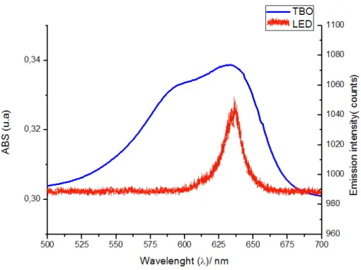
The emission spectrum of red LED and the aqueous TBO absorption spectrum are
shown in Figure 2. A strong absorption band of TBO between 550 and 680 nm and an
absorption peak centered at 632±8 nm were observed. Additionally, a decrease in TBO absorption as a function of irradiation time is shown in Figure 3.
Scanning Electron Microscopy (SEM)
disintegration or an unclear cell wall, were observed (Fig. 5c). In addition, the presence of amorphous masses was found involving individual bacterial cells that merged (Fig. 6c).
Microbiological analysis
Mean and standard deviation of CFU/mg for carious dentin before and after treatments, and log reduction achieved by each treatment are shown in Table 1. Based on paired tests, statistically significant bacterial reduction was found after treatment for groups Control15, LED15, PACT5, PACT10, and PACT15.
The log reduction values ranged from 0.39 to 5.80, with the lowest value for the Control5 group and the highest for the PACT15 group. Statistically significant differences between the groups are described in Table 1. The groups treated with PACT presented statistically higher log reduction values when compared to the respective control groups, except for those groups irradiated, but not sensitized by TBO, indicating that LED resulted in some cytotoxicity.
Temperature change
For ∆T variation, all groups treated with photodynamic processes presented temperature rises lower than 2oC degree (Table 1). For pulp temperature, the increase ranged from 0.9 to 1.7, and exhibited a statistically significant difference for only the PACT5 and PACT15 groups. With regard to periodontal data, the values ranged from 0.3 to 0.6 and no statistically significant differences were found, as summarized in Table 1.
DISCUSSION
The in vivo eradication of bacteria in microbiota within or over the dentinal tubules of
potential remineralizing processes to proceed more rapidly. The lack of excessive instrumentation, unwarranted dentin removal, and use of caustic chemicals or antibiotics on remaining dentin may improve the long-term prognosis for the repaired tooth. Thus, characterization of standardization of photodynamic processes is an important clinical issue.
According to our results, the groups Control15, LED15, PACT5, PACT10, and PACT15 were capable of reducing S. mutans in carious dentin. These results confirm several studies, which have previously demonstrated that cariogenic bacteria in culture baths and biofilms are susceptible to PACT [3,7,13,15–20,30]. However, there are few studies that have investigated the use of this approach on different substrates. Recent investigations have evaluated the bacterial effectiveness of photodynamic therapy in an environment similar to carious tooth (collagen matrix, ex vivo carious dentin, and carious bovine dentin) [23,27]. Although these studies have demonstrated promising results for using photodynamic approaches in dentin, there have been limitations to these investigations, such as absence of required control groups, utilization of different calculations to verify efficacy, and use of bovine dentin [23] as well as bacterial irradiation through the dentin and not inside the tissue tubules [27]. In this context, our research demonstrated for the first time that S. mutans hosted in carious human dentin is susceptible to photodynamic therapy using TBO and a LED with energy densities ranging from 47 to 187 J/cm2.
The results of this study also indicated that the red LED operating with 187 J/cm2 may
cause an inhibitory effect in bacterial cells of S. mutans present in carious dentin formed in
vitro. This inhibition is thought to have occurred as a result of the length of the irradiation
hosted in dentin were not protected from dryness by the presence of the polysaccharide matrix and higher amounts of water.
The use of high energy density (187 J/cm2) to exhibit an antimicrobial effect in bacteria located inside dentin tubules has been suggested by Burns et al. [7]. Nevertheless, the significant reduction in viable counts after exposing bacteria to air for 15 min without light activation indicates that the time of treatment should not exceed 10 min for analysis of S.
mutans viability in the current in vitro caries lesion model. The long irradiation time to
achieve higher energy densities was one limitation of the chosen LED source used in this work. Additionally, group PACT15 demonstrated no contamination after treatment, which is not a realistic situation since the group Control15, which involved exposure of S. mutans the air for 15 min, was also able to disinfect dentin tissue without total eradication of the bacteria as PACT15. Consequently, the antimicrobial effect on the group PACT15 was improved by time of irradiation, resulting in a more effective therapy.
According to the UV-vis spectroscopy analyses, the photosensitizing mechanism of the LED on TBO follows the same pattern as the majority of other light sources that generate singlet oxygen. Therefore, the use of an LED source is advantageous, considering that the best previous were obtained using conventional lasers to perform this therapy, although the power output can still be a limiting factor in their widespread application [34-36]. Use of this experimental methodology may result in technology simplification and the lower cost of
treatment in comparison to complex laser systems. Moreover, LED sources provide more than a monochromatic form of irradiation, which is a special characteristic compared to lasers that increases the overlay of the spectrum of LED irradiation and the light absorption by TBO.
bacteria decreased and very few aggregated colonies were observed as the light dose increased.
Zach and Cohen in 1965 [40] discovered that an increase in temperature of 3.3°C resulted in reversible variations in pulpal histology, but that when the temperature increase exceeded 5.6°C, approximately 15% of the examined teeth had a loss of pulpal vitality. This possibility of temperature increase by diffusion of heat across the remaining dentin and the potential effect on pulpal tissue damage during the photodynamic process led to an additional investigation. The elevation of the temperature on the surface of the dentin depends on the thermal conductivity of this tissue, but also on the thickness and mass of these structures [38]. As the thickness of the dentin is the least in deeper cavity, with 1 mm or less of remaining dentin, the use of higher energy density should be careful to not exceed safe parameters.
Nevertheless, the results of this study demonstrated a low range of temperature variation. Difference related to the increase of pulpal or periodontal temperature during irradiation for the selected time periods was only accepted for the intrapulpal temperature, since the PACT15 group presented a more significant increase compared to the PACT5 group for the pulp temperature. However, all groups presented small temperature increases, not exceeding 2°C even for the highest LED density applied (187 J/cm2) and the increase was less than 1°C for the periodontal area, which is an even safer range. Thus, these findings indicate that PACT can be applied to carious dentin in deep cavity preparations without exposing pulp
vitality to danger.
In conclusion, these results demonstrated that S. mutans housed within lesions of in
vitro caries were susceptible to LED exposure in the presence of TBO, suggesting that this
REFERENCES
1. Ogushi K, Fusayama T. Electron microscopic structure of the two layers of carious
dentin. J Dent Res 1975;54:1019–26.
2. Zavgorodniy AV, Rohanizadeh R, Swain MV. Ultrastructure of dentine carious lesions.
Arch Oral Biol 2008;53:124–32.
3. Burns T, Wilson M, Pearson GJ. Killing of cariogenic bacteria by light from a gallium
aluminium arsenide diode laser. J Dent 1994;22:273–78.
4. Kidd EA, Joyston-Bechal S. Essentials of dental caries: the disease and its management,
Oxford:Oxford University Press, 1997.
5. Banerjee A, Watson TF, Kidd EA.Dentine caries excavation: a review of current clinical
techniques. Br Dent J 2000;188:476-82.
6. Mertz-Fairhurst EJ, Curtis JW Jr., Ergle JW, Rueggeberg FA, Adair SM.
Ultraconservative and cariostatic sealed restorations: Results at year 10. J Am Dent
Assoc 1998;129:55–66.
7. Burns T, Wilson M, Pearson GJ. Effect of dentine and collagen on the lethal
photosensitization of Streptococcus mutans. Caries Res 1995;29:192–97.
alternative approach to antimicrobial drugs. J Photochem Photobiol B 1990;5:281–93.
9. Bhatti M, MacRobert A, Meghji S, Henderson B, Wilson M. Effect of dosimetric and
physiological factors on the lethal photosensitization of Porphyromonas gingivalis in
vitro. Photochem Photobiol 1997;65:1026–31.
10.Castano AP, Demidova TN, Hamblin, MR. Mechanisms in photodynamic therapy: part
one-photosensitizers, photochemistry and cellular localization. Photodiagn Photodyn
Ther 2004;1:279–93.
11.Akilov OE, O’Riordan K, Kosaka S, Hasan T. Photodynamic therapy against intracellular
pathogens: problems and potentials. Med Laser Appl 2006;21:251–60.
12.Hamblin MR, Hasan T. Photodynamic therapy: a new antimicrobial approach to
infectious disease? Photochem Photobial Sci 2004;3:436–50.
13. Wood S, Metcalf D, Devine D, Robinson C. Erythrosine is a potential photosensitizer for
the photodynamic therapy of oral plaque biofilms. J Antimicrob Chemother
2006;57:680–84.
14.Jori G, Fabris C, Soncin M, Ferro S, Coppellotti O, Dei D, Fantetti L, Chiti G, Roncucci
G, Photodynamic therapy in the treatment of microbial infections: basic principles and
15.Jori G, Coppellotti O. Inactivation of pathogenic microorganisms by photodynamic
techniques: mechanistic aspects and perspective applications. Anti-Infective Agents in
Medicinal Chemistry 2007;6:119–31.
16.Dobson J, Wilson M. Sensitization of oral bacteria in biofilms to killing by light from a
low-power laser. Arch Oral Biol 1992;37:883–87.
17.Wilson M, Yianni C. Killing of methicillin-resistant Staphylococcus aureus by
low-power laser light. J Med Microbiol 1995;42:62–66.
18.Griffiths MA, Wren BW, Wilson M. Killing of methicillin-resistant Staphylococcus
aureus in vitro using aluminium disulphonated phthalocyanine, a light-activated
antimicrobial agent. J Antimicrob Chemother 1997;40:873–76.
19.Zanin, IC, Brugnera Jr A, Gonçalves RB. In vitro study of bactericidal effect of low level
laser therapy in the presence of photosensitizer on cariogenic bacteria. Proceedings of
SPIE, Lasers in Dentistry VIII 2002;4610:154–161.
20.Chabrier-Roselló Y, Foster TH, Pérez-Nazario N, Mitra S, Haidaris CG. Sensitivity of
Candida albicans germ tubes and biofilms to photofrin-mediated phototoxicity.
Antimicrob Agents Chemother 2005;49:4288–295.
21.Zanin IC, Goncalves RB, Brugnera Jr A, Hope CK, Pratten J. Susceptibility of
Streptococcus mutans biofilms to photodynamic therapy: an in vitro study. J Antimicrob
22.Zanin IC, Lobo MM, Rodrigues LK, Pimenta LA, Hofling JF, Gonçalves RB.
Photosensitization of in vitro biofilms by toluidine blue O combined with a light-emitting
diode. Eur J Oral Sci 2006;114:64–69.
23.Giusti JS, Santos-Pinto L, Pizzolito AC, Helmerson K, Carvalho-Filho E, Kurachi C,
Bagnato VC. Antimicrobial photodynamic action on dentin using a light-emitting diode
light source. Photomed Laser Surg 2008;26:279–85.
24.Stringer GJ, Bird PS, Walsh LJ. Lethal laser photosensitization of Streptococcus mutans
with a visible red diode laser. Aust Dent J 2000;45:Suppl S-22.
25.Paulino TP, Ribeiro KF, Thedei G Jr, Tedesco AC, Ciancaglini P. Use of hand held
photopolymerizer to photoinactivate Estreptococcus mutans. Arch Oral Biol
2005;50:353–9.
26.Williams JA, Pearson GJ, Colles MJ, Wilson M. The effect of variable energy input from
a novel light source on the photoactivated bactericidal action of toluidine blue O on
Streptococcus mutans. Caries Res 2003;37:190–3.
27.Williams JA, Pearson GJ, Colles MJ, Wilson M. The photo-activated antibacterial action
of toluidine blue O in a collagen matrix and in carious dentine. Caries Res 2004;38:530–
28.Bolliger CT, Zellweger JP, Danielsson T, van Biljon X, Robidou A, Westin A,
Perruchoud AP, Säwe U. Smoking reduction with oral nicotine inhalers: double blind,
randomised clinical trial of efficacy and safety. BMJ 2000;321:329-33.
29.Pantera EA Jr., Schuster GS. Sterilization of extracted human teeth. J Dent Educ
1990;54:283–5.
30.O’neill JF, Hope C, Wilson M. Oral bacteria in multi-species biofilms can be killed by
red light in the presence of toluidine blue. Lasers Surg Med 2002;31:86–90.
31.Bevilacqua IM, Nicolau RA, Khouri S, Brugnera A Jr, Teodoro GR, Zângaro RA,
Pacheco MT. The impact of photodynamic therapy on the viability of Streptococcus
mutans in a planktonic culture. Photomed Laser Surg 2007;25:513–8.
32.Prates RA, Yamada AM Jr, Suzuki LC, Eiko Hashimoto MC, Cai S, Gouw-Soares S,
Gomes L, Ribeiro MS. Bactericidal effect of malachite green and red laser on
Actinobacillus actinomycetemcomitans. J Photochem Photobiol B 2006; 86:70-6.
33.Yazici AR, Khanbodaghi A, Kugel G. Effects of an in-office bleaching system (ZOOM)
on pulp chamber temperature in vitro. J Contemp Dent Pract 2007;8:19–26.
34.Brancaleon L, Moseley H. Lasers and non-lasers light sources for photodynamic therapy.
Lasers Med Sci 2002;17:173-86.
photobactericidal agents. J Chemother 2003;15:329–34.
36.Soukos NS, Mulholland SE, Socransky SS, Doukas AG. Photodestruction of human
dental plaque bacteria: enhancement of the photodynamic effect by photomechanical
waves in an oral biofilm. Lasers Surg Med 2003.33:161-8.
37.O’Riordan K, Akilov OE, Hasan T. The potential for photodynamic therapy in the
treatment of localized infections. Photodiagn Photodyn Ther 2005;2:247-262.
38.Lee BS, Lin YW, Chia JS, Hsieh TT, Chen MH, Lin PC, Lan WH. Bactericidal effects of
diode laser on Streptococcus mutans after irradiation through different thickness of
dentin. Lasers Surg Med 2006;38:62–9.
39.Sharma M, Visai L, Bragheri F, Cristiani I, Gupta PK, Speziale P. Toluidine
blue-mediated photodynamic effects on staphylococcal biofilms. Antimicrob Agents
Chemother 2008;52:299–305.
40.Zach L, Cohen G. Pulp response to externally applied heat. Oral Surg Oral Med Oral
Table 1:Mean and standard deviation of CFU per milligram of carious dentin before and after treatments, p value according paired tests, log reduction achieved by each treatment and temperature increase caused by photodynamic therapy treatments.
*Means statistically significant different by Paired t test, α = 5%.
** Means statistically significant different by Wilcoxon matched pairs test, α = 5%.
Log Reduction means followed by distinct letters statistically differ by Tukey test, α = 5%.
Groups Pre-treatment Post-treatment p value
(Paired test)
Log Reduction
Temperature rise
(CFU/mg x 106) Pulp
chamber
Periodontal area
Control5 4.58±4.72 3.18±3.36 0.311 0.39±0.45f - -
Control10 2.61±2.72 10.7±34.5 0.370 0.58±0.59f - -
Control15 55.4±73.7 1.67±2.88 0.006* 1.75±1.50cd - -
TBO 29.1±5.08 9.32±23.5 0.137 0.77±0.78def - -
LED5 9.11±9.98 9.54±16.8 0.915 0.68±0.89ef - -
LED10 62.9±7.54 26.6±74.8 0.125 1.91±1.21bcd - -
LED15 28.6±38.5 0.30±0.57 0.010* 3.47±2.02ab - -
PACT5 53.2±76.2 9.25±26.2 0.036* 1.80±2.01cde 0.9±0.8a 0.4±0.6a
PACT10 50.7±78.1 5.30±13.5 0.023* 2.48±2.02bc 1.3±0.9ab 0.3±0.6a
Figure 1- Schematic diagram illustrating preparation of dentin slabs and representation of the
devices used in the model to produce caries lesions in vitro. A – Sound tooth and removal
from the enamel of the occlusal face. B – Cut of the slabs. C - Slab prepared. D – Isolation of
the occlusal area. E- Fixation of the slabs in the device. F - Inoculation of the culture broth.
G-Device immersed in BHI broth. H-Carious slabs after 5 days. I- First collection of sample:
before. J- Treatment according the group. L- Last collection of sample:after. M-
Figure 2: Spectrum of the red LED light source system and the absorbance spectrum of the
Figu
show
ure. 4a. Sam wn at lower
mple of Con magnificati
ntrol10 group ion).
(4a)
p b: Sample
e of PACT110 (Single fflora of S. m
(4b)
Figu
rod s deseg
ure. 5. a: Sa shape. b: S gregations.
ample of Co Sample of P
c. Sample o
ontrol10: no PACT5: sm of PACT10
(5a)
(5c)
ormal morph mall alterati
: some cells
hology of S.
ions in mor s appear shr
. mutans ch
rphology, s riveled or fu
haracterized such same d
used.
(5b)
Figu
displ fusio
ure. 6 a: S layed elong on with amo
Sample of gated forms orphous mas
Control10 s with lost sses.
(6a)
(6c)
at higher connection
resolution. ns. c: PAC
b: Sample CT10: exhib
e PACT5: bited disinte
(6b)
S. mutans
egration or
s
4 CONCLUSÃO GERAL
Da avaliação dos resultados obtidos neste trabalho, pode-se concluir que:
- Nas condições desse estudo in vitro, o uso da terapia fotodinâmica foi capaz de
atuar na viabilidade do Streptococcus mutans e de promover sua redução em cárie dental produzida artificialmente;
- A aplicação da terapia fotodinâmica apresentou parâmetros seguros em relação à temperatura não promovendo alterações que possam levar a danos pulpares com as densidades estudadas;
REFERÊNCIAS
ALLISON, R, MOTA, H, BAGNATO, V, SIBATA, C. Bio-nanotechnology and photodynamic therapy—State of the art review. Photodiagn. Photodyn. Ther., v.5, n.1, p.19-28, 2008.
ASHKENAZI, H, NITZAN, Y, GÁL, D. Photodynamic effects of antioxidant substituted porphyrin photosensitizers on gram-positive and negative bacteria. Photochem. Photobiol., v.77, n.2, p. 186-189, 2003.
BAGNATO, V. S; KURACHI, C.; FERREIRA, J.; MARCASSA, L. G., SIBATA, C. H., ALLISON, R. R. PDT experience in Brazil: a regional profile. Photodiagn. Photodyn. Ther., v.2, n.2, p.107-118, 2005.
BONSOR, S. J.; PEARSON, G. J. Current clinical applications of photo-activated disinfection in restorative dentistry. Dent. Update, v.33, n.3, p.143-150, 2006.
BURNS, T.; WILSON, M.; PEARSON, G. J. Effect of dentine and collagen on the lethal photosensitization of Streptococcus mutans. Caries Res., v. 29, n. 3, p. 192-197, 1995.
BURNS, T.; WILSON, M.; PEARSON, G. J. Killing of cariogenic bacteria by light from a gallium aluminium arsenide diode laser. J. Dent. Res., v. 22, n. 5, p. 273-278, Oct. 1994.
CHAVES, S.C.; VIEIRA-DA-SILVA, L.M.. As práticas preventivas no controle da cárie dental: uma síntese de pesquisas. Cad. Saúde Pública v. 18, n. 1, p. 129-139, 2002.
CHAMBRIER-ROSELLÓ, Y.; FOSTER, T. H.; PÉREZ-NAZARIO, N.; MITRA, S.; HAIDARIS, C. G. Sensitivity of Candida albicans germ tubes and biofilms to photofrin-mediated phototoxicity. Antimicrob. Agents. Chemother., v. 49, n.10, p. 4288-4295, 2005.
DOBSON, J.; WILSON, M. Sensitization of oral bacteria in biofilms to killing by light from a low-power laser. Archs. Oral Biol., v. 37, n. 11, p. 883-887, 1992.
GIUSTI, J. S.; SANTOS-PINTO, L.; PIZZOLITO, A. C.; HELMERSON, K.; CARVALHO-FILHO, E.; KURACHI, C.; BAGNATO, V. C. Antimicrobial Photodynamic Action on Dentin Using a Light-Emitting Diode Light Source. Photomed. Laser Surg., v. 26, n. 4, p. 279–285, 2008.
GRIFFITHS, M. A.; WREN, B. W.; WILSON, M. Killing of methicillin-resistant
Staphylococcus aureus in vitro using Aluminium disulphonated phthalocyanine, a
light-activated antimicrobial agent. J. Antimicrob. Chemother., v. 40, n. 6, p. 873-876, 1997.
HAMBLIN, M. R.; HASAN, T. Photodynamic therapy: a new antimicrobial approach to infectious disease? Photochem. Photobial. Sci., v. 3, p. 436-456, 2004
JORI, G.; BROWN, S. B. Photosensitized inactivation of microorganisms. Photochem. Photobial. Sci.,v. 3, n.5, p. 403-405, 2004.
JORI, G.; COPPELLOTTI, O. Inactivation of Pathogenic Microorganisms by Photodynamic Techniques:Mechanistic Aspects and Perspective Applications. Anti. Infect. Agents. Med. Chem.,v. 6,n.2, p. 119-131, 2007.
JORI, G.; FABRIS, C.; SONCIN, M.; FERRO, S.; COPPELLOTTI, O.; DEI, D.; FANTETTI, L.; CHITI, G.; RONCUCCI, G. Photodynamic therapy in the treatment of microbial infections: basic principles and perspective applications. Lasers Surg. Med., v. 38, n. 5, p. 468–481, 2006.
KIDD, E. A. M. Caries removal and the pulpo-dentinal complex. Dent. Update, v. 27, p. 476–482, 2000.
KUBLER, A. C. Photodynamic therapy. J. Med. Laser Appl., v. 20, p. 37-45, 2005.
LEE, B. S.; LIN, Y. W. ; CHIA, J. S.; HSIEH, T. T.; CHEN, M. H; LIN. P. C.; LAN, W.H. Bactericidal Effects of Diode Laser on Streptococcus mutans After Irradiation through Different Thickness of Dentin. Lasers Surg. Med.,v. 38, p. 62–69, 2006.
LUKŠIENĖ, Z. Photodynamic therapy: mechanism of action and ways to improve the efficiency of treatment. Medicina (Kaunas), v. 39, n. 12, p. 1137-1150, 2003.
MAISCH, T. Anti-microbial photodynamic therapy: useful in the future? Lasers Med. Sci., v. 22, n. 2, p. 83-91, 2007.
MITRA, S. Photodynamic therapy: biophysical mechanisms. and molecular responses. 2004. 260 f. Thesis (Doctor of Philosophy) - Department of Biochemistry and Biophysics in the School of Medicine and Dentistry, University of Rochester, Rochester, New York, 2004. Disponívelem:<http://www.urmc.rochester.edu/smd/Rad/foster/people/Soumya_PhDthesis.pd f>. Acesso em: 9 Jul.2007.
MOUNT, G. J.; NGO, H. Minimal intervention: A new concept for operative dentistry. Quintessence Int., v. 31, p. 527–533, 2000.
MOUNT, G. J. A new paradigm for operative dentistry. Aust. Dent. J., v. 52, n. 4, p. 264-270, 2007.
NUNEZ, S. Estudo da dinâmica de fotodegradação e agregação das fenotiazinas azul de metileno e azul orto-toluidina com relação à eficiência fotodinâmica. 2007. 157 f. Tese (Doutorado) - Instituto de Pesquisas Energéticas e Nucleares, Universidade de São Paulo, São Paulo, 2007. Disponível em:<http://www.teses.usp.br/teses/disponiveis/85/85134/tde-29112007-172514/pdf >. Acesso em: 3 jan. 2008.
O’NEILL J. F., HOPE C., WILSON M. Oral bacteria in multi-species biofilms can be killed by red light in the presence of toluidine blue. Lasers. Surg. Med., v. 31, n. 2, p. 86-90, 2002.
PERUSSI, J. R. Inativação fotodinâmica de microorganismos. Quim. Nova, v. 30, n. 4, p. 988-994, 2007.
QIN, Y., LUAN, X., BI, L., HE, G., BAI, X., ZHOU, C., ZHANG, Z. Toluidine blue-mediated photoinactivation of periodontal pathogens from supragingival plaques. Lasers Med. Sci.,v. 23, n.1, p. 49-54, 2008.
SIMPLÍCIO, F. I.; MAIONCHI, F.; HIOKA, N. Terapia fotodinâmica: aspectos farmacológicos, aplicações e avanços recentes no desenvolvimento de medicamentos. Quim.
Nova, v. 25, n. 5, p. 801-807, 2002.
WILLIAMS, J. A.; PEARSON, G. J.; COLLES, M. J.; WILSON, M. The effect of variable energy input from a novel light source on the photoactivated bactericidal action of toluidine blue O on Streptococcus Mutans. Caries Res., v. 37, n. 3, p. 190-193, 2003.
WILSON, M.; BURNS, T.; PRATTEN, J. Killing of Streptococcus sanguis in biofilms using a light-activated antimicrobial agent. J. Antimicrob. Chemother., v. 37, p. 377-381, 1996.
WILSON, M. Lethal photosensitization of oral bacteria and its potential application in the photodynamic therapy of oral infections. Photochem. Photobiol. Sci., v. 3, n. 5, p. 412-418, 2004.
WILSON, M. Photolysis of oral bacteria and its potential use in the treatment of caries and periodontal disease. J. Appl. Bacteriol., v. 75, n. 4, p. 299-306, 1993.
WILSON, M.; YIANNI, C. Killing of methicillin-resistant Staphylococcus aureus by low-power laser light. J. Med. Microbiol., v. 42, n. 1, p. 62-66, 1995.
WOOD, S.; METCALF, D.; DEVINE, D.; ROBINSON, C. Erythrosine is a potential photosensitizer for the photodynamic therapy of oral plaque biofilms. J. Antimicrob. Chemother., v. 57, n. 4, p. 680-684, 2006
YAVARI, N. Optical spectroscopy for tissue diagnostics and treatment control. 2006. 140 f.Thesis (Doctoral) – Department of Physics and Technology, the University of Bergen, Bergen, 2006. Disponível em:<http://bora.uib.no/bitstream/1956/1684/6/Main%20Thesis.pdf >. Acesso em: 14 jun. 2008.
ZACH, L.; COHEN, G. Pulp response to extemally applied heat. Oral Surg. Oral Med. Oral Patol., v. 19, p. 515-530, 1965.
ZANIN, I. C.; BRUGNERA, A.; GONÇALVES, R. B. In vitro study of bactericidal effect of low level laser therapy in the presence of photosensitizer on cariogenic bacteria. Proceedings of SPIE: Lasers in Dentistry VIII, v. 4610, p. 154-161, 2002.
ZANIN I. C. J., GONCALVES R. B., BRUGNERA-JR A., HOPE C. K., PRATTEN J. Susceptibility of Streptococcus mutans biofilms to photodynamic therapy: an in vitro study. J Antimicrob. Chemother., v. 56, n. 2, p. 324-330, 2005.
APÊNDICE A
TERMO DE CONSENTIMENTO LIVRE E ESCLARECIDO
Título da pesquisa: DETERMINAÇÃO DE PARÂMETROS EFETIVOS E SEGUROS PARA O USO DA TERAPIA FOTODINÂMICA ANTIMICROBIANA EM DENTINA
CARIADA: ESTUDO IN VITRO
Esta pesquisa tem o objetivo de avaliar a ação antimicrobiana da terapia fotodinâmica sobre dentes humanos. As amostras utilizadas nos experimentos serão de dentes que têm indicação para extração, conforme diagnóstico e planejamento do profissional responsável pelo atendimento clínico. Portanto, não existe nenhum risco ao paciente que fará a doação destes dentes para a pesquisa, já que os dentes serão utilizados apenas em testes laboratoriais. A identidade dos doadores dos dentes não será divulgada por qualquer meio, e o material recolhido será utilizado unicamente para a presente pesquisa. Qualquer esclarecimento adicional poderá ser obtido com os responsáveis pela pesquisa: Dra Mary Anne Sampaio de Melo (3254 38 41- 88641812); Dra Lidiany Karla Azevedo Rodrigues (3366 84 10-3366 84 26), ou com o comitê de Ética em pesquisa da UFC (4009 83 38)
Eu, ___________________________________________________________, após ter sido devidamente esclarecido (a) dos objetivos da pesquisa acima mencionada, aceito voluntariamente participar, através da doação aos pesquisadores dos meus dentes que foram extraídos, e concordo que os mesmos sejam utilizados para os fins a que se propõe a pesquisa. Fortaleza, _____-_______-________
____________________________________ Testemunha
____________________________________
Mary Anne Sampaio de Melo Pesquisadora responsável
____________________________________ __
Assinatura paciente – doador __________________________________
APÊNDICE B
DECLARAÇÃO DE DOAÇÃO
Declaro ao Comitê de Ética em Pesquisa da Universidade Federal do Ceará (UFC), a
concessão de _____________dentes humanos ao pesquisador(a) Mary Anne Sampaio de Melo, a fim de viabilizar a execução do Projeto de Pesquisa intitulado: “DETERMINAÇÃO DE PARÂMETROS EFETIVOS E SEGUROS PARA O USO DA TERAPIA FOTODINÂMICA ANTIMICROBIANA EM DENTINA CARIADA: ESTUDO IN
VITRO”. Os dentes foram extraídos em
____________________________________________, previamente à Resolução 196/96, do Conselho Nacional de Saúde e armazenados em solução fisiológica, sem identificação dos doadores.
Afirmo que a indicação da extração fundamentou-se em exames clínicos e/ou radiográficos, não tendo relação com o desenvolvimento da pesquisa científica.
As informações acima mencionadas são verdadeiras e de minha inteira responsabilidade, sendo que estou ciente das suas eventuais repercussões cíveis e penais.
Fortaleza, ___ de ___________________ de _______.
Nome: ________________________________ Assinatura: ____________________________ CRO: _________________________________