Article
ISSN 0102-695X http://dx.doi.org/10.1590/S0102-695X2011005000179 Received 4 Aug 2010 Accepted 16 Feb 2011 Available online 30 Sep 2011
Cliomar A. Santos,
1Ailane M. P. R. Passos,
1Fernando C.
Andrade,
2Enilton A. Camargo,
1Charles S. Estevam,
3Márcio
R. V. Santos,
2Sara M. Thomazzi
*,11Laboratório de Farmacologia da Inflamação, Departamento de Fisiologia,
Universidade Federal de Sergipe, Brazil;
2Laboratório de Farmacologia Cardiovascular, Departamento de Fisiologia,
Universidade Federal de Sergipe, Brazil;
3Laboratório de Bioquímica, Departamento de Fisiologia, Universidade Federal de
Sergipe, Brazil.
Abstract: Caesalpinia pyramidalis Tul., Fabaceae, is a plant with an anti-inflammatory activity that is used in folk medicine. To evaluate the mechanism of action of this plant, studies were performed on its antinociceptive and anti-inflammatory properties using an ethanol extract (EE) made from the inner bark. Oral treatment of mice with the EE (100, 200, and 400 mg/kg) decreased their acetic acid-induced abdominal writhes (p<0.001) and their formalin-induced paw licking in both the first and second phases (p<0.001). This treatment increased the reaction time of mice on the hot-plate test (400 mg/kg, p<0.05); however, it did not alter their performance on the Rotarod performance test. The carrageenan-induced paw edema in the rats and the leukocyte migration into the peritoneal cavity of the mice were also reduced by the EE given at a dose of 400 mg/kg (p<0.05). In addition, the EE (100-400 mg/kg, v.o.) did not alter the arterial pressure of non-anesthetized rats. In conclusion, the EE of C. pyramidalis shows antinociceptive and anti-inflammatory activities in rodents, supporting the usage of this plant to treat various inflammatory diseases for which it has traditionally been used.
Keywords:
Anti-inflammatory antinociceptive
Caesalpinia pyramidalis
Fabaceae
Introduction
Inl ammation is one of the most important processes involved in the defense of an organism, however, it often progresses to painful and sometimes chronic diseases needing pharmacological treatment. Unfortunately, the therapies currently available to treat inl ammation and pain are associated with unwanted side effects and low efi cacy. There has been a resurgence of interest in herbal medicines in Western countries (Phillipson & Anderson, 1989) as alternative sources of drugs for often intractable diseases.
Previous studies of species of the genus Caesalpinia, Fabaceae, report remarkable biological activities, such as antimicrobial (Saeed & Sabir, 2001), antidiabetic (Sharma et al., 1997) (C. bonducella), antimalarial (Deharo et al., 2001; Kuria et al., 2001) (C. volkensii, C. pluviosa), and anti-inl ammatory (Hikino et al., 1977; Carvalho et al., 1996) (C. sappan, C. ferrea) activities.
Caesalpinia pyramidalis Tul. is an endemic tree of northeastern region of Brazil and is one of the predominant species in the “caatinga” vegetation. The
plant C. pyramidalis, known as “catingueira,” is a plant
species used in folk medicine to treat cough, bronchitis, respiratory infection, inl uenza, asthma, gastritis, colic, fever, heartburn, l atulence, diarrhea, collision, injury, diabetes, and stomach ache and is used as an aphrodisiac and expectorant (Albuquerque et al., 2007).
In this study, we evaluate the antinociceptive and anti-inl ammatory effects of the ethanol extract (EE)
made from C. pyramidalis inner bark.
Materials and Methods
Plant material and extraction of Caesalpinia pyramidalis inner bark
The inner bark of Caesalpinia pyramidalis
Herbarium (number ASE 13,164). The inner bark was dried at 40 ºC in a forced air oven for two days and subsequently powdered (2.840 g) and extracted by maceration at room temperature with 90% ethanol for ive days. The extract was iltered in vacuum, and the solvent was removed using a rotary evaporator (45 °C). The percentage of yield of the EE was 2.6% (73.8 g).
Phytochemical screening
The methods of Matos (1997) were used to
screen the EE of C. pyramidalis inner bark used in this
study for its chemical constituents.
Animals
Young adults Wistar rats (120-180 g) and Swiss mice (20-30 g) of both sexes were obtained from the Central Biotery of the Federal University of Sergipe (São Cristóvão, Brazil). Animals were maintained at controlled room temperature (21±2 °C) with free access to food (Purina®) and water, under a 12 h light/dark cycle. All the experimental procedures were carried out during the light period of the day (8:00 a.m. to 5:00 p.m.) and complied with the guidelines on animal care of the Federal University of Sergipe Ethics Committee for Animal Use in Research (CEPA/UFS 05/09).
Acetic acid-induced abdominal writhes
Abdominal writhes were induced by
intraperitoneal (i.p.) injection of acetic acid (0.6%, 0.1 mL/10 g) in mice (Koster et al., 1959). Animals were pre-treated orally (p.o.) with C. pyramidalis EE (100-400 mg/kg), vehicle (0.2% Tween 80, 0.1 mL/10 g), or acetylsalicylic acid (ASA, 300 mg/kg) 60 min before initiating the algesic stimulation (n=6/group). The abdominal writhes were observed for a period of 20 min and began 5 min after the injection of the nociceptive agent.
Formalin test
The formalin test was conducted according to the method of Hunskaar & Hole (1987). Mice were pre-treated with the C. pyramidalis EE (100-400 mg/kg, p.o. 60 min before the start of the experiment), vehicle (0.2% Tween 80, p.o., 60 min before the start of the experiment),
morphine (10 mg/kg, i.p., 30 min before the start of the
experiment), or ASA (300 mg/kg, p.o., 60 min before the
start of the experiment). An intraplantar injection of 2% formalin solution (20 μL) was given to the right hindpaw of the animal (n=6/group). The time that the animal spent licking or biting its paw was measured during the irst-phase (0-5 min) and the second-irst-phase (15-30 min) of the
test.
Hot-plate test
Mice were pre-treated with C. pyramidalis EE (100-400 mg/kg, p.o.), vehicle (0.2% Tween 80, p.o.), or morphine (3 mg/kg, i.p.), and after 30 min, they were placed on a metallic plate warmed to 55.0±0.5 °C (n=6/ group). In another set of experiments, naloxone (5 mg/ kg, i.p.) was injected 30 min prior of the EE (400 mg/ kg) or morphine (3 mg/kg) treatment (n=6/group). The time that elapsed between the start of the experiment and the appearance of reactions (latency, in seconds) to the thermal stimulus, such as lifting or licking the paws, was recorded as an index of nociception (Woolfe & Macdonald, 1944). Measurements were performed 0, 30, 60, 90, and 120 min after the irst thermal stimulus. To avoid damage to the animals, the maximal time standing on the plate was limited to 30 s.
Motor function assay: rotarod
To evaluate the possible non-speciic muscle-relaxant or sedative effects of EE, mice were submitted to the Rotarod Performance Assay (Duham & Miya, 1957). The rotarod apparatus (AVS, Brazil) consisted of a bar with a diameter of 3 cm, subdivided into ive compartments. Animals were treated with EE (100-400 mg/kg, v.o.), vehicle (0.2% Tween 80, p.o.), or diazepam (1.5 mg/kg, i.p.), and after 60 min, the animals were placed on the rotating rod (7 rpm, n=6/group). The latency to falling was measured for up to 180 s. The results are expressed as the average time (s) that the animals remained on the rotarod.
Measurement of edema and myeloperoxidase (MPO) activity in rat paws
The anti-inlammatory activity of the EE was studied using the paw edema induced by carrageenan (1%, 0.1 mL), which was administered into the subplantar region of the right hindpaw of the rat (Winter et al., 1962). C. pyramidalis EE (100-400 mg/kg, p.o.), dexamethasone (2 mg/kg, s.c.), or vehicle (0.2% Tween 80, p.o.) were administrated 1 h before the edematogenic agent was injected (n=6/group). The paw edema was measured plethysmographically (model 7150, Ugo Basile, Varese, Italy) at 1, 2, 3, and 4 h after the carrageenan was administered. The data obtained were expressed in mL. The percentage inhibition was calculated based on the area under the time-course curves (AUC0-4h).
Myeloperoxidase activity was measured in paw tissue samples obtained from animals after the end (4 h) of edema measurement. These samples were homogenized in 50 mM phosphate buffer (pH 6.0) containing 0.5%
homogenates were incubated for 2 h at 60 °C to inactivate endogenous catalases. The supernatants were mixed to a solution of o-dianisidine dihydrochloride (0.167 mg/mL, in 50 mM phosphate buffer) containing 0.005% of H2O2. The changes of absorbance at 460 nm were measured with a microplate reader (Labsystem Multiskan, Helsinki, Finland). The results were expressed as units of MPO (UMPO)/mg tissue, where one UMPO is deined as the amount of enzyme that degrades 1 μmol of H2O2/min (Bradley et al., 1982).
Leukocyte migration into the peritoneal cavity of mice
The leukocyte migration was induced by injection of carrageenan (1%, 250 μL, i.p.) into the peritoneal cavity of mice (n=6/group) 1 h after the
administration of the EE (100-400 mg/kg, p.o.), vehicle
(0.2% Tween 80, p.o.), or dexamethasone (2 mg/kg,
s.c.) as previously described by Mendes et al. (2010). The mice were euthanized 4 h after the carrageenan injection, and 3.0 mL of saline containing EDTA (1.0 mM) was injected into the peritoneal cavity. The peritoneal lavages were collected and centrifuged at 1000 x g, and the cell pellets were resuspended in 1 mL of saline. The total number of cells was counted in a Neubauer chamber, and cytospin preparations were stained with May-Grunwald-Giemsa for the differential leukocyte counts. The results were expressed as the number of leukocytes/mL.
Measurement of blood pressure in non-anesthetized rats
To measure the mean arterial pressure (MAP), the rats were anesthetized with sodium thiopental (50
mg/kg, i.p.). A polyethylene catheter was inserted
into the abdominal aorta via the left femoral artery for pressure recordings. The catheter was illed with heparinized saline and placed under skin, exiting between the scapulae. Twenty-four hours after the surgery, the rats were placed in large individual cages, and the experiments were performed in non-anesthetized rats. The arterial catheter was connected to a pre-calibrated pressure transducer (Edwards Lifescience, Irvine, CA, USA), and pressure outputs were recorded in an ampliier-recorder (BioData, Model BD-01, PB, Brazil) connected to a personal computer equipped with an analog-to-digital converter board (DI148U, DATAQ Instruments, OH, EUA) (Menezes et al., 2007). After the hemodynamics parameters had stabilized, the MAP was recorded before (baseline values) and 1, 2, 3, and 4 h after oral administration of C. pyramidalis EE (100, 200, and 400 mg/kg, p.o.).
Statistical analysis
The results are presented as the means±SEM of n animals per group. Statistical evaluation of the data was performed using one-way analysis of variance (ANOVA)
followed by Tukey’s test. p values lower than 0.05 were
considered signiicant.
Results
Phytochemical screening
Phytochemical screening showed that the EE of C. pyramidalis inner bark contains lavonoids, phenols, saponins, steroids, tannins, and triterpenes.
Acetic acid-induced writhing in mice
The writhes evoked by injection of acetic acid in the abdominal cavity were markedly reduced by the pre-treatment with C. pyramidalis EE at 100, 200, and 400 mg/kg (p<0.001, Table 1). ASA (300 mg/kg) also signiicant inhibited (p<0.001) the writhes induced by acetic acid (Table 1).
Table 1. The antinociceptive effect of Caesalpinia pyramidalis
EE on acetic acid-induced writhing.
Treatment Dose (mg/kg) Number of writhes Inhibition (%)
Vehicle -- 31.2±0.9
--EE 100 24.7±0.7a 20.9
200 18.2±0.3a 41.7
400 9.5±0.7a 69.5
ASA 300 7.5±0.5a 75.9
ap<0.001 vs. vehicle (n=6/group).
Formalin reaction time in mice
The intraplantar injection of the formalin solution produced nociceptive behavior in both the irst and second phases (105±3 and 122±6 s, respectively,
Figure 1). C. pyramidalis EE produced marked inhibition
of formalin-induced neurogenic (45.5, 31.9, and 42.0% at 100, 200, and 400 mg/kg, respectively, p<0.001) and inlammatory (92.8, 81.0, and 93.0% at 100, 200, and 400 mg/kg, respectively, p<0.001) phases (Figure 1). Similarly, morphine (10 mg/kg) caused signiicant inhibition of 75.9% and 96.3% of formalin-induced nociceptive behavior in the irst and second phases, respectively (p<0.001, Figure 1). ASA (300 mg/kg) caused inhibition of 90.6% in the second phase of formalin-induced nociception (p<0.001, Figure 1).
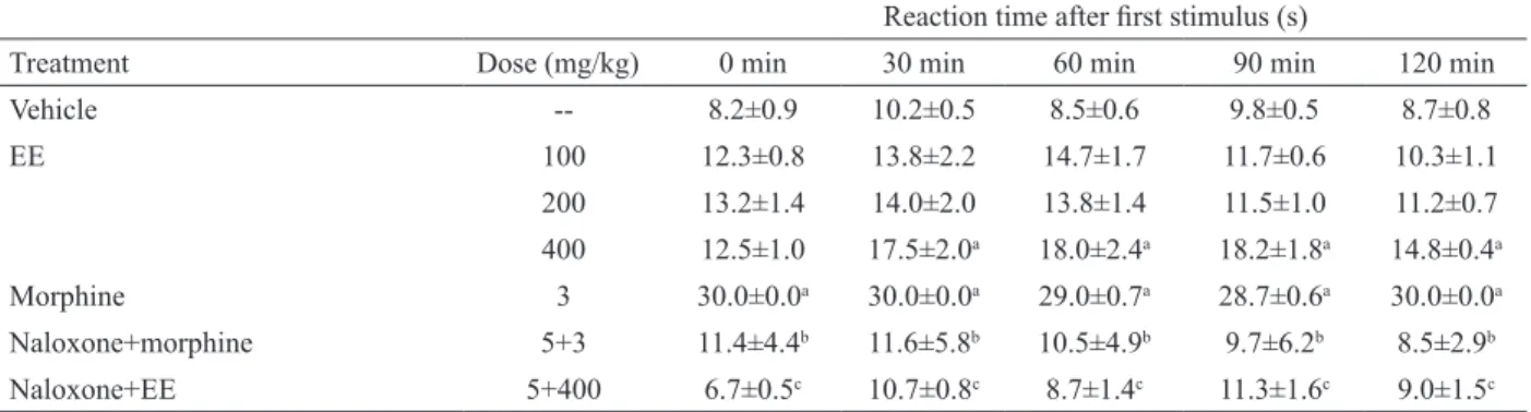
Hot-plate reaction time in mice
test (55 °C) at all time points analyzed (p<0.05, Table 2). Similarly, morphine (3 mg/kg) caused a signiicant and marked increase in the reaction time of mice (p<0.05, Table 2). Naloxone (5 mg/kg) signiicantly prevented the
antinociceptive effect caused by both C. pyramidalis EE
(400 mg/kg) and morphine (3 mg/kg) at all time points observed (p<0.05, Table 2).
0 30 60 90 120 150
200 100 C
C. pyramidalis (mg/kg)
400 ASA *
Morph First phase Second phase
* * *
* *
* * *
Licking/biting time (s)
Figure 1. The effect of Caesalpinia pyramidalis EE on formalin-induced nociception. Mice were pre-treated with vehicle (C), morphine (Morph, 10 mg/kg), acetylsalicylic acid (ASA, 300
mg/kg), or EE (100-400 mg/kg) before a formalin injection.
*p<0.001 vs. the respective control group (n=6/group).
Motor performance
In the rotarod test, EE-treated mice did not show any signiicant motor performance alterations with the 100, 200, and 400 mg/kg dose (180±0, 180±0, and 180±0 s, respectively) compared to control mice (180±0 s). As expected, the injection of the diazepam (1.5 mg/kg) reduced the time the mice were on the rotarod after the treatment (31±3 s, p<0.001).
Carrageenan-induced edema and MPO activity in rat paws
As observed in Figure 2, the single oral treatment of C. pyramidalis EE at the 400 mg/kg dose was capable of reducing (p<0.05) the edema induced by carrageenan at 2, 3 and 4 h after the injection of the phlogistic agent. Likewise, dexamethasone (2 mg/kg) inhibited (p<0.01) the edematogenic response evoked by carrageenan in rats at 2, 3 and 4 h (Figure 2).
Based on AUC0-4h values, the EE at the 400 mg/
kg dose caused a 41.2% (p<0.05) inhibition of the edema response compared to carrageenan-treated group (4.6±0.7 mL x hour). Dexamethasone (2 mg/kg) caused an inhibition of 54.4% (p<0.001).
C. pyramidalis EE (400 mg/kg) also produced a marked inhibition (p<0.05) of carrageenan-induced MPO activity in the paws of rats compared to vehicle-treated controls (4.5±0.5 and 7.1±0.9 UMPO/mg tissue, respectively). Similarly, dexamethasone (2 mg/kg) caused signiicant inhibition of carrageenan-induced
MPO activity (2.6±0.3 UMPO/mg tissue, p<0.001).
0.0 0.4 0.8 1.2 1.6 2.0 2.4
EE 100 mg/k g EE 200 mg/k g EE 400 mg/k g
Edema (mL)
0 1 2 3 4 time (h) ** *
Control
Dexa 2 mg/k g
*
** **
**
Figure 2. The effect of Caesalpinia pyramidalis EE on rat paw edema. Animals were pre-treated with vehicle (control), dexamethasone (Dexa, 2 mg/kg), or EE (100-400 mg/kg) before
a carrageenan injection. *p<0.05 and **p<0.01 vs. the control group (n=6/group).
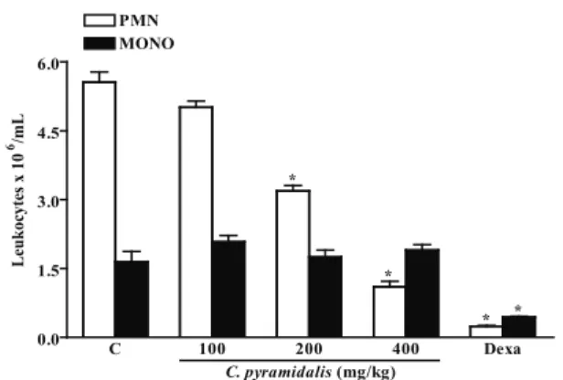
Carrageenan-induced peritonitis in mice
The carrageenan injection in control animals induced leukocyte migration into the peritoneal cavity after 4 h (7.22±0.99 x 10⁶ leukocytes/mL). C. pyramidalis
EE (400 mg/kg) signiicantly inhibited this response (2.63±0.23 x 10⁶ leukocytes/mL, p<0.01), but 200 or 100 mg/kg EE did not (4.87±0.69 x 10⁶ and 7.09±1.03
x 106 leukocytes/mL, respectively). The dexamethasone
injection also (2 mg/kg) inhibited (0.68±0.40 x 10⁶
leukocytes/mL, p<0.001) the carrageenan-induced
leukocyte migration.
The PMN migration evoked by carrageenan was reduced by 200 and 400 mg/kg EE by 42.6% and 80.2% (p<0.001), respectively (Figure 3). Dexamethasone (2 mg/kg) exhibited signiicant inhibition (95.7%, p<0.001) of the PMN migration in the control group (Figure 3).
0.0 1.5 3.0 4.5 6.0
400 200
C
C. pyramidalis(mg/kg)
Dexa 100
* PMN
MONO
* *
*
Leukocytes x 10
6/mL
between the 15th and 30th min after formalin injection and
is putatively caused by the release of pro-inlammatory mediators such as adenosine, bradykinin, histamine, PG, and 5-HT (Reeve & Dickenson, 1995). The treatment with the EE was capable of diminishing the nociceptive response in both the neurogenic and inlammatory phases. This result suggests that the EE of C. pyramidalis might possess anti-inlammatory activity. However, the inhibition presented in the irst phase suggests a disruption of either the production or the release of some central neurotransmitters.
Another interesting result of the current study was the fact that EE of C. pyramidalis produced a signiicant antinociceptive effect after the mice were exposed to a thermal stimulus. The hot-plate test, which is performed at a constant temperature, produces two kinds of behavioral responses: paw licking and jumping. Both of these are considered to be supraspinally integrated responses (Chapman et al., 1985). The present results lead us to the conclusion that the opioid system is involved because the pre-treatment with naloxone, a non-selective opioid receptor antagonist, reversed the antinociceptive effect caused by EE of C. pyramidalis. Although the hot-plate test is commonly used to assess the effect of narcotic analgesics, some sedatives, muscle relaxants, and psychotomimetics have also shown activity in this test (Eddy & Leimbach, 1953). This could indicate that this EE may have non-speciic central actions, but this hypothesis was proven unlikely by the observation that EE did not inluence the performance of mice in the rotarod test, indicating that EE actions may not be due to a motor impairment.
To complement the results obtained in the second phase of the formalin-induced licking response, the EE of C. pyramidalis was tested on models of inlammation (paw edema and peritonitis) induced by carrageenan.
In this model, carrageenan-induced rat paw edema occurs as a non-immune reaction and is used to evaluate the effects of anti-inlammatory drugs. The edema formed is a multi-mediated phenomenon divided in two phases. The irst phase (which last up to 2h after carrageenan MAP in non-anesthetized rats
In non-anesthetized normotensive rats, the oral
administration of C. pyramidalis EE (100, 200, and 400
mg/kg) did not show any signiicant MAP alterations at any of the time points analyzed (data not shown).
Discussion
This study evaluated the effects of Caesalpinia
pyramidalis Tul., Fabaceae, EE using several acute models of nociception and inlammation in rodents. Our data revealed that C. pyramidalis EE signiicantly diminished the nociceptive and inlammatory responses induced by various agents.
In the abdominal constriction assay, acetic acid acts indirectly causing the release of nociceptive endogenous mediators, such as bradykinin, serotonin (5-HT), histamine, sympathomimetic amines, prostaglandins (PG), and pro-inlammatory cytokines (Ribeiro et al., 2000; Ikeda et al., 2001). Acetic acid can also directly activate non-selective cation channels located at primary afferent pathways (Julius & Basbaum, 2001). This nociceptive effect can be prevented by non-steroidal anti-inlammatory drugs, opioids, analgesics with central actions, sympatholytic agents, and substances that reduce intestinal motility (Duarte et al., 1988; Reichert et al., 2001). Our results demonstrated that the
EE of C. pyramidalis was able to signiicantly diminish
the abdominal writhing induced by acetic acid, likely by interfering with the generation or mechanisms of action of the inlammatory mediators.
In addition, the formalin-induced paw licking assay was employed to evaluate the antinociceptive effect of the EE of C. pyramidalis. This test allowed the evaluation of two distinct phases: the irst (also called neurogenic) and second (also called inlammatory) phases. The former occurs during the irst 5 min after the formalin injection and is characterized by the direct stimulation of nociceptors presents on afferent C and in part by Aδ ibers (glutamate and substance P release). The latter occurs
Table 2. The antinociceptive effect of Caesalpinia pyramidalis EE using the hot-plate test.
Reaction time after irst stimulus (s)
Treatment Dose (mg/kg) 0 min 30 min 60 min 90 min 120 min
Vehicle -- 8.2±0.9 10.2±0.5 8.5±0.6 9.8±0.5 8.7±0.8
EE 100 12.3±0.8 13.8±2.2 14.7±1.7 11.7±0.6 10.3±1.1
200 13.2±1.4 14.0±2.0 13.8±1.4 11.5±1.0 11.2±0.7
400 12.5±1.0 17.5±2.0a 18.0±2.4a 18.2±1.8a 14.8±0.4a
Morphine 3 30.0±0.0a 30.0±0.0a 29.0±0.7a 28.7±0.6a 30.0±0.0a
Naloxone+morphine 5+3 11.4±4.4b 11.6±5.8b 10.5±4.9b 9.7±6.2b 8.5±2.9b
Naloxone+EE 5+400 6.7±0.5c 10.7±0.8c 8.7±1.4c 11.3±1.6c 9.0±1.5c
injection) is due to liberation of histamine, 5-HT, and bradykinin in paw tissue, while the second phase (3 and 4 h after carrageenan treatment) is sustained by liberation of PG (Di Rosa, 1972). In this study, EE of C. pyramidalis effectively reduced the edematogenic responses evoked by carrageenan between 2 and 4 h after the injection. These effects may be related to a reduction in the release or actions of histamine, 5-HT, bradykinin, or PG on local tissue.
The EE of C. pyramidalis also signiicantly
decreased the elevated paw MPO activity, which is currently used as an indicator of neutrophil presence in inlamed tissues, suggesting that the inhibition of neutrophil iniltration may be another characteristic of the anti-inlammatory actions of this EE. This was further investigated using carrageenan as a stimulus to produce an acute inlammatory response in the peritoneal cavity of mice, which is a widely accepted model for induction of a massive inlux of leukocytes (mainly neutrophils) to this cavity. The EE of C. pyramidalis inhibited both the total migration and the PMN migration induced by carrageenan at the same dose that inhibited the paw edema formation. The mechanism of action of carrageenan on peritonitis involves synergistic action
between PG, leukotriene B4, and other chemotactic
agents, which promote an increase of the vasodilatation, exudation, and recruitment of leukocytes (Foster et al., 1986). As these anti-edematogenic and anti-chemotatic actions of EE could be strongly inluenced by the possible vasoconstrictor effects of this extract, we conducted experiments to evaluate this possibility by measuring the mean arterial pressure of non-anesthetized rats after the
administration of EE. We found that the doses of EE of C.
pyramidalis used did not alter the mean arterial pressure, indicating that the anti-inlammatory activity of EE is not linked to alterations in the blood supply to the local of injury and might instead be related to the interference of the generation or mechanisms of action of inlammatory mediators. Furthermore, this hypothesis is supported by the antinociceptive effects of this extract, such as those observed in the second phase of the formalin-induced nociceptive test and the acetic acid-induced writhing test.
In summary, the data reported in this work conirmed the anti-inlammatory indications of C. pyramidalis, which have been observed in traditional medicinal practices. Additionally, this study suggests, for the irst time, that C. pyramidalis has relevant antinociceptive properties in acute pain-like behavioral
animal models. The mechanisms by which the EE of C.
pyramidalis exerts its actions require further studies.
Acknowledgements
This study was supported by Conselho Nacional
de Desenvolvimento Cientíico e Tecnológico. CA Santos and AMPR Passos received grants from CNPq.
References
Albuquerque UP, Medeiros PM, Almeida ALS, Monteiro JM, Lins Neto EMF, Melo JG, Santos JP 2007. Medicinal plants of the caatinga (semi-arid) vegetation of NE Brazil: A quantitative approach. J Ethnopharmacol 114: 325-354.
Bradley PP, Priebat M, Christensen M, Rothstein G 1982.
Measurement of cutaneous inlammation: estimation
of neutrophil content with an enzyme marker. J Invest Dermatol 78: 206-209.
Carvalho JCT, Teixeira JRM, Souza PJC, Bastos JK, Santos Filho D, Sarti SJ 1996. Preliminary studies of analgesic
and anti-inlammatory properties of Caesalpinia ferrea
crude extract. J Ethnopharmacol 53: 175-178.
Chapman CR, Casey KL, Dubner R, Foley KM, Graceley RH,
Reading AE 1985. Pain measurement: an overview.
Pain 22: 1-31.
Deharo E, Bourdy G, Quenedo C, Muñoz V, Sauvin MA 2001. A search for natural bioactive compounds in Bolivia through a multidisciplinary approach. Part V. Evaluation of the antimalarial activity of plants used by the Tacana Indians. J Ethnopharmacol 77: 91-98.
Di Rosa M 1972. Biological properties of carrageenan. J Pharm Pharmacol 24: 89-102.
Duarte ID, Nakamura M, Ferreira SH 1988. Participation of the
sympathetic system in acetic acid-induced writhing in mice. Braz J Med Biol Res 21: 341-343.
Duham NW, Miya TS 1957. A note on a simple apparatus for
detecting neurological deicit in rats and mice. J Am Pharm Assoc 46: 208-209.
Eddy NB, Leimbach D 1953. Synthetic analgesics. II. Dithienylbutenyland dithienylbutylamines. J Pharmacol Exp Ther 107: 385-393.
Foster SJ, McCormick ME, Howarth A, Aked D 1986. Leukocyte
recruitment in the subcutaneous sponge implant model
of acute inlammation in the rat is not mediated by
leukotriene B4. Biochem Pharmacol 35: 1709-1717.
Hikino H, Tagushi T, Fujimura H, Hiramatsu Y 1977. Antiinlammatory principles of Caesalpinia sappan
wood and of Haematoxylon campechianum wood.
Planta Med 31: 214-220.
Hunskaar S, Hole K 1987. The formalin test in mice: dissociation between inlammatory and non-inlammatory pain. Pain 30: 103-114.
Ikeda Y, Ueno A, Naraba H, Oh-Ishi S 2001. Involvement of
vanilloid receptor VR1 and prostanoids in the acetic acid-induced writhing response of mice. Life Sci 69: 2911-2919.
Julius D, Basbaum AI 2001. Molecular mechanism of nociception. Nature 413: 203-210.
analgesic screening. Fed Proc 18: 418-420.
Kuria KAM, De Coster S, Muriuki G, Masengo W, Kibwage
I, Hoogmartens J, Laekeman GM 2001. Antimalarial
activity of Ajuga remota Benth (Labiatae) and
Caesalpinia volkensii Harms (Caesalpiniaceae): conirmation of ethnopharmacological use. J Ethnopharmacol 74: 141-148.
Matos FJA 1997. Introdução a Fitoquímica Experimental.
Fortaleza, Brazil: Edições UFC.
Mendes SS, Bomim RR, Jesus HCR, Alves PB, Blank AF,
Estevam CS, Antoniolli AR, Thomazzi SM 2010.
Evaluation of the analgesic and anti-inlammatory
effects of the essential oil of Lippia gracilis leaves. J Ethnopharmacol 129: 391-397.
Menezes AC, Moreira IJA, Carvalho AA, Antoniolli AR, Santos MRV 2007. Cardiovascular effects of the aqueous extract from Caesalpinia ferrea: Involvement of ATP-sensitive potassium channels. Vasc Pharmacol 47: 41-47. Phillipson JD, Anderson LA 1989. Ethnopharmacology and
Western Medicine. J Ethnopharmacol 25: 61-72.
Reeve AJ, Dickenson AH 1995. The roles of spinal adenosine
receptors in the control of acute and more persistent nociceptive response of dorsal horn neurons in the anaesthetized rat. Brit J Pharmacol 116: 2221-2228. Reichert JA, Daughters RS, Rivard R, Simone DA 2001.
Peripheral and preemptive opioid antinociception in a mouse visceral pain model. Pain 89: 221-227.
Ribeiro RA, Vale ML, Thomazzi SM, Paschoalato AB, Poole S,
Ferreira SH, Cunha FQ 2000. Involvement of resident
macrophages and mast cells in the writhing nociceptive response induced by zymosan and acetic acid in mice.
Eur J Pharmacol 387: 111-118.
Saeed MA, Sabir A 2001. Antibacterial activity of Caesalpinia bonducella. Fitoterapia 72: 807-809.
Sharma SR, Dwivedi SK, Swarup D 1997. Hypoglycaemic,
antihyperglycaemic and hypolipidemic activities of
Caesalpinia bonducella seeds in rats. J Ethnopharmacol 58: 39-44.
Winter CA, Risley EA, Nuss GW 1962. Carrageenin-induced edema in hind paw of the rat as an assay for
anti-inlammatory drugs. Proc Soc Exp Biol Med 111: 544-547.
Woolfe G, Macdonald AD 1944. The evaluation of the analgesic action of penthidine hydrochloride (Demerol). J Pharmacol Exp Ther 80: 300.
*Correspondence
Sara Maria Thomazzi
Departamento de Fisiologia, Universidade Federal de Sergipe Av Marechal Rodon s/n, 49100-000 São Cristóvão-SE, Brazil, [email protected]