*Corresponding author.
E-mail: [email protected] (A.Sowemimo) A R T I C L E I N F O
Article history:
Received 19 August 2013 Accepted 26 October 2013
Keywords
Blepharis maderaspatensis Ant-inflammatory Antinociception Phenol
Flavonoid
A B S T R A C T
Blepharis maderaspatensis (L.) B. Heyne ex Roth, Acanthaceae, is a procumbent or scrambling perennial herb used traditionally for treatment of snakebites, wounds, edema and gout. The anti-inflammatory and anti-nociceptive properties of the ethanol extract of the whole plant of B. maderaspatensis was investigated using carrageenan-induced paw edema in rats, xylene-induced edema in mice, mouse writhing and tail clip tests respectively. The effect of the extract on inflammatory mediators, serotonin and histamine, using the most active dose (75 mg/kg) was also carried out. The results showed that the extract of B. maderaspatensis in carrageenan-induced test caused a significant inhibition (84.5%, 90 min) of paw edema at a dose of 75 mg/kg while the xylene-induced test caused a significant inhibition (62.65%) at 50 mg/kg. The histamine-induced test showed significant inhibition (90.9%, 90 min) while serotonin-induced test showed moderate inhibition (54.10%, 180 min). In the mouse writhing and tail clip tests, the extract produced a significant inhibition of 66.21% and 15.81% at 75 mg/kg, respectively. These results collectively demonstrate that the ethanol extract of B. maderaspatensis possesses anti-inflammatory and anti-nociceptive properties, and this supports the ethnopharmacological use of the plant in the treatment of inflammation.
© 2013 Elsevier Editora Ltda. All rights reserved.
Original Article
Studies on the anti-inflammatory and anti-nociceptive
properties of
Blepharis maderaspatensis
leaves
Abimbola Sowemimo
a,*, Monsurat Onakoya
a, Muyiwa S. Fageyinbo
b, Titilayo Fadoju
a aDepartment of Pharmacognosy, Faculty of Pharmacy, University of Lagos, College of Medicine Campus, Idi-Araba, Nigeria, Lagos bDepartment of Pharmacology, Faculty of Basic Medical Sciences, University of Lagos, College of Medicine Campus, Idi-Araba, Nigeria, LagosIntroduction
Inflammation is a fundamental defensive reaction of the body to an invasion of pathogens or injury. It involves a complex array of enzyme activation; mediators release, extravasation, cell migration, tissue breakdown and repair (Vane and Bolting, 1995). The classic signs of inflammation such as pain, redness, swelling and loss of function are produced by inflammatory agents such as nitric oxide, prostaglandins, bradykinin,
Blepharis maderaspatensis (L.) B. Heyne ex Roth (BM), a member of the family Acanthaceae, is a procumbent or scrambling perennial or rarely annual herb with stems up to 2.5 m long. It can be found in a wide variety of grassland, shrub and woodland habitats as well as forest-merging in the savannah zone, from Senegal to South Nigeria (Burkill, 1985). Traditionally it is used in treatment of swellings, edema and gout. A paste of the leaves is mixed with black gram powder, crushed onion and white egg yolk and the mixture is applied topically over fractured bones in Nigeria (Burkill, 2004). The whole plant is burnt to ash and mixed with oil for rubbing onto swollen legs after it has been washed in warm water in Tanganyika (Burkill, 1985).
Although the cytotoxic activity against colon cancer cells, wound healing and antioxidant activities of the plant have been reported (Baskar et al., 2012; Rajasekaran et al., 2012), the anti-inflammatory and anti-nociceptive activities have not been scientifically confirmed. Hence, the present study aimed to evaluate the anti-inflammatory and anti-nociceptive activities of the ethanol extract of Blepharis maderaspatensis.
Materials and methods
Plant material and extraction
Fresh samples of Blepharis maderaspatensis (L.) B. Heyne ex Roth, Acanthaceae (whole plant), were collected from Odofin village in Ikire town, Osun State, Nigeria, and authenticated at the Herbarium of the Department of Botany and Microbiology where a voucher specimen was deposited (LUH 4592). The plant was dried in a hot air oven at 40 ºC and grounded in a mechanical grinder. The powdered material (500 g) was macerated with absolute ethanol for 72 h at room temperature. The extract was filtered and evaporated to dryness in a water bath at 40 °C. The percentage yield was 6.52% (w/w).
Animals
All the animals (male Wistar rats (100-200 g) and Swiss albino mice (20-30 g) used in this study were obtained from the National Agency for Food and Drugs Administration and Control, Yaba. The animals were kept in hygienic and well ventilated compartments and maintained under standard laboratory conditions (12 h light/dark cycle at 22 ± 2 °C) as approved by the Experimentation and Ethics Committee of the College of Medicine, University of Lagos (CM/COM/08). All animals were acclimatized for one week, fed rodent diet (Livestock Feeds PLC, Ibadan, Oyo State, Nigeria) and had free access to drinking water. They fasted for at least 12 h prior to experimentation.
Acute toxicity test
The method of Lorke (1983) was used with minor modifications for this study. A single oral dose of the extract (5 g/kg) was administered to a group of six healthy male mice the control group only received the vehicle (10 ml/kg, p.o.). The animals were observed for general behavioural changes, physiological
function and mortality at 1, 4 and 24 h after treatment. The mice were further observed for up to seven days for any signs of delayed toxicity and mortality.
Anti-edematogenic models
Carrageenan-induced rat paw edema
In this assay, the rats were divided into seven groups (n = 6 each), which were orally treated with distilled water (control) or indomethacin (reference drug, 10 mg/kg), or the extract (12.5, 25, 50, 75 and 100 mg/kg). After 1 h after oral administration, 0.1 ml of 1% suspension of carrageenan in normal saline was injected into the sub-plantar tissue of the right hind paw to induce edema (Winter et al., 1962). The paw edema was measured at 30 min interval for 3 h after carrageenan injection. The linear paw circumference was measured using the cotton thread method of Bamgbose and Naomesi (1981). The difference between the initial and subsequent values gave the actual oedema value, which was compared to control. The inhibition of inflammation was calculated using the following formula:
% inhibition=100
(
Vc- Vt)
Vcwhere Vc represents mean edema in control and Vt mean edema in group treated with standard/extract.
Histamine and serotonin-induced rat paw oedema
Three groups of six rats each were treated with the extract (75 mg/kg, p.o.), indomethacin (10 mg/kg, p.o.), and distilled water (10 ml/kg, p.o.). Paw edema was induced after 1 h by sub-plantar administration of 0.1 ml histamine or serotonin (10-3
mg/ml) on the right hand paw (Amann et al., 1995). The linear paw circumference was measured at 0 min and thereafter every 30 min for 3 h.
% inhibition=100
(
Vc- Vt)
Vcwhere Vc represents mean edema in control and Vt mean edema in group treated with standard/extract.
Xylene-induced ear edema
The animals were divided into five groups of six mice each. Edema was induced by applying 0.03 ml of xylene to the inner surface of the right ear 30 min after oral administration of distilled water (10 ml/kg), extract (12.5-100 mg/kg) and dexamethasone (1 mg/kg) respectively. The left ear was considered as control. Fifteen minutes after the application of xylene, the mice were killed under ether anesthesia and both ears were cut off and weighed (Nunez Guillen et al., 1997). The mean of the difference between the right and left ears was calculated.
% inhibition=100
(
Vc- Vt)
VcAnti-nociceptive activity
Mouse writhing test
Mice divided into seven groups of six animals each were pre-treated with distilled water (10 ml/kg, p.o.); extract (12.5-100 mg/kg, p.o.) and acetylsalicylic acid (10 mg/kg, p.o.). After 1 h, the mice were injected with acetic acid (0.6% v/v in saline, 10 ml/kg, i.p.). The number of writhing and stretching movements was counted for a period of 30 min (Koster et al., 1959). The percentage of anti-nociceptive activity was calculated as:
% inhibition=100
(
Vc- Vt)
Vcwhere Vc represents mean number of writhing in control and Vt mean number of writhing in group treated with standard/extract.
Haffner’s tail clip test
In this model, a initial sensitivity test was carried out by applying a metal artery clip to the base of the tail to induce pain, animals that failed to attempt to dislodge the clip in 10 s were discarded. The responsive mice were then divided into seven groups of six animals each. The pre-treatment reaction time of all mice was determined and treatment was then administered using distilled water (10 ml/kg, p.o.); extract (12.5-100 mg/kg, p.o.) and the standard drug, morphine (10 mg/ kg, s.c.). The reaction time of each animal was determined 60 min treatment for oral administration and 30 min post-treatment for subcutaneous administration (Adeyemi et al., 2004; Bianchi and Franceschini, 1954). A post-treatment cut-off time of about 60 s was used.
100 x latency) treatment Pre
time off -(Cut
latency) treatment (Pre
-latency) treatment
-(Post Inhibition
% =
Quantitative analysis
Determination of total phenolic content
The concentration of total phenolics in the ethanol extract of
Blepharis maderaspatensis was determined by the Folin-Ciocalteu colorimetric method (Slinkard and Singleton, 1977). Briefly, 500 µl of the solution (containing 1 mg/ml) extract in methanol was added to 2.5 ml Folin-Ciocalteu reagent (diluted with water 1:1) and 2 ml (75 g/l) sodium carbonate. After 30 min of incubation at 40 °C, the resulting absorbance was measured at 760 nm using a spectrophotometer (T80 spectrometer, PG Instrument Ltd.). A calibration curve, using gallic acid in a concentration range of 0.01-0.05 mg/ml was prepared. Measurements were carried out in triplicate. The total phenolic content was expressed as milligram gallic acid equivalent (GAE) per gram of sample.
Determination of total flavonoid content
The total flavonoid content of the extract was determined using the method of (Ordonez et al., 2006). Quercetin was used as standard. Solution of 2% AlCl3 in ethanol (1 ml) was added to 1 ml of the extract. The absorbance was measured at 420 nm after 1 h of incubation at room temperature. Measurements were carried out in triplicate. Total flavonoid content was expressed as milligram quercetin equivalent (QE) per gram of sample.
Statistical analysis
Data were presented at mean ± SEM. The data were analysed using GraphPad Prism for windows version 5 (GraphPad Software, San Diego, CA, USA). Values were considered significant when p ≤ 0.05.
Results
Acute toxicity test
Oral administration of BM extract at 5 g/kg did not produce any mortality. The extract did not produce significant changes in behavior during the time of observation.
Anti-inflammatory activity
Carrageenan-induced rat paw edema
The subplantar injection of carrageenan produced edema development which increased progressively with time in the control group. Peak edema development was observed at 180 min (2.53 ± 0.15). The administration of BM (12.5, 25, 50 and 75 mg/kg, p.o.) showed significant (p < 0.05) inhibition from 30 min after carrageenan injection with peak effect (84.5%) produced at the dose of 75 mg/kg at 90 min. Oral administration of indomethacin (10 mg/kg) also significantly (p < 0.01) reduced the edema with 86.7% inhibition at 180 min (Table 1). However pro-inflammatory activity was observed for the plant extract at 100 mg/kg.
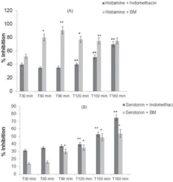
Histamine and serotonin-induced rat paw edema
A significant (p < 0.001) reduction in the edema of mice treated with the extract was observed in the histamine-induced paw edema model (Fig. 1). The maximum inhibition was observed at 90 min (90.9%).
In the serotonin-induced paw edema model, the extract had low effect on the inhibition of edema. The maximum inhibition was observed at 180 min (54.1%) (Fig. 1).
Xylene-induced ear edema
The effect of B. maderaspatensis was dose-dependent with peak effect (62.65% inhibition) produced at the dose of 50 mg/kg (Table 2). This effect was greater than but not significantly (p < 0.05) different from that produced by dexamethasone (50.2% inhibition). Pro-inflammatory activity was also observed for the extract at 100 mg/kg.
Anti-nociceptive activity
Mouse writhing test
In this model, the oral administration of B. maderaspatensis
Table 1
Effect of extract of BM on carrageenan induced rat paw oedema.
Trt
Dose mg/kg
Increase in paw circumference (cm)
T0 T30 T60 T90 T120 T150 T180
BM 12.5 1.93 ± 0.03 2.18 ± 0.03 (53.7) 2.22 ± 0.01 (42.3) 2.25 ± 0.03 (44.8) 2.28 ± 0.01b (59.1) 2.28 ± 0.03c (23.9) 2.32 ± 0.01 (48.0)
BM 25 1.95 ± 0.02 2.15 ± 0.04 (63.0) 2.22 ± 0.01 (48.1) 2.25 ± 0.04 (48.2) 2.28 ± 0.05 (50.0) 2.3 ± 0.05 (23.9) 2.32 ± 0.03 (50.7)
BM 50 1.92 ± 0.04 2.05 ± 0.02a (75.9) 2.03 ± 0.05b (78.9) 2.13 ± 0.05a (69.0) 2.21 ± 0.05 (56.1) 2.25 ± 0.06 (28.3) 2.25 ± 0.08a(56.0)
BM 75 1.97 ± 0.05 2.08 ± 0.07 (81.4) 2.07 ± 0.03ª (82.7) 2.07 ± 0.03a (84.5) 2.15 ± 0.04a (74.2) 2.22 ± 0.05 (47.8) 2.32 ± 0.04b(66.7)
BM 100 1.74 ± 0.02 2.33 ± 0.04 (-9.3) 2.13 ± 0.03 (25.0) 2.33 ± 0.06 (-1.7) 2.4 ± 0.05 (0.0) 2.29 ± 0.09 (-19.6) 2.3 ± 0.05 (25.33) Indn 10 1.92 ± 0.05 2.14 ± 0.08 (59.3) 2.18 ± 0.06 (50.0 ) 2.18 ± 0.06 (55.2) 2.13 ± 0.05b (68.2) 2.05 ± 0.03 (71.7) 2.02 ± 0.01c (86.7)
D.W 10ml/kg 1.78 ± 0.03 2.32 ± 0.05 2.30 ± 0.07 2.36 ± 0.1 2.44 ± 0.1 2.24 ± 0.1 2.53 ± 0.15
Figures in parenthesis represent percentage inhibition of edema development.
Trt, Treatments; Indn, Indomethacin; D.W, Distilled water; BM, Blepharis maderaspatensis. Values are expressed as mean ± SEM, n =6.
ap < 0.05. bp < 0.01.
cp < 0.0001 compared to the control.
Table 2
Effect of extract of BM on xylene induced edema in mice.
Treatment Dose (mg/kg) Ear swelling (g) Inhibition (%)
BM 12.5 0.025 ± 0.004 12.84
BM 25 0.002 ± 0.004a 21.40
BM 50 0.004 ± 0.004b 62.65
BM 75 0.009 ± 0.002a 30.74
BM 100 0.018 ± 0.002a -37.98
Dexamethasone 10 0.007 ± 0.003a 50.20
Distilled water 10 ml/kg 0.013 ± 0.006 _
BM, Blepharis maderaspatensis. Values are expressed as mean ± SEM, n = 6.
ap < 0.05.
bp < 0.01 compared to the control..
Fig. 2 -Effect of Blepharis maderaspatensis on mouse writhing
test in mice. Values are expressed as mean ± SEM (n=6). *p < 0.05 compared to the control.
Haffner’s tail clip test.Haffner’s tail clip t.Haffner’s tail clip
The effect of BM on the tail clip test is shown in Table 3. The animals of the control group elicited reactions towards clip removal with pre-treatment latency being 4.43 ± 1.13 s and post-treatment latency being 5.79 ± 2.74 s. BM showed a significant (p < 0.05) dose-dependent increase in reaction latency. Maximum inhibition was produced at 75 mg/kg (15.81%). This effect was however less than that elicited by morphine (75.67%) at 10 mg/kg.
Fig. 1 - The effects of Blepharis maderaspatensis and
indomethacin on rat’s hind paw edema induced by (a) histamine and (b) serotonin. Data represented as mean ± S.E.M (n=6). *p < 0.01. **p < 0.0001 (Two-way ANOVA followed by Bonferroni’s test).
Quantitaive analysis
Discussion
Modern medicine has gained immensely from the research of natural products as a number of the therapeutic drugs currently in use have been derived from pharmacologically active agents obtained from plants (Shu, 1998). However, in spite of this progress, the potential of higher plants as sources for new drugs is still largely unexplored.There is also an urgent need for effective anti-inflammatory and anti-nociceptive agents in medicine.
The result from the acute toxicity test carried out in this work indicated that the ethanol extract of Blepharis maderaspatensis (L.) B. Heyne ex Roth, Acanthaceae, has low oral toxicity as the 5 g/kg dose did not cause death in the animals even after seven days of administration.
The evaluation of the anti-inflammatory activity of B. maderaspatensis in this study was carried out using the carrageenan, histamine, serotonin and xylene-induced edema tests. It has been reported that the carrageenan-induced rat paw model is a suitable in vivo model to study anti-inflammatory effects of natural products since it involves several mediators (Woldesellassie et al., 2011). Three phases have been postulated for carrageenan induced edema and these include; the initial phase (between 0 and 1.5 h) which is attributed to the action mediators such as histamine and serotonin; a second phase (1.5-2.5 h) mediated by bradykinin and a third phase (2.5-6 h) mediated by prostaglandins (Di Rosa et al., 1971; Suba et al., 2005). In this investigation, B. maderaspatensis showed significant inhibition of rat paw edema in the initial phase. This suggests that the extract (75 mg/kg,
p.o.) plays an important role as a protective factor against carrageenan-induced acute inflammation and possibly acts by inhibiting the release of/ and the action of histamine and/ or serotonin.
Histamine and serotonin are potent vasodilator substances and are known to increase vascular permeability (Skidmore and Whitehouse, 1967). In order to confirm the results obtained from the carrageenan-induced edema test, the effect of the extract at the most effective concentration (75 mg/kg) was
Table 3
Effect of BMon Haffner’s tail clip reflex in mice.
Treatment mg/kgDose Pre-treatment(s) treatment(s)Post- % Inhibition
BM 12.5 3.35 ± 0.69 5.39 ± 1.14a 3.60
BM 25 3.49 ± 0.54 6.65 ± 1.43a 5.59
BM 50 2.89 ± 1.18 10.22 ± 3.14a 12.83
BM 75 3.20 ± 0.70a 12.18 ± 2.50a 15.81
BM 100 2.86 ± 0.55a 4.81 ± 0.53a 3.41
Morphine 10 5.96 ± 0.96 46.85 ± 7.56a 75.67
Distilled water
10 ml/kg 4.43 ± 1.13 5.79 ± 2.74 2.45
BM, Blepharis maderaspatensis. Values are expressed as mean ± SEM (n = 6).
investigated using histamine and serotonin-induced edema models. The results showed that the extract effectively suppressed the edema produced by histamine but had a low effect on the edema produced by serotonin indicating that the extract exhibits its anti-inflammatory action by inhibiting the synthesis, release or action of histamine.
The xylene-induced edema model is useful for the screening of anti-inflammatory agents. It is characterized by fluid accumulation and edema. Suppression of this response is taken as an indication of antiphlogistic effect (Atta and Alkofahi, 1998). The effectiveness of B. maderaspatensis (50 mg/kg, p.o.) in this model may suggest the inhibition of phospholipase A which is involved in the pathophysiology of inflammation due to xylene (Lin et al., 1992).
In this study, the anti-nociceptive activity of B. maderaspat-ensis was investigated using the mouse writhing and Haffner’s tail clip tests. The mouse writhing test is used for the evalu-ation of peripherally acting drugs and the induction of pain occurs by the release of endogenous substances as well as other pain mediators such as arachidonic acid via cyclooxygen-ase, and prostaglandin biosynthesis (Franzotti et al., 2000). The dose-dependent inhibition of writhing of the extract observed in this study, suggests a peripherally mediated anti-nociceptive activity based on the association of the model with stimulation of peripheral receptors (Bentley et al., 1983).
The tail clip test is a confirmatory test to show the centrally acting component of pain mechanism (Richardson et al., 1998). Centrally acting anti-nociceptive drugs are known to elevate the pain threshold of rodents to pressure and heat (Singh and Majumdar, 1995). In this study, B. maderaspatensis
extract elicited a very low activity (< 20%). This suggests that the extract does not have centrally acting anti-nociceptive properties.
The results obtained from the inflammatory and anti-nociceptive tests suggest that B. maderaspatensis is effective against inflammatory and nociceptive pains with a more pronounced effect in the former. It was also observed that in most of the models investigated, the highest inhibtion was at 75 mg/kg after which there was a reduction in activity. This may be due to the filling of the opioid receptors (Bentley et al., 1983).
Previous studies have shown that anti-inflammatory and analgesic effects can be a result of the high polyphenol content of plants especially phenolics and flavonoids (Handa et al., 1992; Orhan et al., 2007). Flavonoids are known to prevent the synthesis of prostaglandins. Biochemical investigations on the mechanism of action of flavonoids have shown that these compounds can inhibit a wide variety of enzymes. The release of arachidonic acid is closely related to the cyclooxygenase and 5-lipoxygenase enzyme systems (Middleton et al., 2000; Williams et al., 1995). The polyphenols present in this plant may be responsible for the observed anti-inflammatory and analgesic activities.
Authorship
MO and TF contributed in collecting plant samples and identification, running the laboratory work and analysis of the data. MSF contributed to biological studies and analysis of the data. AS designed the study, supervised the laboratory work and wrote manuscript. All the authors have read the final manuscript and approved submission.
R E F E R E N C E S
Adeyemi, O.O., Okpo, S.O., Okpaka, O., 2004. The analgesic effect of the methanolic extract of Acanthus montanus. J. Ethnopharmacol. 90, 45-48.
Amann, R., Schuligoi, R., Lanz, I., Donnerer, J., 1995. Histamine-induced edema in the rat paw - effect of capsaicin denervation and a Cgrp receptor antagonist. Eur. J. Pharmacol. 279, 227-231.
Atta, A.H., Alkofahi, A., 1998. Anti-nociceptive and anti-inflammatory effects of some Jordanian medicinal plant extracts. J. Ethnopharmacol. 60, 117-124.
Bamgbose, S.O., Noamesi, B.K., 1981. Studies on cryptolepine. II: Inhibition of carrageenan induced oedema by cryptolepine. Planta Med. 41, 392-396.
Banasik, C., 2000. Inflammation and immunity, in: Banasic, J.L. (Ed.), Pathophysiology: Biological and Behavioral perspectives. W.B. Saunders Company, Pennsylvania, pp. 197-201.
Baskar, A.A., Numair, K.S.A., Alsaif, M.A., Ignacimuthu, S., 2012. In vitro antioxidant and antiproliferative potential of medicinal plants used in traditional Indian medicine to treat cancer. Redox Rep. 17, 145-156.
Bentley, G.A., Newton, S.H., Starr, J., 1983. Studies on the anti-nociceptive action of -agonist drugs and their interaction with opoid mechanisms. Brit. J. Pharmacol. 79, 125-134. Bianchi, C., Franceschini, J., 1954. Experimental observation
on Haffner’s method for testing analgesic drugs. Brit. J. Pharmacol. 9, 280-284.
Burkill, H.M., 1985. The Useful Plants of West Tropical Africa. Royal Botanic Gardens, Kew.
Burkill, H.M., 2004. The Useful Plants of West Tropical Africa. Royal Botanic Gardens, Kew.
Chandrasoma, P., Taylor, C.R., 2005. Part A. General Pathology, Section II. The Host Response to Injury, Chapter 3. The Acute Inflammatory Response, sub-section Cardinal Clinical Signs, Concise Pathology. McGraw-Hill, New York.
Di Rosa, M., Giroud, J.P., Willoughby, D.A., 1971. Studies on the mediators of the acute inflammatory response induced in rats in different sites by carrageenan and turpentine. J. Pathol. 104, 15-29.
Franzotti, E.M., Santos, C.V.F., Rodrigues, H.M.S.L., Mourao, R.H.V., Andrade, M.R., Antoniolli, A.R., 2000. Anti-inflammatory, analgesic activity and acute toxicity of Sida cordifolia L. (Malva-branca). J. Ethnopharmacol. 72, 273-278. Handa, S.S., Chawla, A.S., Sharma, A.K., 1992. Plants with
anti-inflammatory activity. Fitoterapia. 63, 3-31.
Koster, R., Anderson, M., De Beer, E.J., 1959. Acetic acid for analgesic screening. Federation Proceedings. 18, 412.
Lin, L.L., Lin, A.Y., Knopf, J.L., 1992. Cytosolic phospholipase-A2 is coupled to hormonally regulated release of arachidonic-acid. P. Natl. Acad. Sci. USA. 89, 6147-6151.
Lorke, D., 1983. A new approach to practical acute toxicity testing. Arch. Toxicol. 54, 275-287.
Middleton, E., Kandaswami, C., Theoharides, T.C., 2000. The effects of plant flavonoids on mammalian cells: Implications for inflammation, heart disease, and cancer. Pharmacol. Rev. 52, 673-751.
Nunez Guillen, M.E., Emim, J.A., Souccar, C., Lapa, A.J., 1997. Analgesic and anti-inflammatory activities of the aqueous extract of Plantago major L. Int. J. Pharmacogn. 35, 99-104. Ordonez, A.A.L., Gomez, J.D., Vattuone, M.A., Isla, M.I., 2006.
Antioxidant activities of Sechium edule (Jacq.) Swartz extracts. Food Chem. 97, 452-458.
Orhan, D.D., Hartevioglu, A., Kupeli, E., Yesilada, E., 2007. In vivo anti-inflammatory and anti-nociceptive activity of the crude extract and fractions from Rosa canina L. fruits. J. Ethnopharmacol. 112, 394-400.
Rajasekaran, A., Sivakumar, V., Darlinquine, S., 2012. Evaluation of wound healing activity of Ammannia baccifera and Blepharis maderaspatensis leaf extracts on rats. Rev. Bras. Farmacogn. 22, 418-427.
Richardson, J.D., Kilo, S., Hargreaves, K.M., 1998. Cannabinoids reduce hyperalgesia and inflammation via interaction with peripheral CB1 receptors. Pain 75, 111-119.
Shu, Y.Z., 1998. Recent natural products based drug
development: A pharmaceutical industry perspective. J. Nat. Prod. 61, 1053-1071.
Singh, S., Majumdar, D.K., 1995. Analgesic activity of fixed oil of Ocimum sanctum Linn (Tulsi) and its possible mechanism of action. Int J Pharmacogn 33, 188-192.
Skidmore, I., Whitehouse, M., 1967. Biochemical properties of antiinflammatory drugs X: the inhibition of serotonin formation in vitro and inhibition of the esterase activity of α-chymyotrysin. Biochem. Pharmacol. 16, 737-751. Slinkard, K., Singleton, V.L., 1977. Total phenol analysis:
Automation and comparison with manual methods. Am. J. Enol. Vitic. 28, 49-55.
Suba, V., Murugesan, T., Kumaravelrajan, R., Mandal, S.C., Saha, B.P., 2005. Antiinflammatory, analgesic and antiperoxidative efficacy of Barleria lupulina Lindl. extract. Phytother. Res. 19, 695-699. Talwar, S., Nandakumar, K., Nayak, P.G., Bansal, P., Mudgal, J.,
Mor, V., Rao, C.M., Lobo, R., 2011. Anti-inflammatory activity of Terminalia paniculata bark extract against acute and chronic inflammation in rats. J. Ethnopharmacol. 134, 323-328. Vane, J.R., Bolting, R.M., 1995. New insights into the mode of
action of anti-inflammatory drugs. Inflam. Res. 44.
Williams, C.N., Honet, J.R.S., Harbone, J.B., Greenham, J., Eeagles, J., 1995. A biologically active lipophilic flavonols from Tanacetum parthenium. Phytochemistry 38, 267-270. Winter, C., Risley, E., Nuss, O., 1962. Carrageenin-induced
inflammation in the hind limb of the rat. Federation Proceedings. 46, 118-126.