ISSN 0102-695X DOI: 10.1590/S0102-695X2013005000026 Received 18 Sep 2012 Accepted 23 Jan 2013 Available online 26 Mar 2013
activity of hexanic extract, cyperenoic acid, and
jatrophone terpenes from
Jatropha ribifolia roots
Elaine de S. Fernandes,
1Fátima A. Rodrigues,
1Danilo Tófoli,
2Paulo M. Imamura,
3João E. de Carvalho,
4Ana L. T. G. Ruiz,
4Mary A. Foglio,
4Sandro Minguzzi,
1Rogério C. L. Silva
*,11Departamento de Química, Unidade Universitária de Naviraí, Universidade
Estadual de Mato Grosso do Sul, Brazil,
2Departamento de Química, Universidade Federal de Mato Grosso do Sul, Centro
de Ciências Exatas e Tecnologia, Universidade Federal de Mato Grosso do Sul, Brazil,
3Departamento de Química, Universidade Estadual de Campinas, Brazil,
4Centro Pluridisciplinar de Pesquisas Químicas, Biológicas e Agrícolas,
Universidade Estadual de Campinas, Brazil.
Abstract:The cytotoxicity of a hexanic fraction produced from the ethanolic crude extract, obtained from Jatropha ribifolia (Pohl) Baill, Euphorbiaceae, roots was evaluated against ten human cancer cell lines (MCF-7, NCI-ADR/RES, OVCAR-3, PC-3, HT-29, NCI-H460,786-O, UACC-62, K-562, U251) compared with doxorrubicine as positive control. Compounds jatrophone and cyperenoic acid were isolated from the hexanic extract and characterized by spectroscopic techniques (NMR of 1H, 13C and IR). The in vitro antiproliferative activity of jatrophone
showed selectivity in a concentration dependent way with Total Inhibition growth
of: glioma 0.57 μg mL-1 (U251), breast cancer 9.2 μg mL-1 (MCF-7),
adriamycin-resistant ovarian cancer 0.96 μg mL-1 (NCI-ADR/RES), kidney 4.2 μg mL-1 (786-0),
prostate cancer 8.4 μg mL-1 (PC-3), colon cancer 16.1 μg mL-1 (HT29) and leukemia
0.21 μg mL-1 (K-562).
Keywords:
cytotoxic activity diterpene Euphorbiaceae Jatropha ribifolia sesquiterpene
Introduction
Bioactive terpenes have a signii cant importance for the pharmaceutical industry which covers a wide range of diverse chemical compounds, providing opportunities for synthesis (Wang & Bidigare, 2005). The genus Jatropha is recognized as an important source of secondary metabolites, with mainly terpenes that are fairly known to this genus (Can-Aké et al., 2004).
Jatropha ribifolia (Pohl) Baill (Euphorbiaceae) is found throughout the Brazilian northeastern region, popularly known as “pinhão-de-purga” (purgin nut). The latex is used by folkmedicine for treatment of snake bites and to treat upper tract decongestions. No previous reports were found describing the phytochemical studies of this species that is considered endemic in the state of Mato Grosso do Sul and known in Navirai as “minâncora-do-campo” (Souza & Rodal, 2010). Herein we report the isolation and structure elucidation of the compounds jatrophone and cyperenoic acid with in vitro antiproliferative activity.
Materials and Methods
Plant material
Material was collected at Naviraí, Mato Grosso do Sul, Brazil. Voucher specimen (CGMS 31.481) was deposited at the Herbarium of the Institute of de Botany of São Paulo at the University of São Paulo, Brazil.
Isolation procedures
Fresh roots (1.5 kg) of Jatropha ribifolia (Pohl) Baill., Euphorbiaceae, were ground, and extracted with EtOH at room temperature. The solvent was removed under reduced pressure yielding 386 g (25.7%) of crude ethanol extract. The crude extract was further partitioned with n-hexane and MeOH-H2O (9:1). The crude dried n-hexane-soluble fraction (7.75 g) was purii ed by silica gel column chromatography using gradients of n-hexane/ EtOAc and EtOAc. Fractions eluted with n-hexane:EtOAc (20 and 25%) were grouped and purii ed by column chromatography yielding jatrophone compound 1 (431.3
Rev. Bras. Farmacogn. Braz. J. Pharmacogn. 23(3): May/Jun. 2013 mg). Cyperenoic acid was isolated from the crude n-hexane extract obtained from freshly picked roots (660 g) further extracted with n-hexane under relux. After removal of the organic solvent under reduced pressure, 8 g of the extract was puriied by silica gel column chromatography affording 475.8 mg of compound 2. The active compounds were monitored by the in vitro antiproliferative assay.
Spectroscopic analysis
The structures determination were based mainly on spectroscopic investigation of 1H and 13C NMR studies including HMBC (hetero nuclear multiple bond correlation) and COSY (proton-proton correlation) experimental. Infrared (IR) spectroscopy was utilized for identifying absorption band and gas chromatography coupled mass spectrometry for the structure mass-charge (m/z).
Biological tests
In vitro antiproliferative assay in human cancer cell lines
For the in vitro antiproliferative activity screening, ten cell lines were selected from human tumors, designated as: strains K562 (leukemia), MCF-7 (breast), NCI-ADR/RES (ovarian phenotype of resistance to multiple drugs), UACC-62 (melanoma), NCI-H460 (lung), PC 3 (prostate), HT29 (colon), OVCAR-3 (ovarian), 786-0 (kidney) and U251 (glioma), and a normal cell lines: VERO (monkey kidney) later replaced by HaCat (human keratinocyte). All cell lines were provided by the National Cancer Institute (NCI), USA. The experimental procedures were performed according with the literature (Skehan et al., 1990; Monks et al., 1991; Rubinstein et. al., 1990).
Stock solution of the n-hexanic extract, jatrophone (1) and cyperenoic acid (2) compounds (100 mg mL-1) were prepared in dimethylsulfoxide (DMSO). Cell culture DMSO was diluted 400 times in RPMI/FBS/ gentamicin, to avoid toxicity. After dilution, 100 μL of medium containing the extract and compounds to be tested were added to a 96 compartments (except for the control) at concentrations of 0.25, 2.5, 25 and 250 μg mL-1. The plates were incubated for 24 h at 37 °C in a 5% CO2 in a humid environment.
Cells were cultured in RPMI-1640 medium (10 mL), supplemented with 5% fetal calf serum (SFB). The vials were centrifuged (2000 g) by 4 min at 4 oC. The supernatant was collected and discarded and precipitated of cells were resuspended in 5 mL culture medium. After 48 h, the plate T0 was ixed by adding 50 μL trichloroacetic acid (TCA) 50 % determining the actual amount of cells present at the time the samples were applied. After 48 h
treatment 50 μL of TCA (50%) was added and incubated for 1 h at 4 oC. Then the plates were subjected to four successive washes with water to remove residues of TCA, medium, SFB and secondary metabolites, and then stored at room temperature until complete drying with further adition of 50 μL of sulforhodamine B (SRB) to 0.4% (weight/volume) dissolved in 1% acetic acid, and incubated at room temperature for 30 min. After wards the plates were washed four times consecutively with a solution of 1% acetic acid for complete removal of residues of SRB. After complete drying the plates at room temperature, the protein bound dye was solubilized by adding 150 μL of 10 μM Trizma Base in pH 10.5. The cells were then ixed with 50% trichloroacetic acid. Cell proliferation was determined by spectrophotometric quantiication at 540 nm using the sulforhodamine B as dyeing reagent. Doxorubicin chloridate (0.025 to 25 μg mL-1) was used as positive control (Table 3).
Date analysis
The averages of the absorbance were calculated discounting the value the white and total growth inhibition (TGI) was determined by the equation: if T>C the drug stimulated the growth and showed not IC; if T≥To and <C, the drug was cytostatic and the equation used was 100 x [(T-To)/(C-To)]; is T<To and drug was cytocidal equation used was 100 x [(T-To)/(To)], being T the absorbance of treated cell, C the cell control and To control cells on day of addition of the drugs, the results was subtracted from 100% to yield the percentage of growth. The absorbance data were analyzed and compiled in constructing graphs using the Origin 7.5 software correlating the percentage of inhibition or cell death at the concentration of extract and terpenes.
Results and Discussion
Isolation and structural identiication
was obtained as colorless crystals with mp 162-164 °C. The GC/MS spectrum gave an [M-H] ion at m/z 234.1 corresponding to the molecular formula C15H22O2. The FTIR spectrum showed a broad absorption at 3430 cm-1 due to the presence of OH group and carbonyl group at 1674 cm-1 indicating the presence of carboxylic acid. The 1H and 13C NMR spectroscopy and 2D NMR technique (Table 2)
were compared with the data reported by Pertino (2006) for cyperenoic acid, conirming the chemical structure. This is the irst time where the presence of this compound was described in this specie. The NOESY spectrums of jatrophone and cyperenoic acid showed the H groups in proximity (Figure 1 and 2) conirming the stereochemical aspects observed by X-ray, for both compounds. (Pertino et. al., 2006; Jacobs et al., 1987).
O O CH3 H3C
CH3
CH3 CH3
O Hb
Ha
1 2
3 4
15
14 13
12 20
11 19
10
9 8 7
6 5
17
18
16 H
a Hb
Figure 1. NOESY correlations for jatrophone (1).
In vitro antitumour activity
This is the irst report exploring the in vitro antiproliferative activities effect of J. ribifolia extract and two isolated terpenic compounds, jatrophone and cyperenoic acid against ten human cells lines and normal monkey cells (VERO). A range of J. ribifolia roots extract and terpenes concentration (0.25 μg mL-1 to 250 μg mL-1) were used to investigate the relative degree of TGI against the following cell line: glioma (U251), melanoma (UACC-62), breast cancer (MCF7), adriamycin-resistant ovarian cancer (NCI-ADR/RES), kidney (786-0), non-small lung cancer (NCl-H460), ovarian cancer (OVCAR-3), prostate cancer (PC-3), colon cancer (HT29), leukemia (K-562) and normal green monkey kidney cells (VERO). Figure 3 shows the curves of the concentration response for the doxorrubicin control drug, hexanic extract, cyperenoic acid and jatrophone terpenes against human tumor cells. Table 3 presents the results determined by the in vitro antiproliferative assay with human cancer cell lines.
The activity of hexanic extract was eficient for seven cell lines tested, when compared with doxorrubicine, with an in vitro antiproliferative activity ranging from 1.2 to 26.5 μg mL-1. The hexanic extract’s cytocidal activity against tumoral cells growth was signiicantly eficient
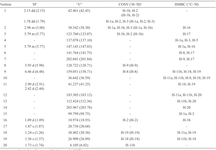
Table 1. NMR data for jatrophone in CDCl3, with instrument operating at 250 MHz for NMR 1H and NMR 13C and 500 MHz for 2D
COSY and HMBC.
Position 1Hb 13Cb COSY (1H-1H)b HMBC (13C-1H)
1 2.15 dd (2.15) 42.461 (42.45) H-1b, H-2
(H-1b, H-2)
-1.78 dd (1.79) H-1a, H-2, H-3 (H-1a, H-2, H-3)
2 2.98 m (3.00) 38.342 (38.30) H-1a, H-1b, H-3 (H-1a, H-1b) H-16
3 5.79 m (5.77) 123.760 (123.07) H-1b, H-2 (H-1b) H-17
4 - 137.078 (137.10) - H-1a, H-3, H-5
5 5.79 m (5.77) 147.143 (147.03) - H-1a, H-16
6 - 141.764 (141.75) - H-8, H-17
7 - 202.041 (201.84) - H-9, H-17
8 5.95 d (5.98) 128.722 (128.71) H-9 (H-9)
-9 6.46 d (6.48) 159.051 (158.71) H-8 (H-8) H-11b, H-18, H-19
10 - 36.642 (36.59) - H-11a, H-11b, H-8, H-18, H-19
11 2.90 d (2.91) 2.42 d (2.44)
41.237 (41.25) - H-18, H-19
12 - 183.305 (183.12) - H-11a, H-11b, H-20
13 - 112.424 (112.36) - H-11b, H-20
14 - 203.967 (203.78) - H-20
15 - 99.799 (99.75) - H-1a, H-3
16 1.09 d (1.09) 18.974 (18.93) H-2 (H-2) H-1b
17 1.87 s (1.87) 20.738 (20.68) -
-18 1.24 s (1.26) 30.402 (30.38) H-19 (H-19) H-11a, H-19
19 1.36 s (1.37) 26.909 (26.89) H-18 (H-18) H-11b, H-18
20 1.73 s (1.74) 6.105 (6.02) H-11b
Rev. Bras. Farmacogn. Braz. J. Pharmacogn. 23(3): May/Jun. 2013
(normal cell) with 1.7 μg mL-1. The activity on tumoral cells was also observed for the crude hexane extract due to the presence of jatrophone and cyperenoic acid. The terpenes when isolated also showed signiicant results in the bioassay under study. The cytocidal activity for cyperenoic acid against tumoral growth cells was eficient for three tested cells. The TGI for tumoral cells were 11.4 μg mL-1 (U251), 25.1 μg mL-1 (PC-3) and 10.5 μg mL-1 (K-562). For VERO cells 9.5 μg mL-1 was the concentration determined. Better results were noted for jatrophone (1) with selectivity in a concentration dependent way determined as 0.57 μg mL-1 (U251), 9.2 μg mL-1 (MCF-7), 0.96 μg mL-1 (NCI-ADR/RES), 4.2 μg mL-1 (786-0), 8.4 μg mL-1 (PC-3), 16.1 μg mL-1 (HT-29) and 0.21 μg mL-1 (K-562) and VERO (normal cells). Doxorubicin drug was eficient to total growth inhibition in concentration 25 μg mL-1.
Table 2. NMR data for cyperenoic acid in CDCl3, with instrument operating at 250 MHz for NMR 1H and NMR 13C and 500 MHz
for 2D COSY and HMBC.
Position 1Hb 13Cb COSY (1H-1H) HMBC (13C-1H)
1 - 68.20 (68.23) - H-2a, H-2b, H-10
2 1.78 m (1.78) 1.58 m (1.58)
25.71 (25.74) H-2b, H-3b
H-2a, H-3a, H-14
H-16 3 2.83 m (2.83) 2.75 m
(2.75)
36.31 (36.33) H-2b, H-3b
H-2a, H-3a
H-2a, H-2b,
4 - 123.07 (123.19) -
-5 - 173.09 (173.10) -
-6 2.78 m (2.77) 2.28 brd (2.28)
31.30 (31.34) H-6b, H-7
H-6a
-7 2.00 m (2.00) 48.15 (48.20) H-6a, H-8B H-9a, H-12, H-13
8 1.91 m (1.92) 1.42 m (1.41)
26.91 (26.96) H-8b, H-9b
H-7, H-8a, H-9a, H-9b
H-9b, H-13 9 1.55 m (1.55) 1.17 m
(1.17)
27.86 (27.90) H-9b, H-8b, H-10 H-8a, H-8b, H-9a
H-10, H-14
10 2.10 m (2.10) 35.96 (36.00) H-9a H-2a, H-2b, H-9a, H-9b, H-14
11 - 41.70 (41.73) - H-2a, H-10, H-12, H-13
12 0.85 s (0.86) 26.19 (26.22) - H-13
13 1.03 s (1.03) 19.26 (19.28) - H-12
14 0.88 d (0.90) 17.96 (17.99) H-2b H-10, H-9 b
15 0.85 s (0.86) 170.71 (171.05) -
-bData in parenthesis from literature (Pertino et al., 2006).
Table 3. Eficacy of organic extract and terpenes obtained from roots of Jatropha ribifolia and positive control doxorubicin against human tumor cells - Total Growth Inhibition (TGI).
TGI (µg.mL-1)
2 m a 7 4 p o h k v
Hexanic extract 1.2 15.5 9.5 9.1 >250 12.3 37.5 26.5 2.4 1.7
Cyperenoic acid 11.4 86.9 41.2 41.0 >250 25.1 65.6 57.9 10.5 9.5
Jatrophone 0.57 9.2 0.96 4.2 >250 8.4 55.8 16.1 0.21 0.21
Doxorrubicin 25 >25 >25 >25 >25 25 >25 >25 25 >25
2: U251 (glioma, SNC); m: MCF-7 (breast); a: NCl-ADR/RES (ovarian phenotype resistance to multiple drugs); 7: 786-0 (kidney); 4: NCl-H460 (lung); p: PC-3 (prostate); o: OVCAR-3 (ovarian); h: HT-29 (colon); k: K562 (leukemia); v: Vero (monkey kidney epitelial cell).
Figure 2. NOESY correlations for cyperenoic acid (2).
CH3
HO2C
1 2 3
14
4 5
15
13
12
6
7 8
9 10
CH3 Hb Ha Hb Ha
Hb
Ha Hb Ha Ha Hb
11
Further studies are necessary to better understand the toxic effects toward normal cells. These terpenes were isolated in other Euphorbiaceae species (Pertino et al., 2006), nevertheless no antiproliferative activities test were reported for cell lines mentioned herein. Previous authors evaluated activities against epithelial gastric cell line (AGS) and gastroprotecion effect (Pertino et al., 2006; Fröhlich et al., 2010). Cyperenoic acid (2) has been also isolated from different species of plants as Sandwithia guyanensis (Jacobs et al., 1987) Croton crassifolius (Boonyaratavej et al., 1988) and from Joannesia princeps which was evaluated for antifungic activity (Fröhlich et al., 2010). The preliminary results presented here show that jatrophone (1) and cyperenoic acid (2) could be a promising molecule to study for the development of a new cancer treatment.
Acknowledgment
The authors are grateful to Dr. Lauro E. S. Barata and the University of Campinas for the chemical analysis.
Authors’ contributions
ESF, FAR, and DT (graduation students)
contributed in collecting plant sample and identiication and isolation, puriication of the extracts of the isolated compounds. PMI contributed to puriication and spectroscopic analysis. JEC, ALTGR and MAF contributed to in vitro antiproliferative assay and interpretation. SM and RCLS contributed to EF student orientation and analysis of the results and writing of the manuscript. All the authors have read the inal manuscript and approved the submission.
References
Boonyaratavej S, Roengsumran S, Bates RB, Ortega RB, Petsom A 1988. X-ray structure of cyperenoic acid from Croton crassifolius. J Nat 51: 769-770.
Can-Aké R, Erosa-Rejón G, May-Pat F, Peña-Rodríguez LM,
Peraza-Sánchez SR 2004. Bioactive terpenoids from roots and leaves of Jatropha gaumeri. Rev Soc Quim Mex 48: 11-14.
Fröhlich JK, Boligon AA, Feltrin AC, Janovik V, Froeder ALF, Athayde ML 2010. Compostos isolados de Jatropha isabelli (Müell Arg.) com atividade gastroprotetora.
Saúde 36: 19-27.
Goulart MOF, Santana AEG, Lima RA, Cavalcante SH, Carvalho
MG, Filho RB 1993. Fitoconstituintes químicos isolados
Figure 3. Citotoxic activity proile of doxorubicin in cultured human tumor cells. Cyperenoic acid (A); jatrophone (B); hexanic
Rev. Bras. Farmacogn. Braz. J. Pharmacogn. 23(3): May/Jun. 2013 de Jatropha ellíptica: atribuição de deslocamentos químicos dos átomos de carbono e hidrogênio dos diterpenos A e B. Quim Nova 16: 95-100.
Jacobs H, Lachmansing SS, Ramdayal F, Mclean S, Puzzuoli
FV, Reynolds WF 1987. Applications of 2D-NMR spectroscopy to phytochemical studies: cyperenol and cyperenoic acid. J Nat Prod 50: 835-842.
Monks A, Scudiero D, Skehan P, Shoemaker R, Paull K, Vistica
D, Hose C, Langley J, Cronise P, Vaigro-Wolff A,
Gray-Goodrich M, Campbell H, Mayo J, Boyd M 1991.
Feasibility of a high lux anticancer drug screen using a
diverse panel of cultured human tumor cell lines. J Natl Cancer I 83: 757-766.
Pertino M, Rodriguez JA, Theoduloz C, Razmilic I, Schmeda-Hirschmann G 2006. Gastroprotective activity and cytotoxic effect of cyperenoic acid derivatives. J Pharm Pharmacol 58: 1507-1513.
Rubinstein LV, Shoemaker RH, Paull KD, Simon RM, Tosini
S, Skehan P, Scudiero DA, Monks A, Boyd MR 1990. Comparison of in vitro anticancer-drug-screening data generated with a tetrazolium assay versus a protein assay against a diverse panel of human tumor cell lines. J Natl Cancer I 82: 1113-1118.
Souza JAN, Rodal MJN 2010. Levantamento lorístico em trecho de vegetação ripária de caatinga no rio Pajeú, loresta/
Pernambuco-Brasil. Rev Caatinga 23: 54-62.
Skehan P, Storeng R, Scudiero D, Monks A, McMahon J, Vistica D, Warren JT, Bokesch H, Kenney S, Boyd MR 1990. New colorimetric cytotoxicity assay for anticancer-drug screening. J Natl Cancer I 82: 1107-1118.
Taylor MD, Smith III AB, Furst GT, Gunasekara SP, Bevelle CA, Cordell GA, Farmworth NR, Kupchan SM, Uchida H, Branfman AR, Dailey RG Jr, Sneden AT 1983. New Antileukemic jatrophone derivatives from Jatropha gossypiifolia: structural and stereochemical assignment through nuclear magnetic resonance spectroscopy. J Am Chem Soc 105: 3177-3183.
Wang G, Tang W, Bidigare RR 2005. Terpenoids as therapeutic
drugs and pharmaceutical agents. In: Zhang L, Demain L
(eds.) Natural Products: Drug Discovery and Therapeutic Medicine. Totowa NJ: Humana Press, p. 197-227. *Correspondence
Rogério Cesar de Lara da Silva
State University Mato Grosso do Sul
Rua Emilio Mascoli, 275, 79950-000 Naviraí-MS, Brazil [email protected]