Bormann RL et al. / Gadoxetic acid in the diagnosis of focal liver lesion
Radiol Bras. 2015 Jan/Fev;48(1):43–51 43
Review Article
The role of gadoxetic acid as a paramagnetic contrast medium
in the characterization and detection of focal liver lesions:
a review
*
O papel do ácido gadoxético como meio de contraste paramagnético na caracterização e detecção da lesão hepática focal: uma revisão
Bormann RL, Rocha EL, Kierzenbaum ML, Pedrassa BC, Torres LR, D’Ippolito G. The role of gadoxetic acid as a paramagnetic contrast medium in the characterization and detection of focal liver lesions: a review. Radiol Bras. 2015 Jan/Fev;48(1):43–51.
Abstract
R e s u m o
Recent studies have demonstrated that the use of paramagnetic hepatobiliary contrast agents in the acquisition of magnetic resonance images remarkably improves the detection and differentiation of focal liver lesions, as compared with extracellular contrast agents. Paramagnetic hepatobiliary contrast agents initially show the perfusion of the lesions, as do extracellular agents, but delayed contrast-enhanced images can demonstrate contrast uptake by functional hepatocytes, providing further information for a better characterization of the lesions. Additionally, this intrinsic characteristic increases the accuracy in the detection of hepatocellular carcinomas and metastases, particularly the small-sized ones. Recently, a hepatobiliary contrast agent called gadolinium ethoxybenzyl dimeglumine, that is simply known as gadoxetic acid, was approved by the National Health Surveillance Agency for use in humans. The authors present a literature review and a practical approach of magnetic resonance imaging utilizing gadoxetic acid as contrast agent, based on patients’ images acquired during their initial experiment.
Keywords: Magnetic resonance imaging; Gadolinium; Liver; Contrast media.
Estudos recentes têm demonstrado que a utilização dos agentes de contraste paramagnéticos hepatobiliares na obtenção das imagens de ressonância magnética hepática melhoram de maneira expressiva a detecção e diferenciação das lesões hepáticas focais, em com-paração com a utilização de meios de contraste de ação apenas extracelular. O uso do meio de contraste hepatobiliar permite uma avaliação inicial da perfusão do tumor, da mesma forma que os agentes de contraste extracelulares, além de uma avaliação tardia da captação pelos hepatócitos funcionantes, fornecendo informações adicionais que permitem uma melhor caracterização das lesões. Além disso, a utilização do agente de contraste hepatobiliar pode aumentar a acurácia do método na detecção de metástases e do carcinoma hepatocelular, especialmente os de pequenas dimensões. Recentemente, foram aprovadas pela Agência Nacional de Vigi-lância Sanitária a utilização e a comercialização de um agente de contraste hepatobiliar, o gadolínio etoxibenzil dimeglumine, conhecido genericamente com ácido gadoxético. Revisamos a literatura atual e apresentamos uma abordagem prática da utilização da ressonância magnética com o ácido gadoxético utilizando exemplos de imagens de pacientes da nossa experiência inicial.
Unitermos: Ressonância magnética; Gadolínio; Fígado; Meios de contraste.
* Study developed at Department of Imaging Diagnosis – Escola Paulista de Medicina da Universidade Federal de São Paulo (EPM-Unifesp), São Paulo, SP, Brazil.
1. MDs, Fellows, Abdominal Imaging, Department of Imaging Diagnosis – Escola Paulista de Medicina da Universidade Federal de São Paulo (EPM-Unifesp), São Paulo, SP, Brazil.
2. MD, Master, Department of Imaging Diagnosis – Escola Paulista de Medicina da Universidade Federal de São Paulo (EPM-Unifesp), São Paulo, SP, Brazil.
3. Associate Professor, Department of Imaging Diagnosis – Escola Paulista de Medicina da Universidade Federal de São Paulo (EPM-Unifesp), São Paulo, SP, Bra-zil.
method with the development of fast sequences and new tech-niques such as diffusion-weighted imaging and, most recently,
the introduction of hepatospecific contrast media(1–4).
In MRI, contrast agents have demonstrated their use-fulness in the imaging of a variety of organs, for improved detection and characterization of several lesions and func-tional abnormalities, since the study performed with the uti-lization of contrast medium adds morphological and func-tional information to non-contrast-enhanced imaging stud-ies(1,5,6). Currently, a variety of contrast agents have been
utilized for liver MRI studies, most of them based on gado-linium ion chelates which have been utilized from late
Renata Lilian Bormann1, Eduardo Lima da Rocha1, Marcelo Longo Kierzenbaum1, Bruno Cheregati
Pedrassa1, Lucas Rios Torres2, Giuseppe D’Ippolito3
INTRODUCTION
Nowadays, in spite of the role played by ultrasonogra-phy (US) and computed tomograultrasonogra-phy (CT) as the main tools in the screening of focal liver lesions (FLL), magnetic reso-nance imaging (MRI) plays a key role in the characteriza-tion of such lesions, thanks to technical advances of this
Mailing Address: Dr. Giuseppe D’Ippolito. Departamento de Diagnóstico por Ima-gem – EPM-Unifesp. Rua Napoleão de Barros, 800, Vila Clementino. São Paulo, SP, Brazil, 04024-012. E-mail: [email protected].
1980s(7,8). The types of gadolinium-based contrast media
currently in the marketplace may be divided into two cat-egories – nonspecific extracellular and specific intracellular agents – with the main difference between the two types being
the chelating molecule that carries the gadolinium(8). The
nonspecific extracellular gadolinium was the first category of MRI contrast agents approved for clinical use, with an excellent safety track record for patients with normal renal
function(1). More recently, specific intracellular contrast
agents have been developed for liver MRI studies, in order to overcome the limitations of extracellular gadolinium
che-lates, thus being called hepatospecific contrast agents(7). The
two main classes of hepatospecific contrast agents are superparamagnetic iron oxide, which presents selective up-take by the reticuloendothelial system, particularly by liver and spleen, and hepatobiliary contrast agents, which are uptaken by the hepatocytes and are excreted by the renal and
biliary tracts(7,9,10). Thus, the hepatobiliary contrast agents
provide, initially, perfusional data similar to those from nonspecific extracellular gadolinium (with renal excretion), and later, hepatocyte-selective data (with biliary excretion), thus allowing for the differentiation between lesions contain-ing hepatocytes and lesions without functional hepato-cytes(9,10).
In Brazil, Agência Nacional de Vigilância Sanitária (Na-tional Health Surveillance Agency) has recently approved the commercialization and utilization of a hepatobiliary contrast agent, gadolinium ethoxybenzyl dimeglumine (Gd-EOB-DTPA, gadoxetate disodium, gadoxetic acid disodium,
Primovist®
), generally known as gadoxetic acid. Such a con-trast agent, already in use in the United States of America
(Eovist®
), Europe (Primovist®
) and Asia, has demonstrated to be useful to improve the detection and characterization of FLLs(4,10–15).
Between October 2012 and February 2013, the authors had the opportunity to perform twenty hepatobiliary MRI studies in their service with the utilization of gadoxetic acid. Some of those cases were selected as being illustrative and useful for the understanding on the behavior, utilization and value of the gadoxetic acid in the investigation of FLLs.
PHARMACOLOGICAL CHARACTERISTICS
Gadoxetic acid is a paramagnetic contrast medium uti-lized in MRI scans, whose enhancement effect is mediated by gadoxetate, an ionic complex formed by gadolinium and the ethoxybenzyl diethylenetriamine pentaacetic acid ligand (EOB-DTPA). Because of the lipophilic property of the ethoxybenzyl component, the gadoxetate disodium provides a biphasic or two-compartmental action: after intravenous Gd-EOB-DTPA injection, the agent distributed within the vessels and in the extracellular spaces (vascular/interstitial space) during the dynamic enhancement phases (arterial, portal and equilibrium or transition phases), and later un-dergoes progressive uptake by normal functional hepatocytes, being completely eliminated by the renal and hepatobiliary
tracts, in similar amounts (50% each), as the functioning of
such organs is normal(11,12,16,17). Because of this action
pro-file, the gadoxetic acid is considered as being a mixed-ac-tion contrast agent: extracellular and hepatobiliary.
Gadoxetic acid is an ionic contrast medium with a lin-ear molecular structure. The uptake by the hepatocytes oc-curs mainly by means of a transportation protein present in the sinusoidal membrane (OATP1B1 and B3), and later the biliary excretion is obtained by means of proteins located in the canalicular membrane (MRP2). On account of such char-acteristics, the Gd-EOB-DTPA behaves similarly to the non-specific (or extracellular) gadolinium chelates during the dynamic phases and provides additional data during the hepatobiliary excretion. In that phase, the normal liver pa-renchyma with functional hepatocytes uptakes or concentrates the contrast medium; the lesions without normal functional hepatocytes do not uptake the contrast medium (for example, metastases), thus allowing for a better evaluation and
char-acterization of the FLL(11,12).
The Gd-EOB-DTPA has a high capability of binding with proteins that significantly increase the gadoxetic acid T1 relaxivity, which provides good enhancement effect of the vessels and the liver, allowing for a reduction of dose as compared with other nonspecific gadolinium-based contrast media. However, the resulting effect of T1 shortening for dynamic images is more subtle, in particular for vascular enhancement, as compared with nonspecific gadolinium chelates, a fact that makes it not ideal for angiographic stud-ies(11,12,18).
The gadoxetic acid must be applied by means of intra-venous (either arterial or intra-venous) bolus injection, at a dose of 0.025 mmol/kg of body weight (0.1 mL/kg), which cor-responds to one-half of the dose for nonspecific
extravascu-lar gadolinium usually utilized in abdominal studies(12).
The gadoxetic acid is not metabolized and, in healthy patients, is also eliminated through the renal and hepato-biliary tracts. In patients presenting with terminal renal dys-function, it can be eliminated by means of dialysis. Although the systemic body exposure to gadolinium is low, consider-ing the small dose and double elimination pathway (renal and hepatobiliary tracts), there is a possibility of occurrence of systemic nephrogenic fibrosis. Therefore, gadoxetic acid can only be utilized in patients with severe renal dysfunc-tion, after a careful risk/benefit evaluation. Its half-life is ap-proximately 2 hours, the peak of accumulation in the hepa-tocyte occurs between 20 and 40 minutes, and the beginning of hepatocytic concentration and biliary excretion occur, re-spectively, after three and ten minutes. The compound does not cross the intact hematoencephalic barrier and diffuses through the placental barrier only in a small concentra-tion(16,17).
manifestations, occasionally causing severe reactions, includ-ing shock. Gadoxetic acid is well tolerated, with side effects similar to those reported in the utilization of nonspecific gadolinium chelate, namely: nausea (1%), headache (0.9%), lumbar pain (0.5%), vertigo (0.4%), vasodilation (0.6%),
dysgeusia and pain at the injection site(7,11,13,15-17,19). In the
authors’ experience, no adverse side effects or limitations to the use of ethoxybenzyl were observed.
There are no data available in the literature about expo-sure to gadoxetic acid during pregnancy. In clinical doses, no effect to the infant is expected, and it may be utilized during breastfeeding period. Dose adjustments are not re-quired in elderly patients (> 65 years) as well as in patients with hepatic dysfunction and renal dysfunction. Increased bilirubin (> 3 mg/dl) or ferritin levels might reduce the
enhancement effect in the liver(16,17).
IMAGING PROTOCOL
The recommended imaging protocol includes non-con-trast-enhanced sequences, T1-weighted gradient-echo in-phase and out-of-in-phase sequences, fast T2-weighted sequences with fat saturation and a phase with intravenous contrast bolus injection utilizing the dose of 0.1 mL/kg of Gd-EOB-DTPA (equivalent to 0.025 mmol/kg), either manual or by means of an automatic infusion pump, at a rate of 1 mL/s, followed
by a 20 mL saline solution flush at the same infusion rate(20).
After the contrast agent injection, a T1-weighted gradient echo sequence with fat saturation is obtained, in the arterial phase (15 to 20 seconds after initiating the intravenous injection), portal phase (50 to 60 seconds), equilibrium or transition phase (120 seconds) and in the hepatobiliary phase (10 and
20 minutes after initiating the intravenous injection)(4,12). The
total scan time is approximately 30 to 40 minutes, but such time may be reduced by performing the T2-weighted se-quence and the diffusion sese-quence between the equilibrium
phase and the hepatobiliary phase(4,21). In liver MRI, the
diffusion sequence is generally added to the routine proto-cols, and is usually performed before the intravenous con-trast injection. However, it has been demonstrated that the diffusion sequence may be performed after the gadoxetic acid injection, before the images acquisition in the hepatobiliary phase, reducing the scan time, without compromising the values of the apparent diffusion coefficient and the contrast/
noise ratio of the lesion(22). Additionally, several studies have
demonstrated that, in non-cirrhotic patients, the hepatobiliary phase may be performed earlier, i.e., 10 minutes after the intravenous contrast injection, without affecting MRI
re-sults(4,21). At the authors’ service, the MRI apparatuses
oper-ate at 1.5 T, with synergy coils (Magnetom Sonata®
; Siemens,
Erlangen, Germany, and Gyroscan Intera®
; Phillips Medical Systems, Best, The Netherlands).
INDICATIONS
The clinical application of this new contrast media must be understood as a new tool to solve problems with patients
with FLL with atypical characteristics, in cases where there is suspicion of either primary or secondary liver tumors not clearly identified with other methods, and to complement the data provided by the utilization of nonspecific
extracel-lular gadolinium chelates or by contrast-enhanced CT(1).
One of the main indications for the utilization of gadoxetic acid is the differentiation between hepatocellular and non-hepatocellular FLLs. In that sense, it may be uti-lized to differentiate lesions containing biliary ducts, such as dysplastic nodules in cirrhotic patients and focal nodular hyperplasia (FNH) in non-cirrhotic patients, from non-hepa-tocellular lesions, such as hepanon-hepa-tocellular carcinoma (HCC), adenoma, metastasis and hemangioma. Table 1 shows the main indications for the utilization of gadoxetic acid in the evaluation of the liver by means of MRI, and adopted e by the authors in their studied cases.
Table 1—Main indications for the utilization of gadoxetic acid in liver MRI scans.
Differentiation between FLL of hepatocellular origin and non-hepatocellular lesions(23)
Differentiation between FNH and adenoma(1,4,24,25)
Detection of small HCC (< 2.0 cm)(26–29)
Pre-transplant evaluation in HCC patients(4)
Detection of liver metastasis(11,13,30)
Note: The numbers between parentheses correspond to the bibliographic refer-ences for each indication.
FNH versus adenoma
FNH and adenoma are, respectively, the second and third
most common benign liver tumors(25) and affect patients with
similar epidemiologic profiles(26). In spite of the fact that
both lesions are considered to be benign, their differentia-tion is important because of possible complicadifferentia-tions associ-ated with adenomas, such as risk for bleeding and malig-nant transformation into HCC, which require appropriate
clinical management(25). Sometimes, the imaging findings
of these tumors overlap, making their differentiation more difficult. In such cases, gadoxetic acid may be useful to dif-ferentiate such entities(1,25).
FNH is defined as a frequently single, well circumscribed liver lesion, characterized by a fibrotic central scar, sur-rounded by hyperplastic hepatocyte agglomerates and small biliary ducts, in a liver with normal histological
appear-ance(31). Due to the fact that this lesion presents with
imma-ture bile canaliculi which do not communicate with biliary ducts of larger caliper, there is a greater uptake of the hepatospecific contrast medium by the lesion than by the adjacent normal liver parenchyma. Additionally, at the im-ages acquired with Gd-EOB-DTPA, an either homogeneous or heterogeneous enhancement of the lesion at the hepato-cellular phase can be observed, depending on the amount of fibrosis and its distribution in the lesion (also known as “cen-tral scar”). Thus, the FNH becomes iso- or hyperintense in relation to the normal liver, allowing for its differentiation from other lesions, even in cases where heterogeneous
Histologically, hepatic adenomas consist of well differ-entiated hepatocyte cords, with absence biliary ducts or
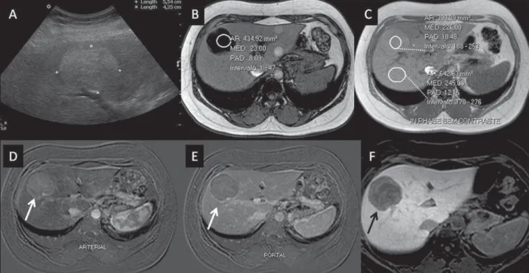
por-tal tracts(10,32). At images with Gd-EOB-DTPA, adenomas
typically present contrast uptake in up to 100% of the cases, but lower than the uptake by the parenchyma in the hepatobiliary phase, due to the absent or quite reduced hepa-tocellular uptake of gadoxetic acid by the lesion (Figure 2). On the other hand, in up to 10% of cases, a similar or greater uptake can occur in relation to the parenchyma in the
hepatobiliary phase(33), or peripheral ring-shaped
enhance-ment, since some hepatocytes maintain the capacity of ab-sorption and excretion in the hepatocellular phase. This may represent a confusing factor in the diagnosis of adenoma,
making it similar to FNH(10,23,33).
The presence of fat in the adenoma allows an easy
dif-ferentiation from FNH, as the latter rarely contains fat(34).
Recently, however, three subtypes of adenomas were described on the basis of histological differences, as follows: steatotic, inflammatory and activated beta-catenin. Adenomas of the inflammatory subtype correspond to approximately 40% of
such tumors and present a low fat content(35). At nonspecific
contrast-enhanced MRI, adenomas present marked arterial enhancement keeping an iso- or subtle hypersignal in rela-tion to the parenchyma at the delayed phases, with difficult differentiation from FNHs. In such cases of diagnostic doubt between FNH and adenoma, gadoxetic acid may be
particu-larly useful(36). On the other hand, adenomas of activated
beta-catenin subtype may present signal hyperintensity in the
hepatobiliary phase, after the hepatospecific contrast injec-tion, remaining as a source of diagnostic doubt.
Hepatic nodules in cirrhotic patients
HCC is the most common malignant primary hepatic neoplasm, generally occurring as a complication of hepatic
cirrhosis, particularly that caused by B and C viruses(10,32).
HCC is the main cause of deaths in cirrhotic patients, and for this reason an early and accurate diagnosis is very im-portant for an appropriate treatment and management of such
patients(29). Nodular lesions in a cirrhotic liver can be divided
into two major groups: a) regenerative and dysplastic
nod-ules; b) neoplastic nodules(32). However, the correct
imag-ing characterization of these lesions still remains a challenge, as frequently pre-neoplastic hepatocellular lesions as well as
dysplastic nodules mimic small HCCs well-differentiated(37).
The diagnosis of non-invasive HCC has been made ac-cording to the “Barcelona Criteria” which adopt imaging methods, particularly CT and MRI with intravenous contrast agent, to characterize as HCC a focal lesion in a cirrhotic
liver; HCCs are those nodules ≥ 2.0 cm in diameter,
hypervascular in the arterial phase (wash-in) and wash-out in delayed phases, at contrast-enhanced axial images (either
CT or MRI)(38). In cases where a liver lesion is between 1.0
and 2.0 cm in diameter, the guidelines by American Asso-ciation for Studies of Liver Diseases recommend that the diagnosis of HCC be based on two dynamic imaging
stud-ies with typical findings(39). However, such typical imaging
findings are not frequently observed in cases of small HCCs (< 2.0 cm), particularly in cases of well-differentiated early HCCs, requiring liver biopsy for diagnosis and close fol-low-up(29).
In cirrhotic patients, the gadoxetic acid may be useful in the identification of small HCCs and in the correct char-acterization of lesions presenting atypical behavior at T1- and T2-weighted images, with a nonspecific enhancement pat-tern(4,29). In the hepatobiliary phase after gadoxetic acid in-jection, regenerative nodules show up iso- or slightly hyperin-tense in relation to the surrounding liver parenchyma, as they are composed of functioning hepatocytes, and are many times identified by the presence of a thin pseudocapsule with
hyposignal resulting from the surrounding fibrous matrix(37).
On the contrary, HCCs present hyposignal in relation to the adjacent liver parenchyma in the hepatobiliary phase, par-ticularly in cases of moderately or poorly differentiated
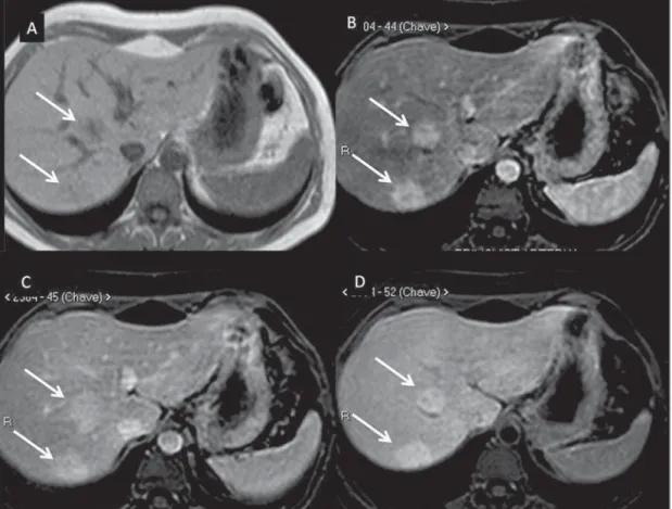
le-sions(29) (Figure 3). Dysplastic nodules represent a common
finding in the cirrhotic liver, and may be classified into low-and high-grade dysplastic nodules, on the basis of the num-ber and type of cellular atypias. High-grade dysplastic nod-ules are considered as being pre-malignant lesions. Even with the utilization of gadoxetic acid, the differentiation between dysplastic nodule and well-differentiated HCC still repre-sents a diagnostic challenge, as in some cases neoplastic cells of well-differentiated HCC may present with preserved hepa-tocellular function, being capable of absorption and metaboli-zation of the contrast medium and, so the malignant nodule can be seen either as iso- or even hyperintense in the
hepato-biliary phase, simulating a regenerative or a dysplastic nod-ule(29,37). On the other hand, some dysplastic nodules are
seen as hypointense in the hepatobiliary phase, mimicking a
HCC(29) (Figure 4).
Recent studies have demonstrated that the Gd-EOB-DTPA uptake by some HCCs is related to the expression of transporting proteins – OATP1B3 – in the hepatocytes
mem-brane of these lesions(40). However, further studies are
nec-essary to confirm such a theory.
Although multidetector CT (MDCT) has achieved a high standard in the detection of HCC, due to the possibil-ity of multiphase scans and a set of high resolution data, MRI is considered the best noninvasive imaging method for de-tecting HCC and for characterizing nodules in cirrhotic pa-tients, because the multiple evaluated parameters, and espe-cially due to the possibility of utilization of hepatospecific
contrast agents(41). Several studies have demonstrated an
increase in the rate of detection of HCC by MRI with gadoxetic acid, as compared with MDCT, especially in cases
of lesions < 1.5 cm(42–44). In 2009, Kim et al. demonstrated
greater accuracy in the diagnosis of HCC by MRI with Gd-EOB-DTPA, with 91.45% sensitivity in the gadoxetic acid group, versus 71.6% in the MDCT group, with a 24.7% higher percentage in the detection of small HCCs (< 1.5 cm). Other investigators have demonstrated that the combi-nation of dynamic study with the hepatocyte phase at gadoxetic acid-enhanced MRI had a better diagnostic per-formance than the dynamic study alone in the characteriza-tion of focal lesions in cirrhotic livers(45,46).
Liver metastasis
Another use for gadoxetic acid contrast media in the study of the liver is the detection of liver metastases, par-ticularly in the follow-up of patients with colorectal carci-noma(4,47). Hepatic metastasis is the most frequent
malig-nant liver lesion. The correct diagnosis is fundamental for the definition of the therapeutic approach as well as to es-tablish the prognosis, hence the relevance of the differentia-tion between such lesions and other benign liver nodules in
cancer patients(32). In the gadoxetic acid-enhanced
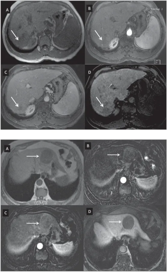
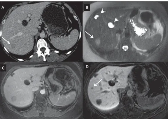
hepato-Figure 3. Infiltrative and undifferentiated HCC (confirmed by percutaneous biopsy). At T1-weighted images before intravenous gadoxetic acid injection (A) and obtained in the arterial and portal contrast phases (B,C), the lesion is poorly defined as com-pared with the image obtained in the hepatobiliary phase (arrows), acquired 20 minutes after the utilization of the contrast medium (D). Notice that, in this phase, contrast uptake by the lesion is lower in relation to the hepatic parenchyma, indicat-ing the absence of normal functional hepa-tocytes.
biliary phase, both hypovascular and hypervascular liver metastases are hypointense in relation to the adjacent paren-chyma due to the absence of functional hepatocytes in such
lesions(30). In that phase, the lesion washout in association
with the enhancement of the surrounding healthy parenchyma improves the liver-tumor contrast, increasing the conspicuity of the lesion. This allows for a significantly higher rate of lesions detection, especially for those < 1.0 cm in diameter, a fact that may impact the therapeutic planning as well as
the surgical approach(15,19,30) (Figure 5). In a prospective
study, Hammerstingl et al.(13) have demonstrated that
gadoxetic acid-enhanced MRI was superior as compared with CT in the evaluation of FLL, considering detection, local-ization, delimitation and management of patients, leading to changes in the therapeutic approach in 14.5% of the pa-tients, allowing for better preoperative planning in cases of liver resection. Another study has also demonstrated a higher rate of detection of liver metastases with the utilization of gadoxetic acid-enhanced MRI, both for lesions smaller and
larger than 1.0 cm(19).
Other liver lesions
For the remaining liver nodules such as the hemangioma, cholangiocarcinoma and other benign lesions (for example: abscesses and hydatid cysts), gadoxetic acid does not seem to be a precise indication, considering that in such cases there is no enhancement of the lesions in the hepatobiliary phase, as such nodules do not have functional hepatocytes. An ex-ception would be benign lesions of biliary ducts, such as Caroli disease, where enhancement of cystic lesions in the hepatobiliary phase is observed because of their
communi-cation with the biliary tree(48).
The enhancement of hemangiomas at Gd-EOB-DTPA-enhanced MRI presents some particularities. The lesion tends to follow the signal from the blood in the abdominal vessels in the extracellular phase. On the other hand, contrary to what occurs as the extracellular contrast medium is utilized, in the hepatobiliary phase, the hemangioma does not present progressive or persistent, and higher or equal enhancement in relation to the liver parenchyma, but rather hyposignal in relation to the liver, because of the absence of hepatocytes, in contrast with the normal adjacent parenchyma, which presents intense Gd-EOB-DTPA uptake in this phase. Such a phenomenon is called liver/lesion enhancement gradient
inversion(4,10,14) (Figure 6).
CONCLUSION
The present study was aimed at reviewing the MRI evalu-ation of liver nodules with the utilizevalu-ation of hepatospecific contrast medium, considering the recent availability of such contrast agent in the market. In their initial experiment,
substantiated by the literature review(1–3,11,13), the authors
could identify different enhancement patterns in lesions stud-ied with gadoxetic acid as a function of their etiology, di-vided into two groups as follows: a group including lesions with functioning hepatocytes, such as FNH and dysplastic nodules, and another group comprising all the remaining liver lesions which do not contain functioning hepatocytes (for example: HCC, adenomas and metastasis). The authors have observed that the utilization of ethoxybenzyl is more useful in the differentiation between FNH and adenoma, in the differentiation between dysplastic nodules and HCC in cirrhotic liver, in the detection of small HCCs (< 2.0 cm diameter) and tiny metastasis, where the hepatospecific
con-Figure 5. Liver metastasis from breast neoplasm (diagnosis based on periodic follow-up). The CT images acquired in the portal contrast phase (A) and the MRI T2-weighted sequence (B) allow for the identification of a single liver nodule in the VII/VIII segment (arrows on A and
trast agents demonstrate greater sensitivity and specificity than extracellular agents.
For such reasons, MRI with the utilization of ethoxy-benzyl is currently considered as the best imaging modality
for the investigation of FLLs(2); but, because of its higher
cost, such type of contrast agent should not be utilized on a routine basis, remaining reserved for selected cases where its usefulness has been proven, as in the case of the above mentioned indications.
REFERENCES
1. Ba-Ssalamah A, Uffmann M, Saini S, et al. Clinical value of MRI liver-specific contrast agents: a tailored examination for a confi-dent non-invasive diagnosis of focal liver lesions. Eur Radiol. 2009; 19:342–57.
2. Holzapfel K, Eiber MJ, Fingerle AA, et al. Detection, classification, and characterization of focal liver lesions: value of diffusion-weighted MR imaging, gadoxetic acid-enhanced MR imaging and the combi-nation of both methods. Abdom Imaging. 2012;37:74–82. 3. Bartolozzi C. MR of the liver: from breakthrough to clinical
appli-cation. Abdom Imaging. 2012;37:154.
4. Tanimoto A, Lee JM, Murakami T, et al. Consensus report of the 2nd International Forum for Liver MRI. Eur Radiol. 2009;19 Suppl 5:S975–89.
5. Bellin MF, Webb JA, Van Der Molen AJ, et al. Safety of MR liver specific contrast media. Eur Radiol. 2005;15:1607–14.
6. Bellin MF, Vasile M, Morel-Precetti S. Currently used non-specific extracellular MR contrast media. Eur Radiol. 2003;13:2688–98. 7. Reimer P, Schneider G, Schima W. Hepatobiliary contrast agents
for contrast-enhanced MRI of the liver: properties, clinical devel-opment and applications. Eur Radiol. 2004;14:559–78.
8. Elias Jr J, Santos AC, Koenigkam-Santos M, et al. Complications from the use of intravenous gadolinium-based contrast agents for magnetic resonance imaging. Radiol Bras. 2008;41:263–7.
9. Semelka RC, Helmberger TK. Contrast agents for MR imaging of the liver. Radiology. 2001;218:27–38.
10. Campos JT, Sirlin CB, Choi JY. Focal hepatic lesions in Gd-EOB-DTPA enhanced MRI: the atlas. Insights Imaging. 2012;3:451– 74.
11. Van Beers BE, Pastor CM, Hussain HK. Primovist, Eovist: what to expect? J Hepatol. 2012;57:421–9.
12. Seale MK, Catalano OA, Saini S, et al. Hepatobiliary-specific MR contrast agents: role in imaging the liver and biliary tree. Radio-graphics. 2009;29:1725–48.
13. Hammerstingl R, Huppertz A, Breuer J, et al. Diagnostic efficacy of gadoxetic acid (Primovist)-enhanced MRI and spiral CT for a thera-peutic strategy: comparison with intraoperative and histopathologic findings in focal liver lesions. Eur Radiol. 2008;18:457–67. 14. Reimer P, Rummeny EJ, Daldrup HE, et al. Enhancement
charac-teristics of liver metastases, hepatocellular carcinomas, and heman-giomas with Gd-EOB-DTPA: preliminary results with dynamic MR imaging. Eur Radiol. 1997;7:275–80.
15. Huppertz A, Balzer T, Blakeborough A, et al. Improved detection of focal liver lesions at MR imaging: multicenter comparison of gadoxetic acid-enhanced MR images with intraoperative findings. Radiology. 2004;230:266–75.
16. Primovist. Bula. Berlim: Bayer Pharma AG; 2011.
17. Tanimoto A, Kadoya M, Kawamura Y, et al. Safety and efficacy of a novel hepatobiliary MR contrast agent, Gd-DTPA-DeA: results of phase I and phase II clinical trials. J Magn Reson Imaging. 2006;23:499–508.
18. Tamada T, Ito K, Sone T, et al. Dynamic contrast-enhanced mag-netic resonance imaging of abdominal solid organ and major vessel: comparison of enhancement effect between Gd-EOB-DTPA and Gd-DTPA. J Magn Reson Imaging. 2009;29:636–40.
19. Bluemke DA, Sahani D, Amendola M, et al. Efficacy and safety of MR imaging with liver-specific contrast agent: U.S. multicenter phase III study. Radiology. 2005;237:89–98.
20. Zech CJ, Vos B, Nordell A, et al. Vascular enhancement in early
dynamic liver MR imaging in an animal model: comparison of two injection regimen and two different doses Gd-EOB-DTPA (gadoxetic acid) with standard Gd-DTPA. Invest Radiol. 2009;44:305–10. 21. Motosugi U, Ichikawa T, Tominaga L, et al. Delay before the
hepa-tocyte phase of Gd-EOB-DTPA-enhanced MR imaging: is it pos-sible to shorten the examination time? Eur Radiol. 2009;19:2623– 9.
22. Choi JS, Kim MJ, Choi JY, et al. Diffusion-weighted MR imaging of liver on 3.0-Tesla system: effect of intravenous administration of gadoxetic acid disodium. Eur Radiol. 2010;20:1052–60. 23. Huppertz A, Haraida S, Kraus A, et al. Enhancement of focal liver
lesions at gadoxetic acid-enhanced MR imaging: correlation with histopathologic findings and spiral CT – initial observations. Radi-ology. 2005;234:468–78.
24. Zech CJ, Grazioli L, Breuer J, et al. Diagnostic performance and description of morphological features of focal nodular hyperplasia in Gd-EOB-DTPA-enhanced liver magnetic resonance imaging: results of a multicenter trial. Invest Radiol. 2008;43:504–11. 25. Purysko AS, Remer EM, Veniero JC. Focal liver lesion detection
and characterization with GD-EOB-DTPA. Clin Radiol. 2011;66: 673–84.
26. Sano K, Ichikawa T, Motosugi U, et al. Imaging study of early hepa-tocellular carcinoma: usefulness of gadoxetic acid-enhanced MR imaging. Radiology. 2011;261:834–44.
27. Sun HY, Lee JM, Shin CI, et al. Gadoxetic acid-enhanced magnetic resonance imaging for differentiating small hepatocellular carcino-mas (< or =2 cm in diameter) from arterial enhancing pseudolesions: special emphasis on hepatobiliary phase imaging. Invest Radiol. 2010;45:96–103.
28. Lee MH, Kim SH, Park MJ, et al. Gadoxetic acid-enhanced hepatobiliary phase MRI and high-b-value diffusion-weighted im-aging to distinguish well-differentiated hepatocellular carcinomas from benign nodules in patients with chronic liver disease. AJR Am J Roentgenol. 2011;197:W868–75.
29. Kogita S, Imai Y, Okada M, et al. Gd-EOB-DTPA-enhanced mag-netic resonance images of hepatocellular carcinoma: correlation with histological grading and portal blood flow. Eur Radiol. 2010;20: 2405–13.
30. Zech CJ, Herrmann KA, Reiser MF, et al. MR imaging in patients with suspected liver metastases: value of liver-specific contrast agent Gd-EOB-DTPA. Magn Reson Med Sci. 2007;6:43–52.
31. van Kessel CS, de Boer E, ten Kate FJ, et al. Focal nodular hyper-plasia: hepatobiliary enhancement patterns on gadoxetic-acid con-trast-enhanced MRI. Abdom Imaging. 2013;38:490–501. 32. Tiferes DA, D’Ippolito G. Neoplasias hepáticas: caracterização por
métodos de imagem. Radiol Bras. 2008;41:119–27.
33. Denecke T, Steffen IG, Agarwal S, et al. Appearance of hepatocel-lular adenomas on gadoxetic acid-enhanced MRI. Eur Radiol. 2012;22:1769–75.
34. Ferlicot S, Kobeiter H, Tran Van Nhieu J, et al. MRI of atypical
focal nodular hyperplasia of the liver: radiology-pathology correla-tion. AJR Am J Roentgenol. 2004;182:1227–31.
35. Bioulac-Sage P, Laumonier H, Couchy G, et al. Hepatocellular adenoma management and phenotypic classification: the Bordeaux experience. Hepatology. 2009;50:481–9.
36. Grazioli L, Bondioni MP, Haradome H, et al. Hepatocellular ad-enoma and focal nodular hyperplasia: value of gadoxetic acid-en-hanced MR imaging in differential diagnosis. Radiology. 2012;262: 520–9.
37. Bartolozzi C, Battaglia V, Bozzi E. Hepatocellular nodules in liver cirrhosis: contrast-enhanced MR. Abdom Imaging. 2011;36:290–9. 38. Bruix J, Sherman M, Llovet JM, et al. Clinical management of hepa-tocellular carcinoma. Conclusions of the Barcelona-2000 EASL conference. European Association for the Study of the Liver. J Hepatol. 2001;35:421–30.
39. Bruix J, Sherman M; Practice Guidelines Committee, American Association for the Study of Liver Diseases. Management of hepa-tocellular carcinoma. Hepatology. 2005;42:1208–36.
40. Narita M, Hatano E, Arizono S, et al. Expression of OATP1B3 determines uptake of Gd-EOB-DTPA in hepatocellular carcinoma. J Gastroenterol. 2009;44:793–8.
41. Zech CJ, Reiser MF, Herrmann KA. Imaging of hepatocellular car-cinoma by computed tomography and magnetic resonance imag-ing: state of the art. Dig Dis. 2009;27:114–24.
42. Kim YK, Kim CS, Han YM, et al. Detection of hepatocellular car-cinoma: gadoxetic acid-enhanced 3-dimensional magnetic resonance imaging versus multi-detector row computed tomography. J Comput Assist Tomogr. 2009;33:844–50.
43. Di Martino M, Marin D, Guerrisi A, et al. Intraindividual compari-son of gadoxetate disodium-enhanced MR imaging and 64-section multidetector CT in the detection of hepatocellular carcinoma in patients with cirrhosis. Radiology. 2010;256:806–16.
44. Kim SH, Lee J, Kim MJ, et al. Gadoxetic acid-enhanced MRI versus triple-phase MDCT for the preoperative detection of hepatocellu-lar carcinoma. AJR Am J Roentgenol. 2009;192:1675–81. 45. Chou CT, Chen YL, Su WW, et al. Characterization of cirrhotic
nodules with gadoxetic acid-enhanced magnetic resonance imag-ing: the efficacy of hepatocyte-phase imaging. J Magn Reson Imag-ing. 2010;32:895–902.
46. Ahn SS, Kim MJ, Lim JS, et al. Added value of gadoxetic acid-en-hanced hepatobiliary phase MR imaging in the diagnosis of hepato-cellular carcinoma. Radiology. 2010;255:459–66.
47. Chan VO, Das JP, Gerstenmaier JF, et al. Diagnostic performance of MDCT, PET/CT and gadoxetic acid (Primovist(®))-enhanced MRI in patients with colorectal liver metastases being considered for hepatic resection: initial experience in a single centre. Ir J Med Sci. 2012;181:499–509.