Endovascular treatment of scalp cirsoid aneurysms*
Tratamento endovascular de aneurismas cirsoideos do couro cabeludo
Eduardo Wajnberg1
OBJECTIVE: To report results of the application of endovascular techniques in the management of scalp cirsoid aneurysms. MATERIALS AND METHODS: Four patients diagnosed with cirsoid aneurysms were submitted to treatment by endovascular approach. All the four patients included in the present series had arteriovenous malformations and were treated solely by embolization. RESULTS: Three of the patients underwent endovascular treatment by transarterial embolization and one was treated by direct puncture of the venous segment. Both clinical and cosmetic outcomes were satisfactory in all of the patients. Clinical relapse was not observed along the follow-up period. CONCLUSION: The endovascular approach is safe and effective in the management of crisoid aneurysms. Although this technique can be used as an adjuvant or complement to surgery, particularly in cases where deep afferents are involved, complete resolution can be achieved only with endovascular treatment. The choice of treatment method should be based on a range of typical characteristics of the lesion, including angioarchitecture, size and clinical presentation.
Keywords: Arteriovenous fistula; Cirsoid aneurysm; Cerebral angiography; Embolization.
OBJETIVO: Relatar os resultados da aplicação de técnicas endovasculares no tratamento de aneurismas cirsoideos do couro cabeludo. MATERIAIS E MÉTODOS: Quatro pacientes com diagnóstico de aneurismas cirsoideos foram submetidos ao tratamento por via endovascular. Todos os quatro pacientes incluídos nesta série tinham malformações arteriovenosas e foram tratados apenas com embolização. RESULTADOS: Três pacientes foram submetidos a tratamento endovascular mediante embolização transarterial e um foi tratado por punção direta da porção venosa. Os resultados clínicos e cosméticos foram satisfatórios em todos os pacientes. Não houve recidiva clínica durante o período de acompanhamento. CONCLUSÃO: A via endovas-cular é uma alternativa segura e eficaz no tratamento dos aneurismas cirsoideos. Embora possa ser efetiva-mente utilizado como uma alternativa adjuvante ou complementar à cirurgia, especialefetiva-mente quando é neces-sário lidar com aferências profundas, a maioria dos casos pode ser totalmente curada apenas com a terapêu-tica endovascular. A escolha do método de tratamento deve ser baseada em uma variedade de característi-cas próprias da lesão, incluindo sua angioarquitetura, tamanho e apresentação clínica.
Unitermos: Fístula arteriovenosa; Aneurisma cirsoideo; Angiografia cerebral; Embolização. Abstract
Resumo
* Study developed at Department of Radiology – Hospital Universitário Clementino Fraga Filho, Universidade Federal do Rio de Janeiro (HUCFF-UFRJ), Rio de Janeiro, RJ, Brazil.
1. Master, MD, Radiologist at Department of Radiology – Hos-pital Universitário Clementino Fraga Filho, Universidade Federal do Rio de Janeiro (HUCFF-UFRJ), Rio de Janeiro, RJ, Brazil.
Mailing address: Dr. Eduardo Wajnberg. Rua Lopes Quintas, 100/602, Jardim Botânico. Rio de Janeiro, RJ, Brazil, 22460-010. E-mail: [email protected]
Received April 26, 2010. Accepted after revision June 18, 2010.
As a result, angiography may demonstrate different findings such as the presence of a nidus or even an associated hemangioma in congenital cases(1,4–8).
Considering that each lesion presents a range of typical angioarchitectural charac-teristics, the treatment should be tailored to each patient’s circumstance. In some cases of major malformations with high-output fistulas, a combined endovascular/surgical approach and reconstruction of the scalp may be the best treatment method(8).
The author reports a series of four pa-tients with arteriovenous malformations of the scalp treated by endovascular approach. All the patients shared a common previous history of blunt head trauma progressing with the development of a pulsatile mass. All of them were treated with embolization either by transarterial approach or by direct
Wajnberg E. Endovascular treatment of scalp cirsoid aneurysms. Radiol Bras. 2010;43(4):224–228.
life. Sixty percent of affected individuals are male(1).
The location of scalp cirsoid aneurysms is more or less evenly distributed among the frontal, temporal and parietal regions(1).
The main complaint of the majority of pa-tients with such vascular lesions is the pres-ence of a pulsatile mass causing scalp de-formity. In some cases, bruit or focal head-ache is reported. At clinical examination, most patients present a noticeable pulsatile tumor-like lesion surrounded by dilated vessels and an audible bruit at auscultation. However, the primary abnormality is a fis-tula that is clinically and angiographically evidenced by the secondary dilation of its draining veins. Arteriovenous fistulas originate in the embryonic vascular devel-opment and vary according to the phase where an abnormal growth takes place(2,3). INTRODUCTION
puncture of the lesion with the utilization of liquid-type adhesives. No recurrence was observed during the follow-up period.
MATERIALS AND METHODS
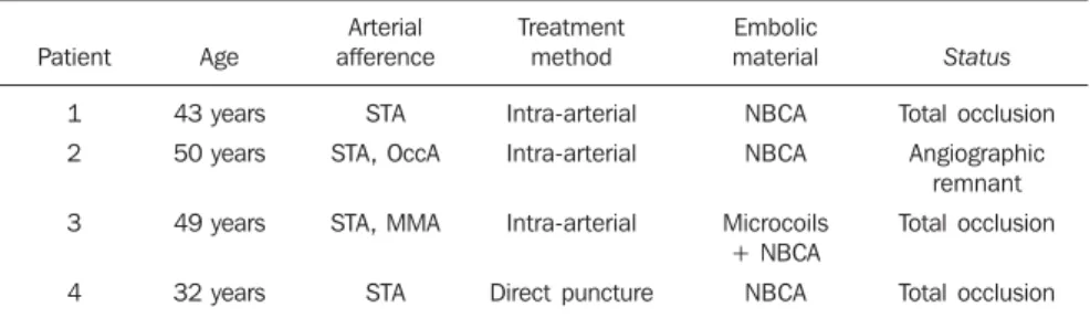
Four patients with scalp cirsoid aneu-rysms were assisted by the group of Interventional Radiology at the Hospital Universitário Clementino Fraga Filho from the Federal University of Rio de Janeiro, RJ, Brazil, in the period from August/2004 to January/2009. The clinical findings and treatment methods are summarized on Table 1. All four patients presented a pre-vious history of blunt cranial trauma and slow development of a pulsatile mass on the scalp. All the lesions were diagnosed with basis on their clinical characteristics and confirmed by angiography. Angio-graphically, all the patients presented cir-soid aneurysms consisting in direct, high-output arteriovenous fistulas, without the presence of any plexiform component or nidus. The patients were submitted to com-plete cerebral angiography with bilateral, selective injections into the internal and external carotid arteries to demonstrate the size and location of the afferent arteries and draining veins. All of the lesions were sup-plied by combinations of the superficial temporal, occipital and middle meningeal arteries.
Embolization techniques
Direct puncture embolization – The procedure was performed under sedation and local anesthesia. Following selective angiography, the lesions were punctured with a 21G Teflon needle at the level of the variceal dilatation proximal to the fistula. Angiography was performed with and without manual compression of the venous drainage in the region. In order to achieve a decrease in the circumferential flow around the lesion, a sterile, round metal ring was maintained in place on the dilated collector vein. Technically, direct venogra-phy should not demonstrate distal leakage of contrast medium through the metal ring borders indicating an appropriate venous compression. As a higher pressure is ap-plied on the metal ring, the arterial flow can also be reduced, so that a complete flow reduction is achieved in the desired region.
With the ring in place, N-butyl-cyanoacry-late (NBCA) mixed with lipiodol was in-jected into the collector vein. Then, the ring remains in place for some minutes and later is gradually removed. The NBCA concen-tration mixed with iodized oil was adjusted in accordance with the flow observed at angiography. Repeated punctures and in-jections were performed as a residual le-sion was observed on a postembolization angiogram.
Transarterial embolization – Transar-terial embolization was performed with the standard microcatheterization technique, by femoral puncture and placement of a 6Fr system with a guide catheter in the exter-nal carotid artery. An Ultraflow 1.5Fr microcatheter (MTI Microtherapeutics; Irvine, CA, USA) was utilized for cy-anoacrylate injection, and the embolization was performed with NBCA mixed with lipiodol at a concentration compatible with the flow velocity in the fistula.
RESULTS
All of the four patients included in the present series had malformations with high-output fistulas and were treated solely with embolization. In none of the reported cases, the presence of nidus in the malfor-mations was observed, and in all of the cases the visualized angioarchitecture pre-sented large arterial afferences in high-out-put fistulas towards large caliber varicose veins. One patient presented intracranial drainage through diploic veins towards the superficial middle cerebral vein and petro-sal sinuses.
Three patients were submitted to endovascular treatment by transarterial embolization and only one was treated by means of direct puncture of the venous
segment. The selection of the method was based on the presence of deep afferent ves-sels to the fistula which were more easily managed by transarterial approach or, in cases where the venous dilatation was not so large, making direct puncture more dif-ficult to be performed. For each patient in the present series, the elimination of the fistula was confirmed by angiography at the treatment conclusion and no recurrence was clinically recognized. All the patients presented satisfactory clinical and cosmetic outcomes and were clinically followed-up (follow-up period ranging between six and 32 months; mean, 20 months) to detect re-currence manifest as increase in volume or audible bruit. Follow-up angiography was not considered until a clinical suspicion of recurrence was raised, therefore none of the patients required such procedure.
Cases description
Patient 1 – A 43-year-old woman pre-sented with a 5 × 6 cm pulsatile mass on the right frontal region following a trauma in a car accident. Bilateral angiography of external carotid arteries demonstrated the large caliber of the superficial temporal artery and respective branches, with a di-rect fistula with the superficial temporal vein and superficial frontal veins. The fis-tula was easily demonstrated at angiogra-phy (Figure 1A). The treatment was per-formed by means of superselective microcatheterization of the superficial tem-poral artery with a flow-guided catheter and embolization with 1.5 ml NBCA at 33%. Total occlusion of the fistula and drainage vein was achieved (Figure 1B).
Patient 3 – A 49-year-old man reported the presence of a mass on the preauricular region in association with pulsatile tinni-tus, observed six months after a blunt
in-Table 1 Treatment demographics, type and outcomes.
Patient
1
2
3
4
Age
43 years
50 years
49 years
32 years
Arterial afference
STA
STA, OccA
STA, MMA
STA
Treatment method
Intra-arterial
Intra-arterial
Intra-arterial
Direct puncture
Embolic material
NBCA
NBCA
Microcoils + NBCA
NBCA
Status
Total occlusion
Angiographic remnant
Total occlusion
Total occlusion
onstrated a large fistula (Figure 2D) that was occluded with microcoils and NBCA. The fistula and respective drainage vein were completely occluded during embo-lization by arterial approach. The patient reported improvement of the tinnitus.
DISCUSSION
The management of scalp cirsoid aneu-rysms is complex. Therapeutic success is achieved with complete elimination of the abnormal arteriovenous communication with occlusion of all the drainage veins, otherwise recurrence is inevitable. Addi-tionally, the treatment must be focused on the improvement of the cosmetic disfigure-ment that in most of cases is the patients’ main preoccupation.
Only 10% to 20% of these arterio-venous fistulas develop after penetrating cranial trauma. In 90% of the patients, the superficial temporal artery is the main af-ferent vessel to the fistula(9). In the
remain-ing cases both the superficial temporal and occipital arteries are generally involved(9). Figure 1. A: Pretreatment digital angiography of external carotid artery demonstrating arteriovenous
fis-tula with draining vein dilatation, supplied by branches of the superficial temporal artery and, at a lower degree, by the left occipital artery. B: Digital angiography of external carotid artery following treatment with NBCA, demonstrating total occlusion of arteriovenous fistula and respective draining vein.
A B
Figure 2. A: Magnetic resonance angiography with de TOF tech-nique demonstrating marked su-perficial veins ectasia. B: Mag-netic resonance angiography demonstrating dilated extracra-nial veins, without any detectable alteration in the intracranial circu-lation. C: Anteroposterior digital angiography of external carotid artery, at the venous phase, dem-onstrating the intracranial drain-age of the fistula bilaterally through the cavernous and pet-rosal sinuses. D: Anteroposterior digital angiography of the external carotid artery, at the arterial phase, demonstrating arterio-venous fistula of the superficial temporal artery, with develop-ment of a large venous aneurysm in the preauricular region. B
A
C D
jury in the region preceding the symptoms onset. The clinical examination demon-strated a pulsatile mass on the left preau-ricular region. Magnetic resonance angiog-raphy demonstrated the presence of a sig-nificant venous engorgement (Figures 2A and 2B) and a nodular lesion with absence of signal in this region. Angiography of the
dem-The management of scalp cirsoid aneu-rysms is complex, particularly because of their high flow. Surgery has been the treat-ment of choice for a long time(2,7,10).
Liga-tion of supply arteries is generally ineffec-tive and should be avoided(2,6,11). With the
developments in materials and endovas-cular techniques(5,12–14), a decrease in
prob-lems with the treatment of these patients has been observed. Previous studies report that embolization can completely occlude large arteriovenous fistulas, determining a significant clinical and even esthetic im-provement in many patients(12–14), although
a standard treatment for these rare lesions is still to be established.
Generally, embolization performed as an adjuvant of surgical therapeutics is suc-cessful in the reduction of blood loss dur-ing the surgical excision of the lesion(5,13).
It is known that embolization can heal an arteriovenous fistula of the scalp without determining its ischemia(14).
Considering that these disfiguring masses are constituted mainly of large draining veins, these veins almost com-pletely disappear after occlusion of the fis-tulous site and of the origin of these venous dilatations. In the present series, the lesions were managed solely by embolization. None of the cases required combined therapy. In the cases treated with direct puncture, NBCA injection was performed under mechanical compression of the draining veins for determining reduction in the blood flow (or even stasis) and to avoid the undesired escape of the liquid embolic material into the jugular vein or even into the pulmonary circulation.
Embolization should be the primary therapy for this type of fistula. Surgical intervention should be reserved either for removal of residual fistulas that could not be occluded with embolization or for cases of more extensive fistulas. Presurgical embolization of arterial afferences is desir-able because it makes the surgical proce-dure safer, reducing the risk for massive hemorrhage. In fact, proximal embolization does reduce the regional blood flow and has been a useful complement to surgery, but is rarely curative since the vascular endothelial growth factor is expressed by these lesions and will be responsible for their continuous growth(15). The selection
of embolic materials must be based on the vascular architecture of the lesion as well as on the skill of the interventional radiolo-gist. A combination of different embolic materials or even of several pathways to approach the nidus may be required(5,13).
Although the etiology of these lesions still remains controversial, the hypothesis of either a congenital or traumatic origin has been generally accepted. In the present series, all the lesions could be directly re-lated to trauma (most of them rere-lated to blunt trauma). Penetrating trauma is well described as cause for these lesions, includ-ing after hair transplant, temporomandibu-lar joint arthroscopy and craniotomy for intracranial procedures(16–18). Congenital
cirsoid aneurysms are actually more fre-quent than the traumatic ones, achieving an incidence of up to 80% in some series(18).
Theories about their occurrence include persistence of arteriovenous fistulas. Al-though extremely rare, familial involve-ment has been described(19).
The diagnosis is clinical in the major-ity of cases. Angiography is required for a better delineation of the lesion and to rule out the presence of an intracranial compo-nent(20). Such component may be consisted
of an enlarged meningeal artery with affer-ent vessels crossing the diploe to feed the malformation. The diagnosis of scalp cir-soid aneurysm as well as its differentiation with sinus pericranii may be difficult and some confusion between these two types of lesion is present in the literature. In a strict sense, sinus pericranii corresponds to a collection of venous blood vessels firmly attached to the outer table of the skull di-rectly communicating with the intracranial venous sinus by diploic veins through a bone defect(21,22).
Indications for treatment include hem-orrhage prevention, tinnitus alleviation or a noticeable presence of a cosmetic defect caused by the pulsatile mass. In the past, the treatment was essentially dependent on surgical excision or proximal ligation of supply arteries(22–24). Ligation of supply
arteries is particularly problematic since this is not a curative technique besides causing loss of the arterial access to the fistula for embolization(25).
Several forms of treatment for scalp cir-soid aneurysms including absolute alcohol
embolization and microcoils have already been reported(13,26). Direct puncture
embo-lization is aimed at occluding the beginning of the venous structure distal to the arterio-venous communication. Thus, with this technique, the occlusion of vascular struc-tures does not involve any risk for ischemic skin complications. Immediate venous oc-clusion allows continuous embolic agent redistribution to the adjacent vascular spaces and an effective devascularization. For a temporary occlusion of the venous flow, the authors utilized a large ring-shaped compression device (the so called cookie cutter technique), described by Duncan et al.(27), avoiding the unnecessary
exposure of the operator’s hand to radia-tion. In the present series, direct angiogra-phy with 21G Teflon needle was many times followed by adjustments of compres-sion points, making the venous occlucompres-sion more effective.
In most of lesions, a high-output arte-riovenous fistula is associated with the venous aneurysm. Preoccupation that the embolic agent may pass to the pulmonary circulation must be always kept in mind. In order to avoid such an event, temporary manual compression during NBCA injec-tion slows down the venous flow, prevent-ing inadvertent embolization. Another measure to be adopted in the transarterial approach is the utilization of a balloon-guiding catheter for management of the proximal blood flow, performing the injec-tion upon flow blockage.
Recent developments in the design of microcatheters and in distal navigation techniques have allowed catheterization of afferent arteries proximal to the fistula. NBCA injection through a microcatheter may lead to complete devascularization of a scalp arteriovenous malformation with-out any risk for ischemia of the healthy adjacent tissue.
CONCLUSIONS
achieved on with the endovascular ap-proach. The selection of the treatment method should be based on the lesion size, angioarchitecture, and clinical presenta-tion. In general, satisfactory aesthetic out-comes can be achieved either by transar-terial embolization or by direct puncture.
REFERENCES
1. Coleman CC Jr, Hoopes JE. Congenital arterio-venous anomalies of the head and neck. Plast Reconstr Surg. 1971;47:354–64.
2. Rappaport I, Yim D. Congenital arteriovenous fis-tulas of the head and neck. Arch Otolaryngol. 1973;97:350–3.
3. Szilagyi DE, Elliott JP, DeRusso FJ, et al. Periph-eral congenital arteriovenous fistulas. Surgery. 1965;57:61–81.
4. Fisher-Jeffes ND, Domingo Z, Madden M, et al. Arteriovenous malformations of the scalp. Neu-rosurgery. 1995;36:656–60.
5. Kaufman SL, Kumar AAJ, Roland JMA, et al. Transcatheter embolization in the management of congenital arteriovenous malformations. Radiol-ogy. 1980;137(1 Pt 1):21–9.
6. Khodadad G. Arteriovenous malformations of the scalp. Ann Surg. 1973;177:79–85.
7. Komatsu Y, Narushima K, Kobayashi E, et al. Congenital arteriovenous malformation of the scalp – case report. Neurol Med Chir (Tokyo). 1989;29:230–4.
8. Marotta TR, Berenstein A, Zide B. The combined role of embolization and tissue expanders in the management of arteriovenous malformations of the scalp. AJNR Am J Neuroradiol. 1994;15: 1240–6.
9. Gurkanlar D, Gonul M, Solmaz I, et al. Cirsoid aneurysms of the scalp. Neurosurg Rev. 2006;29: 208–12.
10. Schultz RC, Hermosillo CX. Congenital arterio-venous malformation of the face and scalp. Plast Reconstr Surg. 1980;65:496–501.
11. Ogawa Y, Inoue K. Electrothrombosis as a treat-ment of cirsoid angioma in the face and scalp and varicosis of the leg. Plast Reconstr Surg. 1982;70: 310–8.
12. Barnwell SL, Halbach VV, Dowd CF, et al. Endo-vascular treatment of scalp arteriovenous fistulas associated with a large varix. Radiology. 1989; 173:533–9.
13. Heilman CB, Kwan ES, Klucznik RP, et al. Elimi-nation of a cirsoid aneurysm of the scalp by di-rect percutaneous embolization with thrombo-genic coils. Case report. J Neurosurg. 1990;73: 296–300.
14. Kasdon DL, Altemus LR, Stein BM. Emboliza-tion of a traumatic arteriovenous fistula of the scalp with radiopaque Gelfoam pledgets. Case report and technical note. J Neurosurg. 1976;44: 753–6.
15. Waga S, Ohtsubo K, Handa J, et al. Extracranial congenital arterio-venous malformations. Surg Neurol. 1974;2:241–5.
16. Williams LR, Robinson JK, Yao JS. Hair trans-plantation producing arteriovenous fistulization. Ann Vasc Surg. 1986;1:241–3.
17. Preisler SA, Koorbusch GF, Olson RA. An ac-quired arteriovenous fistula secondary to tem-poromandibular joint arthroscopy: report of a case. J Oral Maxillofac Surg. 1991;49:187–90. 18. Morioka T, Nishio S, Hikita T. Traumatic
arterio-venous fistulae of the scalp at the area of previ-ous craniotomy. Surg Neurol. 1988;30:404–7.
19. Khodadad G. Familial cirsoid aneurysm of the scalp. J Neurol Neurosurg Psychiat. 1971;34: 664–7.
20. Verbiest H. Results of artificial slow flow angiog-raphy with arteriovenous aneurysms in the sup-ply area of the external or internal carotid arter-ies. Am J Roentgenol Radium Ther Nucl Med. 1972;116:1–15.
21. Lanzino G, Passacantilli E, Lemole GM Jr, et al. Scalp arteriovenous malformation draining into the superior sagittal sinus associated with an in-tracranial arteriovenous malformation: just a co-incidence? Case report. Neurosurgery. 2003;52: 440–3.
22. Vinas FC, Valenzuela S, Zuleta A. Literature re-view: sinus pericranii. Neurol Res. 1994;16:471– 4.
23. Nagasaka S, Fukushima T, Goto K, et al. Treat-ment of scalp arteriovenous malformation. Neu-rosurgery. 1996;38:671–7.
24. Shenoy SN, Raja A. Scalp arteriovenous malfor-mations. Neurol India. 2004;52:478–81. 25. Han MH, Seong SO, Kim HD, et al. Craniofacial
arteriovenous malformation: preoperative embo-lization with direct puncture and injection of n-butyl cyanoacrylate. Radiology. 1999;211:661–6. 26. Mourao GS, Hodes JE, Gobin YP, et al. Curative treatment of scalp arteriovenous fistulas by direct puncture and embolization with absolute alcohol. Report of three cases. J Neurosurg. 1991;75:634– 7.