Revista Brasileira de Anestesiologia 107 Vol. 60, No 1, Janeiro-Fevereiro, 2010
APPLICATIONS OF MAGNESIUM SULFATE IN OBSTETRICS AND ANESTHESIA
Applications of Magnesium Sulfate in
Obstetrics and Anesthesia
Fabiano Timbó Barbosa, TSA, M.D., Luciano Timbó Barbosa, M.D., Mário Jorge Jucá, M.D., Rafael Martins da Cunha, M.D.
INTRODUCTION
Magnesium is the second most abundant intracellular cation1,2 and the fourth when the extracellular medium is also consid-ered1. As a cofactor, it is involved in more than 300 known re-actions, such as3: hormone binding to receptors, flow of trans-membrane ions, regulation of the adenylate kinase system, muscle contraction, neuronal activity, vasomotor tone, cardiac excitability, release of neurotransmitters, and calcium binding to calcium channels.
In 1906, Haulbold and Meltzer reported sensorial and mo-tor blockades in humans after the intrathecal administration of magnesium; in 1950, magnesium was used in anesthesia, mainly to control seizures in gravidas. Currently, it has several applications in anesthesia, obstetrics, and intensive care3-5. The objective of this report was to review the physiology, phar-macology, and reduction in plasma levels of magnesium, as well as some of its applications in obstetric and anesthesia.
PHYSIOLOGY AND PHARMACOLOGIC EFFETCS
Magnesium is an intracellular cation with multiple functions: it par-ticipates in energy metabolism, since it is a cofactor in glucose metabolism, and a cofactor of nucleic acid, protein, and fatty acid degradation enzymes5,6; it regulates the flow of transmembrane ions5; and it mediates the activity of several enzymes5,7.
Magnesium is considered a natural physiologic calcium antago-nist, having several regulatory mechanisms, such as1-3,8-11: com-petitive antagonist affecting type L calcium channels, inhibition of the enzyme Ca2+-ATPase, and it is a cofactor for all enzymes that participate in phosphate transferences that use ATP. In high concentrations, it inhibits the enzyme Na+/K+ -ATPase5.
Magnesium is absorbed in the jejunum and ileum at a propor-tion of 11% to 65% of the ingested amount12. The kidney is the main regulator of the levels of magnesium in the body, being capable of eliminating almost 100% of the filtered magnesium in case of overload5.
Medicine has not elucidated completely the mechanism of ac-tion of magnesium sulfate used therapeutically13. Some of the following propositions have been developed over the years: a) Inhibition of the release of acetylcholine in the
neuromus-cular junction9,14 leading to muscle relaxation, which has been known since the 1950s3. Hypermagnesemia decre-ases the sensitivity of the motor plate to acetylcholine and the amplitude of terminal end-plate potential9.
Bin-108 Revista Brasileira de Anestesiologia Vol. 60, No 1, Janeiro-Fevereiro, 2010 BARBOSA, BARBOSA, JUCÁ ET AL.
ding of this receptor has analgesic, anticonvulsant, and sedative properties5.
c) It can increase the synthesis of prostacyclins and inhibit angiotensin converting enzyme, leading to vasodilation11.
d) It decreases the release of catecholamines after sympa-thetic stimulus1,5,9,11. It has been used in the treatment of
pheochromocytoma-related hypertensive episodes during surgeries or outside the surgical environment5.
e) In asthma patients, it inhibits the release of histamine and acetylcholine5,16, and it potentiates the effects of
beta-adrenergic agents5. It is indicated only in severe cases
because it decreases the rate of hospitalizations and the length of stay in the intensive care unit, but it has little be-neficial effects in moderate and mild cases16.
HYPOMAGNESEMIA
The human body has 21 to 28 grams of magnesium5. It is
distributed as follows: 53% in the bones, 27% in the mus-cles, 19% in the soft tissues, 0.3% in the red blood cells, and 0.3% in the plasma5,6. Fifty-five per cent of plasma
magnesium is ionized and 45% is bound to plasma pro-teins or broken into divalent anions, such as phosphate and sulfate6. Its plasma concentration ranges from 1.6 to 2.3
mg.dL-1 5,6. Since magnesium is an abundant intracellular
ion and it is present in the plasma in very low amounts, measuring its plasma levels is not adequate to evaluate real deficiency or overload5.
Body magnesium stores are better assessed by measuring the urinary excretion in patients without renal failure5,6. Under
normal circumstances, a small amount of magnesium is elimi-nated in the urine6. The urinary retention test is performed by
collecting 24-hour urine after the intravenous infusion of 6 g of magnesium sulfate6. When more than 70% of the amount
administered is recovered from the urine, the presence of de-ficiency is unlike5, but when less than 50% is recovered from
the urine, body stores are probably deficient6.
Hypomagnesemia is seen in 10% to 20% of hospitalized internal medicine patients6, and 60% of patients in
Inten-sive Care Units (ICU)6,7, 7% of admissions for ketoacidosis,
30% of admissions to the neonatal ICU, and up to 70% after coronary revascularization in adults5. The presence of
hypo-magnesemia in the surgical ICU has been associated with increased mortality7. Clinical signs of hypomagnesemia are
non-specific6, and they are associated with cardiac
arrhyth-mia8, reduction in cardiac index8, reduction in neuromuscular
excitability7, disorientation9, seizures9, and psychosis9. It is
the main cause of refractory hypokalemia7.
Critical patients have a tendency to develop hypomagnesemia for several reasons: deficient intake, increased losses, and re-distribution in the body5. The main cause of hypomagnesemia
is the use of diuretics and it is seen in 50% of chronic furo-semide users6. Other possible causes include5: total
paren-teral nutrition; pancreatitis; burns; extracorporeal circulation; use of beta-agonists, aminoglycosides, and amphotericin B; diarrhea; acute tubular necrosis; and hypoparathyroidism.
Treatment consists on correcting the underlying cause, when-ever possible, and replacement of magnesium5. Intestinal
ab-sorption of magnesium is erratic and the intravenous route should be preferred for therapeutic use5,7. Six grams should
be administered in 24 hours5,7 and, in critical patients, serum
levels should be maintained above 2.0 mg.dL-1 7.
OBSTETRIC APPLICATIONS
Magnesium sulfate has been used in obstetrics since 1925 for prevention of seizures in eclampsia17,18, with the advantage
of decreasing peripheral vascular resistance without changing uterine blood flow5.
It has been postulated that the anticonvulsant property of mag-nesium sulfate is due to the blockade of NMDA receptors5,14.
Considerations on the real effects of magnesium in the treat-ment of eclampsia-related seizures have been made, since its effects in the neuromuscular junction can mask the real effects of magnesium in the central nervous system19. Doses used to
depress the activity of the neuromuscular junction have been used in gravidas20, and small alterations or even no changes
on the electroencephalogram have been reported in some studies with women without eclampsia21, with eclampsia22,
and in animal models23. Studies with Doppler flowmetry have
demonstrated cerebral vasodilation2,5 and reversion of
cere-bral vasospasm2,15 after the administration of magnesium.
The therapeutic serum level for the treatment of seizures ranges from 4.2 to 8.4 mg.dL-1 which can be achieved by
the intramuscular administration of 6 g followed by 2 g/hour; intravenous administration of 3 to 4 g (up to 1 g/min) or a combination of both routes19. Two administration schedules of
magnesium are widely used: Pritchard’s and Zuspan’s18.
Prit-chard’s schedule starts with a 14-gram dose, 4 g IV and 5 g in each gluteal region18. Maintenance is achieved with 5 g every
4 hours in the gluteal region18. Zuspan’s schedule begins with
4 g IV18, followed by continuous infusion of 1 g/hour18.
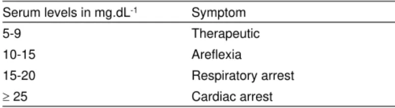
Elevated plasma levels are associated with adverse effects (Table I); therefore, it is necessary to observe some clinical parameters to guarantee the safety of its use17. Those
para-meters include: diuresis of 25 mL.h-1, positive patellar reflex,
respiratory rate greater than 12 bpm, and unchanged vital signs (blood pressure, heart rate, and level of consciousness)17.
Magnesium decreases by 52% of the risk of seizures when compared to diazepam, and 67% when compared to pheny-toin24. This study increased the use of magnesium from 2% to
40% in patients with preeclampsia in the United Kingdom19.
Benzodiazepines are indicated for the treatment of seizures
Table I – Clinical Manifestations of Hypermagnesemia
Serum levels in mg.dL-1 Symptom
5-9 Therapeutic
10-15 Areflexia
15-20 Respiratory arrest
Revista Brasileira de Anestesiologia 109 Vol. 60, No 1, Janeiro-Fevereiro, 2010
APPLICATIONS OF MAGNESIUM SULFATE IN OBSTETRICS AND ANESTHESIA
only postpartum17, in the absence of magnesium sulfate17, or
when treatment with magnesium sulfate has failed2.
Further studies on this area will focus on aquaporin 419, a
wa-ter channel-bound protein found in the final portion of astrocyte axon, whose levels are increased in cerebral edema19.
Mag-netic resonance imaging studies have documented cerebral edema of the white matter of the posterior region of the brain eclampsia patients25. This change has also been documented
in animal models of eclampsia25. The expression of aquaporin
4 is increased in pregnancy26, and the use of magnesium
sul-fate decreases cerebral expression of this protein, which can attenuate cerebral edema in eclampsia patients27.
Magnesium has been used as the standard drug for tocoly-sis during treatment of premature labor, and other drugs have been compared to it28. The mechanism of action has not been
completely elucidated, but it seems to be secondary to calci-um antagonism by competing for the binding site of this ion28.
The loading dose for tocolysis ranges from 4 to 6 g intrave-nous over 15 to 30 minutes, followed by maintenance with 2 to 6 g IV/hour28. Several patients treated with magnesium
devel-op minor adverse reactions, such as: feeling hot, scotomata, nausea, vomiting, blurred or double vision, and lethargy5,28.
Adverse effects can be reverted by the intravenous adminis-tration of 1 g of calcium gluconate5.
APPLICATIONS IN ANESTHESIA
The indications of magnesium sulfate in anesthesia have been increasing over the years to include situations out of the gynecological field5. It has analgesic and sedative properties
with potential neuro- and cardioprotective effects, although it is not know the mechanisms of those actions5,29.
During acute myocardial infarction (AMI), 80% of the patients develop hypomagnesemia in the first 48 hours, probably sec-ondary to the high serum levels of catecholamines6.
Mag-nesium deficiency leads to cell depolarization and promotes tachycardia6. Two studies using magnesium in patients with
AMI, LIMIT 2 and ISIS 4, showed antagonic mortality results5.
Only LIMIT 2 showed a reduction in mortality, but magnesium was used before spontaneous or pharmacologic recovery of the occluded vessel5. The prophylactic use to prevent
hypo-magnesemia during extracorporeal circulation is controversial, although reduction in the incidence of ventricular tachycardia and atrial fibrillation has been shown5.
Neuronal ischemia leads to the outflow of ATP from the cell and inflow of calcium, which triggers the release of toxic metabolites, culminating with cell death5. Blockade of
gluta-mate NMDA receptors inhibits the cellular inflow of calcium and contributes for neuronal protection3,29-31. Other probable
actions for cerebral protection include: reduction in the pre-synaptic release of excitatory neurotransmitters32, blockade of
calcium channels32,33, suppression of anoxic depolarization32,
antioxidant effects31,32, and an increase in cerebral blood
flow29,30,32,33. Besides, cellular energy preservation is also
seen, since magnesium is bound to ATP in the cytosol. Two studies demonstrated antagonic results in patients with
cerebral ischemia, IMAGE and FAST-MAG32. A 90 mg.kg-1
dose reduced infarction volume after middle cerebral artery embolus by 48% when administered in the first six hours32.
It is possible that the doses used in the IMAGE study have not been enough to cause an increase in the concentration of magnesium in cerebral cells15.
Magnesium has been used to attenuate the cardiovascular re-sponse to tracheal intubation5. This effects is, probably,
second-ary to a reduction in the release of catecholamines after sym-pathetic stimulation1,3,5,11,34,35. A 40 mg.kg-1 dose has shown
similar efficacy to that of 10 µg.kg-1 of alfentanil as well as greater
effectivity than 1.5 mg.kg-1 of lidocaine5. It is a complementary
drug in the treatment of hypertensive episodes during the surgi-cal treatment of pheochromocytoma, since it inhibits the release of catecholamines from the adrenal glands5.
Magnesium inhibits the release of acetylcholine in the neuromus-cular junction and behaves as a neuromusneuromus-cular relaxant, poten-tiating the effects of non-depolarizing neuromuscular blockers5.
A 40 mg.kg-1 dose of magnesium reduces the ED
50 of
vecuro-nium by 25%5. When magnesium is administered before
induc-tion, it prevents succinylcholine-induced increase in potassium levels9,36. This drug limits muscular fasciculation, but it does not
interfere with the time of recovery of succinylcholine36.
The analgesic potential of magnesium is partially secondary to the blockade of NMDA receptors, but also to a reduction in the release of catecholamines11. The potential to reduce the
MAC of volatile anesthetics has been confirmed in laboratorial studies with rat models1,5,37, and it can be as high as 60%1.
Schutz-Stubner et al.3 demonstrated a reduction in the need
of remifentanil and fentanyl when one intravenous dose of 50 mg.kg-1 of magnesium was used in humans. Collateral effects
were not observed with this dose3.
The postoperative analgesia of magnesium has been ana-lyzed in a systematic review that used qualitative evaluation methods38. Fourteen randomized clinical assays with 778
pa-tients, 404 of which received magnesium, were included. Mag-nesium levulinate, sulfate, and gluconate were tested. Meta analysis could not be carried out due to methodological het-erogeneity among the studies secondary to a wide variety of magnesium infusion schedules and among the final treatment objectives of each study38. Besides those factors mentioned
by the authors of that study, different age groups and surgi-cal procedures of different subspecialties were also included. The author of that review concluded that the studies included in the analysis did not demonstrate convincing evidence that magnesium was beneficial in the treatment of postoperative pain and reduction in analgesic consumption38.
FINAL CONSIDERATIONS
110 Revista Brasileira de Anestesiologia Vol. 60, No 1, Janeiro-Fevereiro, 2010 BARBOSA, BARBOSA, JUCÁ ET AL.
REFERÊNCIAS – REFERENCES
1. Telci L, Esen F, Akcora D et al. Evaluation of effects of magnesium sulphate in reducing intraoperative anaesthetic requirements. Br J Anaesth 2002;89:594-598.
2. Bahar M, Chanimov M, Grinspun E et al. Spinal anaesthesia induced by intrathecal magnesium sulphate. Anaesthesia 1996;51:627-633. 3. Schutz-Stubner S, Wettmann G, Reyle-Hahn SM et al. Magnesium
as part of balanced general anaesthesia with propofol, remifentanil and mivacurium: a double-blind, randomized prospective study in 50 patients. Eur J Anaesthesiol 2001;18:723-729.
4. Chanimoy M, Cohen ML, Grinspun Y et al. Neurotoxicity after spi-nal anaesthesia induced by serial intrathecal injections of magne-sium sulphate. An experimental study in a rat model. Anaesthesia 1997;52:223-228.
5. Alday Muñoz E, Una Orejón R, Redondo Calvo FJ et al. Mag-nesio en anestesia y reanimación. Rev Esp. Anestesiol Reanim 2005;52:222-234.
6. Marino PL. Magnésio. In: Marino PL. Compêndio de UTI. Porto Ale-gre: Artmed, 2000;529-538.
7. Nácul FE. Distúrbios eletrolíticos em medicina intensiva. In: Nácul FE. Medicina intensiva – abordagem prática. Rio de Janeiro: Revinter, 2004;309-318.
8. Roscoe A, Ahmed AB. A survey of peri-operative use of magnesium sulphate in adult cardiac surgery in the UK. Anaesthesia 2003;58: 363-365.
9. Pascarella PJ e Pineda M. Efectos del sulfato de magnesio en la res-puesta hemodinamica durante la laringoscopia e intubación traqueal. Rev Venez Anestesiol 1998;3:8-12.
10. Kara H, Sahin N, Ulusan V et al. Magnesium infusion reduces perio-perative pain. Eur J Anaesthesiol 2002;19:52-56.
11. Elsharnouby NM, Elsharnouby MM. Magnesium sulphate as a techni-que of hypotensive anaesthesia. Br J Anaesth 2006;96:727-731. 12. Booth CC, Babouris N, Hanna S et al. Incidence of hypomagnesaemia
in intestinal malabsorption. Br Med J 1963;2:141-144.
13. Glover ML, Machado C, Totapally BR. Magnesium sulfate adminis-tered via continuous intravenous infusion in pediatric patients with refractory wheezing. J Crit Care 2002;17:255-258.
14. Ganem EM e Castiglia YMM. Anestesia na pré-eclampsia. Rev Bras Anestesiol 2002;52:481-497.
15. McKee JA, Brewer RP, Macy GE et al. Analysis of the brain bioavail-ability of peripherally administered magnesium sulfate: A study in hu-mans with acute brain injury undergoing prolonged induced hyperma-gnesemia. Crit Care Med 2005;33:661-666.
16. Barbosa FT, Barbosa LT, Cunha RM et al. Uso do sulfato de magné-sio por via venosa e nebulização para o tratamento da asma aguda na emergência. Rev Bras Ter Intensiva 2007;19:369-373.
17. Gonçalves MM. Doença hipertensiva específica da gravidez. In: Rat-ton JLA. Medicina intensiva. São Paulo:Atheneu, 1997;66-76. 18. Cardoso RL, Correa CM. Pacientes obstétricos em UTI. In: Nácul FE.
Medicina intensiva – abordagem prática. Rio de Janeiro: Revinter, 2004;542-548.
19. Euser AG, Cipolla MJ – Magnesium sulfate for a treatment of eclamp-sia: a brief review. Stroke, 2009;40:1169-1175.
20. Ramanathan J, Sibai BM, Pillai R et al. Neuromuscular transmission studies in preeclamptic women receiving magnesium sulfate. Am J Obstet Gynecol 1998;158:40-46.
21. Somjen G, Hilmy M, Stephen CR. Failure to anesthetize human sub-jects by intravenous administration of magnesium sulfate. J Pharma-col Exp Ther 1966;154:652-659.
22. Sibai BM, Spinnato JA, Watson DL et al. Effect of magnesium sulfate on electroencephalographic findings in preeclampsia-eclampsia. Ob-stet Gynecol 1984;64:261-266.
23. Koontz WL, Reid KH. Effect of parenteral magnesium sulfate on pen-icillin-induced seizure foci in anesthetized cats. Am J Obstet Gynecol 1985;153:96-99.
24. The Eclampsia Trial Collaborative Group. Which anticonvulsant for women with eclampsia? Evidence from the collaborative eclampsia trial. Lancet 1995;345:1455-1463.
25. Karumanchi SA, Lindheimer MD. Advances in the understanding of eclampsia. Curr Hypertension Rep 2008;10:305-312.
26. Quick AM, Cipolla MJ. Pregnancy-induced up-regulation of aquaporin-4 protein in brain its role in eclampsia. FASEB J 2005;19:170-175. 27. Ghabriel MN, Thomas A, Vink R. Magnesium restores altered
qua-porin-4 immunoreactivity following traumatic brain injury to a pre-injury state. Acta Neurochir Suppl 2006;96:402-406.
28. Lewis DF. Magnesium sulfate: the first-line tocolytic. Obstet Gynecol Clin North Am 2005;32:485-500.
29. Wadhwa A, Sengupta P, Durrani J et al. Magnesium sulphate only slightly reduces the shivering threshold in humans. Br J Anaesth 2005;94:756-762.
30. Simpson JI, Eide TR, Schiff GA et al. Intrathecal magnesium sulfate protects the spinal cord from ischemic injury during thoracic aortic cross-clamping. Anesthesiology 1994;81:1493-1499.
31. Breen TW, Yang T. The changing role of magnesium sulphate thera-py. Curr Opin Anaesthesiol 1999;12:283-287.
32. Dohi K, Ohtaki H, Shioda S et al. Magnesium sulfate therapy in pa-tients with acute neuronal damage: the problem of intravenous admin-istration. Crit Care Med 2005;33:698-699.
33. Bilotta F, Rosa G. Magnesium sulfate and neuroprotection. Anesth Analg 2003;96:1838.
34. Pivalizza EG. Magnesium sulfate and epidural anesthesia in pheo-chromocytoma and severe coronary artery disease. Anesth Analg 1995;81:414-416.
35. Ramirez Paesano C, Gonzalez O, Rodriguez B et al. Laringoscopia e intubación traqueal: uso de sulfato de magnesio para atenuar la res-puesta cardiovascular refleja. Rev Venez Anestesiol 1998;3:66-71. 36. Stacey MR, Barclay K, Asai T et al. Effects of magnesium sulphate on
suxamethonium-induced complications during rapid-sequence induc-tion of anaesthesia. Anaesthesia 1995;50:933-936.
37. Altan A, Turgut N, Yildiz F et al. Effects of magnesium sulphate and clonidine on propofol consumption, haemodynamics and postopera-tive recovery. Br J Anaesth 2005;94:438-441.
38. Lysakowski C, Dumont L, Czarnetzki C et al. Magnesium as an ad-juvant to postoperative analgesia: a systematic review of randomized trials. Anesth Analg 2007;104:1532-1539.
RESUMEN
Barbosa FT, Barbosa LT, Jucá MJ, Cunha RM – Usos del Sulfato de Magnesio en Obstetricia y en Anestesia.
JUSTIFICATIVA Y OBJETIVOS: El magnesio es un ión
predomi-nantemente intracelular. Su efecto bloqueador del receptor NMDA le confiere características analgésicas y sedativas. El objetivo de este artículo, fue revisar la fisiología, la farmacología y la disminución de la concentración plasmática del magnesio, como también de algunas de sus aplicaciones en obstetricia y en anestesia.
CONTENIDO: El magnesio es un catión intracelular que posee
múl-tiples funciones: es cofactor de enzimas del metabolismo glicídico y de enzimas de la degradación de los ácidos nucleicos, proteínas y ácidos grasos; regula el paso de los iones transmembrana e inter-viene en la actividad de varias enzimas. El paciente en estado crí-tico, presenta una tendencia a la hipomagnesemia, y el tratamiento consiste en corregir la causa cuando es posible, acompañada de la reposición del magnesio. Ya ha quedado demostrada la reducción de la concentración alveolar mínima (CAM), de los agentes inhalatorios en animales y el uso de opioides en humanos bajo anestesia.
CONCLUSIONES: El sulfato de magnesio, ha venido siendo utilizado