115
Rev Bras Med Esporte – Vol. 17, No 2 – Mar/Abr, 2011
Physical Exercise Assessment as An Analgesia
Factor in a Sciatica Experimental Model
Juliana Gaffuri Anamaria Meireles Bruno Pogorzelski Rocha Camila Thieime Rosa Elisangela Lourdes Artifon Lígia Inez Silva
Natalia Boneti Moreira Gladson Ricardo Flor Bertolini
Laboratory of Study of Physiotherapy Injuries and Resources of the State University of Western Paraná (Unioeste), Cascavel Campus – Paraná.
Mailing Address:
Gladson Ricardo Flor Bertolini. Rua Universitária, 2.069, Colegiado de Fisioterapia, Jd. Universitário – 85819-110 – Cascavel, PR. CP 711. E-mail: [email protected]
ABSTRACT
Objective: To evaluate the efficacy of physical exercise (swimming and jumping), with and with-out overload, working in reducing the pain of rats undergone to an experimental model of sciatica. Methods: 24 rats were divided into four groups: Placebo (PG), Swimming Group (SG) Swimming 10% Group (SG10) and Jump Group (JG). All groups were submitted to the experimental sciatica model and assessed for pain post-exercise for the Functional Disability Test and the Von Frey fila-ment. Results: In comparison within groups there were significant differences in the moments after injury with the pre-injury, for both assessment instruments. With Von Frey filament was observed a significant difference in group GN10 and GS in the final moments of evaluation. In comparisons between groups were not statistically significant differences obtained with any assessment in-strument. Conclusion: Treatment with physical exercise was not effective in reducing pain in rats subjected to experimental sciatica model.
Keywords: nerve crush, exercise therapy, analgesia.
INTRODUCTION
The peripheral nerves are constant target of injuries, such as crushing, compression and transection. Such injuries result in pain and reduction, or loss of sensitivity and motricity in the innervated territory. Among the commonest affections, we can mention the ones related to the sciatic nerve, termed sciatica(1).
The lombosciatalgia signs and symptoms include pain on the lumbar region and along the
nerve path, sensory disorders, paresis, gait with intermittent claudication and stiffness(2,3). The main
causes of sciatica are: herniated discs, infections, vascular compression, congenital anomalies,
degenerative neurological diseases, hip trauma dislocation and lumbar canal stenosis(1). Painful
syndrome especially affects individuals between 30 and 50 years of age. It is estimated that worldwide 500,000 new cases are diagnosed every year, out of which 2.8% of these individuals
acquire lifetime incapacities due to the high time of nervous regeneration(4).
The treatments for this dysfunction are countless and depend on the cause. Among these, we can mention initial rest, medication prescription for analgesia and physiotherapeutic techniques as osteopathic treatment, physical exercises, postural orientation, flexibility training and electro-thermophototerapeutical resources. When the conservative treatment fails, surgical treatment is the solution(5,6).
Based on many protocols, therapeutic exercises have been used for sciatica symptoms
relief(7). Although resistance and resisted physical exercises are common among the treatment
modalities, their analgesic effect is contradictory. While some studies demonstrate pain
reduc-tion after exercise, others point out its exacerbareduc-tion after it, especially after resisted exercises(8).
It is supposed that exercise induced analgesia is derived both from the increase of the pain threshold and by the increase of the endogenous opioids level, and the duration of this
analgesia can range according to the intensity, duration and type of exercise characteristics(9).
Experimental models of compression of the sciatic nerve in rats have been used for studies which aim to evaluate the painful situation through the use of different therapeutic
modali-ties due their similarity with the human sciatic nerve(6). Thus, through the sciatica
compres-sion model created by Bennett e Xie(10), it is possible to reproduce in rats the symptomatology
observed in humans.
Thus, considering the great socioeconomical impact derived from sciatica, it is essential that suitable treatments are developed. However, the efficiency of the conservative interventions and
whether a specific exercise is more efficient than another, have not been totally elucidated(5,9). In
addition to this, there are only scant investigations which approach the effect of swimming and LOCOMOTOR APPARATUS IN
EXERCISE AND SPORTS
116 Rev Bras Med Esporte – Vol. 17, No 2 – Mar/Abr, 2011
resisted exercise (jump) on pain derived from compression of the sciatic nerve. Thus, this study has the aim to evaluate he efficiency of physical exercise (swimming and jumping), with and without load, acting in the reduction of the pain of rats submitted to a sciatica experimental model.
MATERIALS AND METHODS
Study and sample characterization
This study was characterized as a transversal experimental assay, with random group division. A sample composed of 24 male Wistar rats, aged 14 weeks and mean weight of 393.70 ± 30.37g was used. During the study period, the animals were kept in polypropylene cages, with free access to water and food ad libitum, with
photope-riod of 12 hours and controlled room temperature (24 ± 1ºC). The
study was conducted according to international norms of ethics on
animal experimentation(11). The research was approved by the Ethics
in Animals Experimentation and Practical Classes Committee of the State University of Western Paraná under protocol number 3.810.
All animals were randomly submitted to the sciatica experimen-tal model in four groups:
Placebo Group (PG, n = 6) – composed of animals submitted only to swimming for time shorter than one minute in each sessions;
Swimming Group (SG, n = 6) – composed of animals submitted to swimming during 30 minutes;
Swimming Group 10% (SG10, n = 6) – composed of animals submitted to swimming, with body overload of 10% of anima’s weight, during 30 minutes;
Jump Group (JG, n = 6) – composed of animals which received intense jumping physical exercise, with overload of 50% of ani-mal’s body weight, performed in four sets of five jumps, as means of therapy.
Sciatica experimental model
The animals were anesthetized with intraperitoneal injection of ketamine chlorhydrate (75mg/kg) and xylazine chlrohydrate (10mg/kg). After sanitation of the site for procedure, incision parallel to the fibers of the biceps femoris of the right thigh of the animal was performed, exposing hence the sciatic nerve.
Fol-lowing the model described by Bennett and Xie(10), compression
in four distinct regions around the sciatic nerve was performed, with approximate distance of 1mm from each other, using a 4.0 chromic catgut cord with the purpose to reproduce chronic pain in the nerve path; plane suture was performed subsequently.
Training protocol
In order to have the training protocols performed, the animals were placed in a tank with 200-liter capacity and 60cm deep, and 30cm of it contained water at 30-32ºC temperature. All animals re-ceived training in five sessions, with intervals of 48 hours, and train-ing prior to the injury took place, specific to each group, for three consecutive days for all groups. The exercise training was initiated only on the third PO day for all groups.
The animals from the swimming groups (SG and SG10) were submitted to swimming during 30 minutes, using (SG10) or not (SG) body overload, according to the group. In order to simulate the body overload, a Velcro band with led weight attached to it with a thread was attached to the abdominal region of the animal, and the number of weight pieces ranged according to the animal’s weight.
Concerning the jump group, the overload was of 50% of the animal’s weight and the protocol consisted in the performance of four sets of five jumps each, with intervals of 30 seconds between sets. For this therapy, the animal with the Velcro band attached to its abdominal region was placed in a cylinder with 30 cen-timeters of diameter and 55 cencen-timeters of height. The animal wearing the overload would submerge and when reaching the bottom of the tank performed an impulse to reach the surface again. Each impulse was counted as one jump.
Pain evaluation
Pain evaluation was performed by the following instruments: functional incapacity test and Von Frey filament.
The functional incapacity test was performed with a metal-lic cylinder in motion and a computer program connected to a metallic boot adapted to the animal‘s paw. This test was originally
described by Tonussi and Ferreira(12). The animals walked during
one minute on the cylinder of approximately 30cm of diameter covered with a stainless steel mesh which performed three ro-tations per minute with an electric motor. Metal boots which conducted the information from the right paw through a cord to the computer program and demonstrated the values of the paw elevation time (PET) of the animal walking were adapted to the hinder legs of the animals. The left hinder limb was also kept connected to a boot, but this one had no information entrance to the computer. Usually, the animals with no alteration present in their gait maintenance of paw on air at around 10 seconds, while the animals with pain due to the sciatica compression
present longer paw elevation times (PET)(12).
The Von Frey filaments were used to assess the nociceptive sensitivity to the mechanical stimulus in the animals (13). The test was performed with the animal held by a thermoplastic restrainer and the Von Frey filament was perpendicularly applied in the region of the surgical procedure of the right thigh, with gradual pressure increase and, as son as the animal removed the paw, the test was interrupted for record of the removal threshold given in grams.
All animals (PG, SG and SG10) were assessed about pain, both by the functional incapacity test and by the Von Frey filament, in the pre-injury prior (AV1) and reassessed on the third PO be-fore (AV2) and after treatment (AV3), eight (AV4), 11th (AV5) and 12th (AV6) days post-injury, always after treatment. After the end of the study, the animals were anesthetized and euthanized by guillotine.
Statistical analysis
The results were analyzed concerning their normality by the Kolmogorov-Smirnov test and later expressed by descriptive sta-tistics (mean and standard deviation) and assessed by inferential statistics, by the analysis of variance (ANOVA) with repeated mea-surements, for comparison within groups and one-way ANOVA for comparison between groups; in both of them Tukey post-test was used and significance level adopted was of 5%.
RESULTS
Functional incapacity test
117
Rev Bras Med Esporte – Vol. 17, No 2 – Mar/Abr, 2011
Figure 2. Pain evolution, demonstrating the mean and standard deviation values of the mechanical pressure (g) supported by the animals in the Von Frey filament application in reevaluations after treatment with physical exercise. *Significant differ-ence when comparing to the pre-injury moment (AV1); #Significant differdiffer-ence when comparing to the eighth PO (AV4); ФSignificant difference when comparing to the third PO after treatment (AV3).
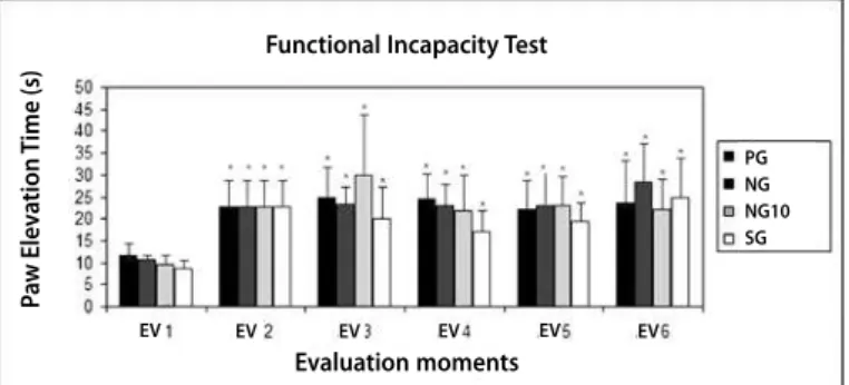
expressed in seconds. It can be observed by the graphic repre-sentation (figure 1) that, in the intragroup comparison, there was significant difference (p < 0.05) in all groups, only when comparing the post-injury moments (AV2, AV3, AV4, AV5 and AV6) with their respective pre-injury moments (AV1), indicating increase of pain-ful episodes after injury. It can be confirmed by the PET increase during the evaluation days. In the intergroup analysis, regardless of the moment of analysis, the differences obtained were not sig-nificant (p > 0.05).
Von frey filament
When intragroup results were compared by the pain evaluation with the Von Frey filament, it was observed that in all groups (PG, SG, SG10 and JG) there was significant dif-ference in the pain level when compared to the post-injury moments (AV2, AV3, AV4, AV5 and AV6) with pre-injury ones (AV1), demonstrating pain increase after injury by sciatic nerve compression (p < 0.05). Moreover, significant differences were observed in SG10 in its 11th (AV5) and 12th PO moments (AV6) in comparison to the eighth PO (AV4). Significant dif-ference was also observed in the JG in its 11th (AV5) and 12th PO moments (AV6) when compared to its third PO after treatment (AV3) (figure 2). These differences were observed due to reduction of the mechanical pressure supported in the evaluation, characterizing pain exacerbation in these groups. Furthermore, in the intergroup comparison by Von Frey filament, significant difference has not been obtained in any moment of the evaluation.
DISCUSSION
During the study period, 24 Wistar rats were submitted to conservative treatment with physical exercise aiming analgesia in the sciatica model. The results obtained suggest that, in the conditions and protocol used, the physical exercise with or without overload was not efficient in functionally reducing the
sciatica symptoms.This fact could be noticed in the functional
incapacity test due to increase of paw elevation time, character-izing claudication and less frequent use of the limb. It could be also observed reduction of mechanical pressure supported by application of the Von Frey filament, with no restoration of the values observed in the pre-injury moment.
Corroborating the results in this study, Miranda et al.(14), when comparing individuals with and without sciatica pain, demon-strated that physical exercise does not affect this
symptomatol-ogy.Faas(15) concludes in similar research that in the acute low
back pain cases, exercise therapy is not efficient, while in chronic low back pain intense exercises can be benefic. This result was also found by Tulder et al.(16) in their systematic review, report-ing that exercise therapy is not more efficient than inactivity for patients with low back pain who present symptoms for less than 12 weeks.
This result of absence of pain relief may be explained by studies which highlight that intense training in rats with sciatica injury promotes deficient regeneration of the injured nervous fibers and tolerance of the opioid receptors, generating hence pain exacerbation(17-19).
In the context concerning with physical exercise intensity, in order to study its effects on the acute inflammatory response
in rats, Lana et al.(20) demonstrated that low intensity physical
exercises on treadmill increase the signs of acute inflammation, a fact not observed with high intensity exercises. According to the authors, it was consequence of the increase in synthesis and secretion of prostaglandins and/or increase in the cytokines plasma levels. However, in the present study it was observed that high intensity physical training produced increase of the pain inflammatory sign.
On the other hand, results by Gonçalves and Luciano(21),
with rats trained on treadmill, showed attenuation of the in-flammatory response induced by carrageenan injection, when compared to sedentary rats. It is worth mentioning that prior to injury the animals were trained, while in the present study the animals were sedentary and performed physical activity
after experimental injury; therefore, they presented different
pain behavior.
Hall et al.(22) highlight the uncertainty concerning the ideal
duration of analgesia induced by physical exercise, but they suggest that the interaction between intensity and duration af-fect analgesia. The authors report that water training programs of shorter duration (2x/week) are less efficient to pain relief. Li et al.(23), in their study to verify neuropatic pain reduction in rats by exercise on the ground (treadmill) and in pool (swimming), they observed that both treatments are significantly efficient in attenuating hyperalgesia and mechanical allodynia.
Koltyn and Arbogast(24) point out that alterations in pain
per-ception after exercise seems to be transitory. According to them, analgesia induced by intense physical exercise lasts a maximum of 15 minutes after it, but pain persists after 15 minutes of
re-PG NG NG10 SG
Evaluation moments Von Frey Filament
Figure 1. Paw elevation time evolution in seconds of the groups according to the pre and post-injury moments, demonstrated in mean and standard deviation values; *Significant difference when comparing to the pre-injury moment (AV1)..
P
aw Ele
v
a
tion
T
ime (s)
Functional Incapacity Test
Evaluation moments
PG NG NG10 SG
EV EV EV EV EV EV
Suppor
ted mechanical
pr
essur
e (g)
118 Rev Bras Med Esporte – Vol. 17, No 2 – Mar/Abr, 2011
sisted exercises. Nevertheless, despite being transitory, it does not explain the fact for lack of analgesia observed in this study, since the animals were reevaluated concerning symptomatology immediately after the end of the training, and this time did not exceed 15 minutes.
Moreover, the kind of exercise performed can also interfere in the painful perception, since some investigations show that resistance training reduces pain more efficiently when compared to resisted training; the former training enables the nervous
re-generation to occur faster than the latter(26). This effect was not
observed in the group which performed resistance exercise (SG and SG10) in this study.
Furthermore, it should be highlighted the cases where exer-cises are performed in water medium. The water heat and float-ing can block nociception by the action of the thermal conditions and on the mecanoreceptors. In addition to that, the water heat increases the blood flow, and consequently helps in the dissipa-tion of catabolites which induce pain(26,27).
Some research highlights the importance of the exercise adaptation to the physical and physiological conditions of the individuals respecting their specific needs in order to reach the
analgesic effect(8). Thus, five days of training may have not been
sufficient to cause adaptation in the animals in this study. Finally, as limitations to this study we can mention the short time of intervention as well as absence of comparison between groups making use of histological analysis. Thus, further investiga-tion with different protocls and exercise intensities are suggested in order to elucidate the results.
CONCLUSION
The treatment with physical exercise, with our without over-load, in the protocols chosen in this study, was not efficient in re-ducing pain of rats submitted to the sciatica experimental model.
ACKNOWLEDGEMENTS
The Unioeste University and the University Hospital of West-ern Paraná (HUOP) partially financed the project. The digital anal-gesimeter was bought with resources from CNPq.
All authors have declared there is not any potential conflict in interests concerning this article
15. Faas A. Exercises: which ones are worth trying, for which patients, and when? Spine. 1996;21:2874-8.
16. Van Tulder M, Malmivaara A, Esmail R, Koes B. Exercise therapy for low back pain: a systematic review within the framework of the Cochrane Collaboration Back Review Group. Spine. 2000;25:2784-96.
17. Gutmann E, Jakoubek B. Effect of increased motor activity on regeneration of the peripheral nerve in young rats. Physiol Bohemoslov. 1993;12:463-8.
18. Mathes WF, Kanarek RB. Wheel running attenuates the antinociceptive properties of morphine and its metabolite, morphine-6- glucuronide, in rats. Physiol Behav. 2001;74:245-51.
19. Smith MA, Lyle MA. Chronic exercise decreases sensitivity to opioids in female rats: correlation with exercise output. Pharmacol Biochem Behav. 2006;85:12-22.
20. Lana AC, Paulino CA, Gonçalves ID. Efeitos dos exercícios físicos sobre o edema inflamatório agudo em ratos Wistar. Rev Bras Med Esporte. 2008;14:33-7.
21. Gonçalves, A L, Luciano E. Respostas inflamatórias em ratos Wistar submetidos a atividade física. Revista Brasileira de Atividade Física e Saúde. 1999;4:39-46.
22. Hall J, Swinkels A, Briddon J, McCabe CS. Does aquatic exercise relieve pain in adults with neurologic or musculoskeletal disease? A systematic review and meta-analysis of randomized controlled trials. Arch Phys Med Rehabil. 2008;89:873-83.
23. Li YT, Li ZY, Hsueh MI, Chun Hung H, Chung Ou H, Wen Chen Y. Treatment with swimming and treadmill exercises reduced neuropathic pain on chronic constriction injury of sciatic nerve of rats. FASEB J. 2010; 24(Suppl):618.15.
24. Koltyn KF, Arbogast RW. Perception of pain after resistance exercise. Br J Sports Med. 1998;32:20-4.
25. Kankaanpa M, Taimela S, Airaksinen O, Hanninen O. The efficacy of active rehabilitation in chronic low back pain. Spine. 1999;24:1034-42.
26. Bender T, Karaglle z, Bálint GP, Gutenbrunner C, Bálint PV, Sukenik S. Hydrotherapy, balneotherapy, and spa treatment in pain management. Rheumatol Int. 2005;25:220-4.
27. Kuphal KE, Fibuch EE, Taylor BK. Extended swimming exercise reduces inflammatory and peripheral neuropathic pain in rodents. J Pain. 2007;8:989-97.
.REFERENCES
1. Stafford MA, Peng P, Hill DA. Sciatica: a review of history, epidemiology, pathogenesis and the role of epidural steroid injection in management. Br J Anaesth. 2007;4:461-73.
2. Rodríguez FJ, Valero-Cabré A, Navarro X. Regeneration and functional recovery following peripheral nerve injury. Drug Discov Today Dis Models. 2004;1:177-85.
3. Kobayashi S, Yoshizawa H, Yamada S. Pathology of lumbar nerve root compression: morphological and immunohistochemical changes of dorsal root ganglion. J Orthop Res. 2004;22:1808.
4. Shabat S, Folman Y, Leitner Y, Fredman B, Gepstein R. Failure of conservative treatment for lumbar spinal stenosis in elderly patients. Arch Gerontol Geriatr. 2007;44:235-41.
5. Hildreth CJ, Lynm C, Glass RM. Sciatica. JAMA. 2009;302:216-17.
6. Ricard F. Estatísticas comparativas en los tratamientos de lumbociáticas por hérnia discal. Fisiot. 2000;22:2030.
7. Tulder M, Malmivaara A, Esmail, R. Exercise therapy for low back pain: a systematic review. Spine. 2000;25:2784-96.
8. Souza JB. Poderia a atividade física induzir analgesia em pacientes com dor crônica? Rev Bras Med Esporte. 2009;15:145-50.
9. Koltyn KF. Analgesia following exercise: a review. Sports Med. 2000;29:85-98.
10. Bennett GJ, Xie YKA. A pheripheral mononeuropathy in rat that produces disorders of pain sensation like those seen in man. Pain. 1988;33:87-107.
11. Andersen ML, D’Almeida V, Ko GM, Kawakami R, Martins PJF, Magalhães LE, et al. Princípios éticos e práticos do uso de animais de experimentação. São Paulo: Unifesp; 2004.
12. Tonussi CR, Ferreira SH. Rat knee-joint carrageenin incapacitation test: an objective screen for central and peripheral analgesics. Pain. 1992;49:421-7.
13. Pedersen,Jl, Galle TS, Kehlet H. Peripheral analgesic effects of ketamine in acute inflammatory pain. Anest. 1998;89:58-66.