ABSTRACT
Sao Paulo Med J. 2008;126(2):126-7.
C
A
SE REPOR
T
Carlos Márcio Nóbrega de JesusJosé Carlos de Souza Trindade Filho
José Goldberg
Late presentation of posterior
urethral valve: two case reports
Faculdade de Medicina de Botucatu, Universidade Estadual de São Paulo
(Unesp), Botucatu, São Paulo, Brazil
CONTEXT: Posterior urethral valve (PUV) is a widely known condition affecting males that generally presents prenatally or at birth. PUVs have also been occasionally described in lit-erature in cases diagnosed during adolescence or adulthood.
CASE REPORT:This report presents two late PUV cases, one in a teenager and the other in an adult. Both cases had had clinical signs of urinary tract infection and obstructive urinary symptoms. The diagnoses were made by means of voiding cystourethrography and urethrocystoscopy. Endo-scopic valve fulguration was the treatment chosen for both. Their follow-up was uneventful. KEY WORDS: Abnormalities. Urethra. Adult. Adolescent. Diagnosis.
INTRODUCTION
Posterior urethral valves (PUVs) are the most common cause of lower urinary tract ob-struction in male neonates, with an incidence of one case per 8,000 to 25,000 live births.1,2 The
diagnosis is usually made prenatally or at birth, when male newborns are evaluated for prenatal hydronephrosis, or during early childhood, but rarely during adolescence or adulthood. The clinical features at these later stages can be confused with many other diseases, thus mak-ing correct diagnosis diffi cult. Here, we describe two cases of late presentation of PUV, which were suspected due to urinary tract infection and voiding dysfunction.
CASE REPORT
Case 1
An 11-year-old white boy presented in-termittent low-pressure voiding with dysuria for one month. He had presented nocturnal enuresis until he was eight years old. His urine sample showed bacteriuria and large numbers of degenerated leukocytes and was positive for nitrite. Urine culturing was positive for
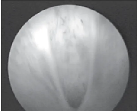
Escherichia coli. After appropriate treatment with antibiotic therapy, this clinical condition appeared again, one month later. Renal and bladder ultrasound scans were normal. During voiding cystourethrography (VCU), dilation and a wide prostatic urethra with a “stop” in the membranous urethra were observed (Figure 1.1). There was no vesicoureteral refl ux. The correct diagnosis was made by cystoscopy, which confi rmed the presence of type I urethral valves (Figure 1.2). Endoscopic valve fulgura-tion was the treatment chosen. This patient evolved successfully and he has not presented any recurrence of urinary symptoms so far.
Case 2
A 40-year-old white man had a history of recurrent urinary tract infection, and his
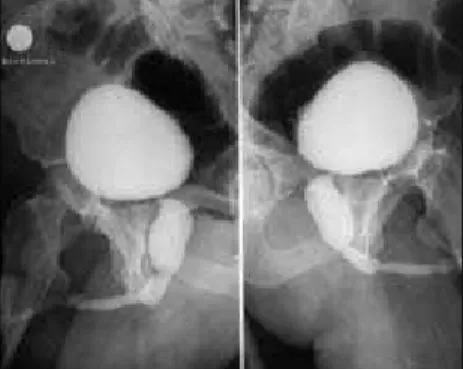
ure-thral discharge had been accompanied by poor, dribbling urine stream since childhood. He sought medical assistance due to a worsening of his urinary symptoms. Digital prostate exami-nation gave normal results. Urine culturing was positive for Escherichia coli. Serum creatinine was within normal limits. Renal ultrasound did not show hydronephrosis. In addition to many stenoses, VCU showed a dilated large posterior urethra and a thick and irregular bladder wall (Figure 2). During endoscopic urethrotomy, type III diaphragmatic posterior urethral valves were observed and fulgurated. This patient is now in a good condition with satisfactory urinary stream.
Figure 1.1. Voiding cystourethrography showing dilation and wide urethra in an 11-year-old boy.
127
Sao Paulo Med J. 2008;126(2):126-7. DISCUSSION
Late presentation of PUV is a rare condi-tion and it has been estimated that it accounts for 10% of PUV cases.3 The usual presentation
is prenatal or at birth. Poor or weak stream, dribbling voiding, repeated urinary tract in-fection and chronic renal failure are the most common clinical pictures in adolescents and adults.4 In most cases, PUV fulguration
im-proves the signs and symptoms of obstructive infravesical syndrome while preserving renal function and renal morphology.5
Many common conditions are superim-posed on late PUV, such as benign prostate hypertrophy, urethral stenosis, prostatitis, urethritis and sphincter-bladder dyssynergy. These other pathological conditions delay or confound the diagnosing of late PUV and, for this reason, we had to consider this condition when the second patient told us about his weak stream and history of urinary tract infection since childhood. Within this scenario, it is essential to include VCU among the diagnostic tools.
Other rare situations could be related to late presentation of PUV, such as retained ejaculation,6 infertility,7 enuresis8 or perineal
pain with dilated Cowper’s glands.9 However,
late presentation of PUV could be a cause of chronic renal failure whether or not it has been treated. Parkhouse at al.10 reported on
long-term follow-up among treated postpu-bertal patients and showed that 26% of them had had chronic or end-stage renal failure.10
Today, 78 late PUV cases have been described in the literature. We believe that this number
is smaller than might have been imagined, because of diagnostic difficulties and the existence of other, similar diseases. In other words, urologists must remain alert to the possibility of late diagnoses of PUV, especially among adults.
In the cases we have reported, the treatment was efficient and led to remission of the illness. However, late PUV is a condition that puts such patients at risk. In a retrospective review of 47 patients who presented late PUV, Bomalaski et al. reported the presence of elevated serum creatinine in 35% and end-stage renal disease in 10%.4 This shows that such patients are at risk
of progression to end-stage renal disease. There-fore, these patients must be treated despite the mild signs and symptoms exhibited when PUV is diagnosed as a late presentation.
CONCLUSION
In conclusion, PUV is not merely a disease of infancy. Boys and men must be investigated if they present voiding complaints, in order to rule out this pathological condition. Urolo-gists must bear this diagnosis in mind and remember that its presentation may be more frequent than might be imagined.
Figure 2. Voiding cystourethrography showing dilation and wide urethra, and several urethral stenoses, in a 40-year-old adult who had had a history of weak urine stream since childhood.
1. Casale AJ. Early ureteral surgery for posterior urethral valves. Urol Clin North Am. 1990;17(2):361-72.
2. Atwell JD. Posterior urethral valves in the British Isles: a mul-ticenter B.A.P.S. review. J Pediatr Surg. 1983;18(1):70-4. 3. Young HH, Frontz WA, Baldwin JC. Congenital obstruction
of the posterior urethra. J Urol. 1919;3:289-365. J Urol. 2002;167(1):265-7; discussion 268.
4. Bomalaski MD, Anema JG, Coplen DE, Koo HP, Rozanski T, Bloom DA. Delayed presentation of posterior urethral valves: a not so benign condition. J Urol. 1999;162(6):2130-32. 5. Martin J, Anderson J, Raz S. Posterior urethral valves in adults:
a report of 2 cases. J Urol. 1977;118(6):978-9.
6. Dutkiewicz S. Posterior urethral valves in an adult male. A case report. Int Urol Nephrol. 1994;26(5):555-8.
7. Woodhouse CR, Reilly JM, Bahadur G. Sexual function and fertility in patients treated for posterior urethral valves. J Urol. 1989;142(2 Pt 2):586-8; discussion 603-5.
8. Dimitriadis G, Hrysogonidis I, Kelidis G, et al. Válvulas congénitas de uretra posterior en pubertad con el único síntoma la enuresis primaria. [Congenital valves of the posterior urethra in puberty with primary enuresis as the only symptom]. Arch Esp Urol. 2002;55(5):539-41. 9. Drouin G, Laperrière J, Grégoire A. Urethral valves as a
cause of dilated Cowper’s glands and perineal pain. J Urol. 1978;120(5):634-5.
10. Parkhouse HF, Barratt TM, Dillon MJ, et al. Long-term outcome of boys with posterior urethral valves. Br J Urol. 1988;62(1):59-62.
Sources of funding: None
Conflict of interest: None
Date of first submission: November 29, 2006
Last received:December 8, 2007
Accepted: March 3, 2008
REFERENCES
AUTHOR INFORMATION Carlos Márcio Nóbrega de Jesus, MD, PhD. Assistant
profes-sor, Department of Urology, Faculdade de Medicina de Botucatu, Universidade Estadual de São Paulo (Unesp), Botucatu, São Paulo, Brazil.
José Carlos de Souza Trindade Filho, MD, PhD. Assistant professor, Department of Urology, Faculdade de Medicina de Botucatu, Universidade Estadual de São Paulo (Unesp), Botucatu, São Paulo, Brazil.
José Goldberg, MD, PhD. Assistant professor, Department of Urology, Faculdade de Medicina de Botucatu, Uni-versidade Estadual de São Paulo (Unesp), Botucatu, São Paulo, Brazil.
Address for correspondence:
Carlos Márcio Nóbrega de Jesus
Departamento de Urologia, Faculdade de Medicina de Botucatu
Distrito de Rubião Junior, s/no Botucatu (SP) — Brasil — CEP 18618-000 Tel./Fax. (+ 55 14) 3811-6271 E-mail: [email protected]
Copyright © 2008, Associação Paulista de Medicina
RESUMO
Apresentação tardia de válvula de uretra posterior: relato de dois casos
CONTEXTO:Válvula de uretra posterior (VUP) é uma conhecida malformação congênita urinária, geralmente diagnosticada em exames ultra-sonográficos pré-natais ou ao nascimento. Raramente, esta doença pode ser encontrada em adolescentes e em adultos.
RELATO DE CASOS:Este artigo mostra dois casos de VUP, encontrados em um adolescente e em um adulto. Ambos apresentavam sinais clínicos de infecção do trato urinário e sintomas obstrutivos infravesicais. Os diagnósticos foram realizados por uretrocistografia miccional e uretrocistoscopia. Fulguração endoscópica das válvulas foi o tratamento de escolha para ambos os casos. O acompanhamento demonstrou melhora importante dos sintomas após o tratamento.