ABSTRACT
Sao Paulo Med J. 2008;126(6):305-8.
ORIGINAL AR
TICLE
Dilermando Pereira de Almeida Filho
Laerte Justino de Oliveira
Vivian Ferreira do Amaral
Accuracy of laparoscopy
for assessing patients with
endometriosis
Center for Health and Biological Sciences, Pontifícia Universidade
Católica do Paraná (PUCPR), Curitiba, Paraná, Brazil
CONTEXT AND OBJECTIVE: Diagnoses of endo-metriosis are based on observation of endometri-otic lesions by means of laparoscopy, along with the pathological fi ndings. The aim of this study was to evaluate the sensitivity and specifi city of the macroscopic fi ndings in relation to the histopathological fi ndings. More specifi cally, we aimed to test the effi cacy of laparoscopy alone for diagnosing endometriosis and to evaluate the laterality of endometriosis among the study population.
DESIGN AND SETTING: Cross-sectional study on women undergoing laparoscopy due to pelvic pain or infertility, in the Gynecology Department of Hospital Santa Cruz in Curitiba, Paraná, Brazil, and Pontifícia Universidade Católica do Paraná.
METHODS: A total of 976 patients underwent laparoscopy and biopsy due to pelvic pain and/ or infertility. We analyzed the laparoscopic and histopathological fi ndings from patients with pelvic endometriosis (n = 468) and patients without endometriosis (n = 508).
RESULTS: In 468 (47.95%) of the cases, the clinical and laparoscopic fi ndings were consis-tent with endometriosis, and this was confi rmed histopathologically in 337 (34.5%). Among the remaining 508 patients, although the laparos-copy was performed for other reasons relating to acute pelvic pain, eight were diagnosed with endometriosis from histopathological examina-tion of the pelvic specimens obtained. Therefore, endometriosis was confi rmed in 345 patients (35.3%). In comparison with the histopathology, laparoscopy alone presented 97.68% sensitivity, 79.23% specifi city, 72% positive predictive value and 98.42% negative predictive value.
CONCLUSION: Laparoscopy should be used in conjunction with histopathology for diagnosing endometriosis.
KEY WORDS: Endometriosis. Pelvic pain. Lap-aroscopy. Infertility. Histology.
INTRODUCTION Endometriosis is described as a benign disease of the female genital system. It is prin-cipally characterized by endometrium-like tissue, consisting of glands and/or stroma, found outside the uterine cavity. Although implanted ectopically, this tissue presents histopathological and physiological respons-es that are similar to the rrespons-esponsrespons-es of the en-dometrium.1
The main symptom of endometriosis is pelvic pain, which is often very intense. Dysmenorrhea and other complaints like dyspareunia and infertility are also seen.2,3
The diagnostic hypothesis of endometrio-sis is based on the clinical history, along with the results from gynecological examinations, laboratory tests and transvaginal ultrasound.4,5
Some clinical characteristics, the physical examination itself, laboratory test results and evidence from imaging examinations may suggest the diagnosis.6 The greatest diffi culty
lies in diagnosing minimal and mild lesions. In these cases, the ideal access for confi rmation is always laparoscopic, since the complementary examinations available do not offer the neces-sary specifi city.7
Diagnosis by means of laparoscopy, which is considered the gold standard, may depend to an as yet unknown degree on confi rmation by means of histopathological assessment. How-ever, the defi nitive diagnosis of the disease can only be obtained through histopathological examination of the biopsy sample.8
Assessment of the accuracy of laparoscopy for diagnosing endometriosis has demon-strated that it is highly precise in ruling out the disease, thereby informing therapeutic decisions.9 Recent studies have shown that
endometriosis is principally diagnosed by laparoscopy combined with histopathological examination, although a negative result does not rule out the possibility of the disease.10
OBJECTIVE The objective of this study was to as-sess the sensitivity and specificity of the macroscopic findings from laparoscopy, in relation to diagnoses of endometriosis based on the results from histopathological examinations. More specifi cally, we aimed to test the effi cacy of laparoscopy alone for diagnosing endometriosis and to evaluate the laterality of endometriosis among the study population.
MATERIALS AND METHODS This was a cross-sectional study. We ana-lyzed 976 women who underwent laparoscopy due to pelvic pain and/or infertility at the Ob-stetrics and Gynecology Department of Hos-pital Santa Cruz between 1994 and 2004. We analyzed the laparoscopic and histopathological fi ndings from all the patients. Of these 976 pa-tients, 468 presented pelvic endometriosis and 508 patients did not present endometriosis (but had other gynecological conditions).
This study was analyzed and approved by the Ethics Committee of Pontifícia Univer-sidade Católica do Paraná (PUCPR), under Ethics Committee Registration No. 530 and protocol No. 056.476.
The criteria for performing laparoscopy were as follows: the subject needed to be in the menacme and presenting pelvic pain, dyspareunia, dysmenorrhea or infertility; and the results from complementary tests such as CA125 determination and ultrasound needed to reveal pelvic masses or blood in the pelvic cavity. Patients who had not yet reached the menarche or had reached the menopause and cases of laparoscopic reintervention were excluded from the laparoscopy performed due to pelvic pain.
306
Sao Paulo Med J. 2008;126(6):305-8. endometriosis, i.e. typical lesions such as
“powder burn”, of reddish color (light or dark), light color (yellow or brown) or dark color (black or blue), or even on fibrotic lesions. The lesions suggestive of endometriosis were biopsied and histopathologically examined in the Pathological Anatomy Department of Hospital Santa Cruz. The endometriosis was staged in accordance with the 1985 American Fertility Society (AFS) classification system, and the staging was compared with the result from the histopathological analysis on the biopsies.11
Pearson’s chi-squared test and Fisher’s exact test were used to assess any propor-tional differences between the groups with and without endometriosis. Differences between the continuous variables were studied using analysis of variance (ANOVA). The signifi-cance level was set at P ≤ 5% for all tests and the power test was 90%.
RESULTS Out of the 976 patients who underwent laparoscopy, 468 (47.95%) were selected for inclusion in the present study, since they presented clinical and laparoscopic profiles suggestive of endometriosis. Among these patients, the clinical and laparoscopic suspicion of endometriosis was confirmed from histopathological analysis in 337 cases (72.0%) (Figure 1).
In the cases of a further eight patients, a positive histopathological diagnosis was made during surgical procedures that were performed due to other causes. Therefore, a total of 345 patients were diagnosed through histopathology.
The mean age of these patients was 30.85 ± 5.54 years. The frequency of endometri-osis of any stage was found to be highest among patients between the ages of 20 and 40 (P = 0.001).
The endometriosis was classified as fol-lows: minimal (15 cases, 4.34%); mild (176 cases, 51.01%); moderate (112 cases, 32.46%); or severe (42 cases, 12.17%).
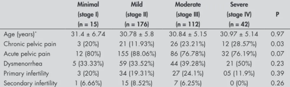
Out of the 345 patients evaluated, 341 (98.84%) presented acute or chronic pelvic pain. Acute pain was more common among the patients presenting the milder stages of endometriosis, whereas chronic pelvic pain was more common in the more severe stages (P = 0.03).
There were 129 patients (37.39%) who complained of dysmenorrhea, 69 (20%) who reported primary infertility and 23 (6.66%) who reported secondary infertility. A tendency towards higher frequency of dysmenorrhea was found among patients with the more severe forms of endometriosis, whereas the frequency of primary or secondary infertility was comparable at all stages of the disease (Table 1).
The histopathological examination confirmed the presence of endometriosis in the right ovary in 77 cases (22.31%) and in the left ovary in 89 cases (25.79%). No statistically significant difference in frequency was observed between the right and left ovaries (22.31% versus 25.79%, P > 0.05). In 29 pa-tients (8.4%), both ovaries were involved.
Endometriosis was identified in the peri-toneum in 260 patients (75.36%) and in the rectovaginal septum in 41 (11.88%).
Endometriosis was confirmed in only one of the biopsied sites in 233 patients (67.53%), in two sites in 102 (29.56%), in three sites in eight (2.31%) and in four sites in two (0.57%). The laparoscopic analysis suggested a diagnosis of minimal endometriosis in 17 patients (12.97%), mild endometriosis in 63 (48.09%), moderate endometriosis in 35 (26.71%) and severe endometriosis in 16 (12.21%).
Taking the histopathological findings to be definitive for the diagnosis of endo-metriosis, the clinical suspicion and laparo-scopic findings presented 97.68% sensitivity, 79.23% specificity, 72% positive predictive value, 98.42% negative predictive value, and 85.75% accuracy (Table 2). False positive results were obtained in 27.99% of the tests, compared with false negative results in 1.57% of the tests.
DISCUSSION Despite the efforts of the scientific com-munity to increase the efficacy of the meth-ods used to diagnose endometriosis, various limitations remain, thus making it difficult to reach a definitive diagnosis.
Table 2. Laparoscopic and histopathological findings (n = 976)
Surgical diagnosis (laparoscopy) Histopathological confirmation Total
Positive Negative
Positive 337 (34.52%) 131 (13.42%) 468 (48.15%)
Negative 8 (0.81%) 500 (51.22%) 508 (52.04%)
Total 345 (35.34%) 631 (64.65%) 976 (100%)
Table 1. Laparoscopic endometriosis staging, by patient age and clinical manifestation
Minimal Mild Moderate Severe
P
(stage I) (stage II) (stage III) (stage IV)
(n = 15) (n = 176) (n = 112) (n = 42)
Age (years)* 31.4 ± 6.74 30.78 ± 5.8 30.84 ± 5.15 30.97 ± 5.14 0.97 Chronic pelvic pain 3 (20%) 21 (11.93%) 26 (23.21%) 12 (28.57%) 0.03 Acute pelvic pain 12 (80%) 155 (88.06%) 86 (76.78%) 32 (76.19%) 0.07 Dysmenorrhea 5 (33.33%) 59 (33.52%) 44 (39.28%) 21 (50%) 0.23 Primary infertility 3 (20%) 34 (19.31%) 27 (24.1%) 05 (11.9%) 0.39 Secondary infertility 1 (6.66%) 15 (8.52%) 7 (6.25%) 0 (0%) 0.26
*Age expressed as mean ± standard deviation; all other values expressed as number and percentage.
Figure 1. Flowchart of the distribution of the patients selected for this study.
Total number of patients: 976
Suspected endometriosis: 468
Endometriosis: 8
Not confirmed by histopathology:
131 Confirmed by
histopathology: 337
307
Sao Paulo Med J. 2008;126(6):305-8.
Clinical parameters such as pelvic pain, dysmenorrhea, dyspareunia and infertility are insufficient to confirm the diagnosis. Likewise, combining laboratory tests such as CA125 level determinations with imaging methods such as ultrasonography, tomography and magnetic resonance provides relative value for reaching a conclusive diagnosis in the initial stages of endometriosis.12-14 Combining
laparoscopy with histopathological examina-tion yields greater sensitivity for the definitive diagnosis of the disease and also decreases the diagnostic errors.15
Among the 976 laparoscopies performed in this study, the frequency of endometriosis was 35.3%. This result is in accordance with findings from previous studies carried out among smaller population samples.16,17
Fur-thermore, our findings corroborate data in the literature regarding the mean age of the patients studied and are in keeping with the results from other studies showing that the on-set of symptoms usually occurs within seven to twelve years after the menarche.18
In the present study, and in accordance with the 1985 AFS system for staging endometriosis,12 4.36% of our patients were
classified as stage I (minimal), 51.01% as stage II (mild), 32.46% as stage III (moderate) and 12.17% as stage IV (severe). In a study involv-ing 44 patients who underwent laparoscopy due to pelvic pain, Petta reported that 50% presented stage I endometriosis, 12.5% pre-sented stage II, 25% prepre-sented stage III and 12.5% presented stage IV.19 These results,
together with others found in the literature, are listed in Table 3.19,20-22
Between these different studies, discrepan-cies can be observed among the stages found. Although one particular macroscopic mapping method for endometriosis was recommended by the American Society for Reproductive Medicine in 1997, the results from many studies differ according to the background and experience of the professional who performed the laparoscopy. Therefore, comparative as-sessments are affected.20 The results from the
present study demonstrate the difficulties ensuing from macroscopic assessments made by various observers. The diversity of the results justifies the use of histopathological analysis for diagnostic confirmation of en-dometriosis.23
The diagnosis of histopathology-con-firmed endometriosis presented a statistically significant association with chronic pelvic pain. However, according to the findings of Wardle and Hull,24 acute pelvic pain,
dys-menorrhea, primary infertility and secondary
Table 3. Staging of endometriosis in the literature
Authors Stage I
(minimal)
Stage II (mild)
Stage III (moderate)
Stage IV (severe)
Petta et al.19 (n = 44) 50% 12.5% 25% 12.5%
Gruppo Italiano per lo Studio dell´ Endometriosi.20 (n = 469)
11.3% 12.2% 51% 21.7%
Bai et al.21 (n = 39) 10% 44% 28% 18%
Chapron et al.22 (n = 209) 13.5% 38.1% 24.2% 24.2%
Almeida Filho, Oliveira & Amaral
(current study) (n = 345) 4.3% 51% 32.4% 12.1%
infertility had no statistically significant influ-ence on the diagnosis of endometriosis.
To date, there is no consensus on the relationship between the extent of endo-metriosis and the intensity of pelvic pain.25
It has been shown that there is a correlation between certain histopathological findings (a well-differentiated pattern or a diagnosis of stromal disease) and the intensity of pelvic pain.26 In the present study, 98.84% of all
patients (regardless of endometriosis stage) reported pelvic pain. Pelvic pain was found to correlate significantly with endometriosis stage (P= 0.03) (Table 3).
In other studies, it was reported that the severity of dysmenorrhea presented no significant association with the stage or loca-tion of endometriosis.20,27 Our results are in
accordance with those of such studies, in that no positive correlation was found between the degree of endometriosis and the intensity of dysmenorrhea.27 We observed dysmenorrhea
in 37.39% of our patients with confirmed endometriosis and in 26.71% of our patients without endometriosis, although the differ-ence was not statistically significant.
Topalski Fistes et al.27 carried out a
com-parative study with a control group of 200 fertile women. They found that the frequency of endometriosis was 32% among infertile women and 5% among fertile women, which was a statistically significant difference (P = 0.001). In the present study, the frequen-cies of primary or secondary infertility were comparable, regardless of the severity of the disease.
When we compared the laterality of ovar-ian involvement in the 345 women evaluated, we found similar frequencies (left ovary versus right ovary: 25.79% versus 22.31%; P > 0.05). Several studies evaluating endometriotic ovar-ian cysts have shown a predisposition towards left-sided lesions.8,28,29 However, this was not
confirmed in our study.
In addition, we observed a greater in-cidence of the disease in the peritoneum (79.3%), regardless of the stage of endometrio-sis, whereas the incidence of peritoneal lesions
described in the literature ranges from 17.5% to 31%.19,20,22
In the present study, the number of bi-opsies testing positive for endometriosis was directly proportional to the severity of the endometriosis. This shows that, whether lap-aroscopy or histopathology is used, it is more difficult to make a definitive diagnosis when the lesions are minimal or mild.
In a study assessing macroscopic findings of anatomical abnormalities and confirmation of endometriosis, it was found that 85.7% of the patients presented pelvic anatomical abnormalities consistent with endometriotic lesions and that 31.1% of them were identified through histopathology as endometriosis.20 In
our study, 468 patients presenting pelvic pain and anatomical abnormalities typical of endo-metriosis were evaluated, and the diagnosis of endometriosis was confirmed in 337 (72%).
Comparison between these studies reveals that, despite the validity of laparoscopy for diagnosing endometriosis, its use without histopathological confirmation gives rise to discrepancies in relation to the macroscopic findings.10 There is a need for an informal
consensus regarding study design, and good surgical practice should be supported by de-tailed documentation in order to systematize the diagnosis.10
The findings from the present study allow us to conclude that endometriosis demonstrated a significant positive correla-tion with chronic pelvic pain, although not with dysmenorrhea or infertility. A greater frequency of peritoneal endometriosis was observed, in comparison with the involvement of other sites, such as the rectovaginal septum or ovaries.
308
Sao Paulo Med J. 2008;126(6):305-8. AUTHOR INFORMATION
Dilermando Pereira de Almeida Filho, MD. Postgraduate Health Sciences program, Pontifícia Universidade Católica do Paraná (PUCPR); responsible for the laparoscopic sector of Hospital Santa Cruz, Curitiba, Paraná, Brazil. Laerte Justino de Oliveira, MD, PhD. Associate professor in
the Department of Gynecology, Irmandade Santa Casa de Misericórdia de Curitiba, Aliança Saúde, Pontifícia Univer-sidade Católica do Paraná (PUCPR), Curitiba, Brazil. Vivian Ferreira do Amaral, MD, PhD. Associate professor in
the Department of Gynecology, Irmandade Santa Casa de Misericórdia de Curitiba, Aliança Saúde, Pontifícia Universidade Católica do Paraná (PUCPR); associate professor at the Center for Health and Biological Sciences, Pontifícia Universidade Católica do Paraná (CCBS-PUCPR), Curitiba, Paraná, Brazil.
Address for correspondence: Vivian Ferreira do Amaral
Centro de Ciências Biológicas e da Saúde, Pontifícia Universidade Católica do Paraná (CCBS-PUCPR) Rua Imaculada Conceição, 1.155 — Prado Velho
Curitiba (PR) — Brasil — CEP 81611-970
Tel./Fax: (+ 55 41) 3271-1657 E-mail: [email protected]
Copyright © 2008, Associação Paulista de Medicina
RESUMO
Eficácia da videolaparoscopia na avaliação de mulheres com endometriose pélvica
CONTEXTO E OBJETIVO: O diagnóstico da endometriose é determinado pela visualização dos implantes à laparoscopia e pela comprovação histológica. O objetivo deste trabalho foi avaliar a sensibilidade e a especificidade dos achados macroscópicos cirúrgicos e histopatológicos. Avaliou-se a eficácia da laparoscopia isoladamente no diagnóstico da endometriose e a lateralidade da doença.
TIPO DE ESTUDO E LOCAL: Estudo transversal realizado no Serviço de Ginecologia do Hospital Santa Cruz em Curitiba, Paraná e na Pontifícia Universidade Católica do Paraná.
MÉTODOS: Foram avaliadas 976 pacientes submetidas à videolaparoscopia por dor pélvica ou inferti-lidade e a biópsia. Foram analisados os achados laparoscópicos e histológicos de 468 pacientes com endometriose pélvica e de 508 pacientes sem endometriose.
RESULTADOS: Foram selecionadas 468 (47,95%) pacientes para inclusão no presente estudo por apre-sentarem quadro clínico e videolaparoscópico de suspeita de endometriose. As 508 (52,04%) pacientes restantes tiveram indicação da cirurgia por outras causas relacionadas à dor pélvica e oito tiveram o diagnóstico de endometriose pelo anatomopatológico. A endometriose foi confirmada em 345 pacientes (35,3%). Ao compararmos a análise histológica com os achados a videolaparoscopia, observou-se sensibilidade de 97,68%, especificidade de 79,23%, valor preditivo positivo de 72%, valor preditivo negativo de 98,42%.
CONCLUSÃO: Laparoscopia deve ser usada em conjunto com histopatologia para o diagnóstico de endometriose.
PALAVRAS-CHAVE: Endometriose. Dor pélvica. Laparoscopia. Infertilidade. Histologia. 1. Jansen RP, Russell P. Nonpigmented endometriosis: clinical,
laparoscopic, and pathologic definition. Am J Obstet Gynecol. 1986;155(6):1154-9.
2. Olive DL, Henderson DY. Endometriosis and mullerian anomalies. Obstet Gynecol. 1987;69(3 Pt 1):412-5. 3. Matorras R, Rodriguez F, Pijoan JI, et al. Are there any clinical
signs and symptoms that are related to endometriosis in infertil-ity women? Am J Obstet Gynecol. 1996;174(2):620-3. 4. Houston DE. Evidence for the risk of pelvic endometriosis by
age, race and socioeconomic status. Epidemiol Rev. 1984;6:167-91.
5. Redwine DB. Age-related evolution in color appearance of endometriosis. Fertil Steril. 1987;48(6):1062-3.
6. Abrão MS, Neme RM, Averbach M. Endometriose de septo retovaginal: doença de diagnóstico e tratamento específicos. [Rectovaginal septum endometriosis: a disease with specific diag-nosis and treatment]. Arq Gastroenterol. 2003;40(3):192-7. 7. Tardif D, Poncelet C, Bénifla JL, Madelenat P. Exploration
paraclinique des endométrioses. [Paraclinical studies of endo-metriosis] Rev Prat. 1999;49(3):263-8.
8. Vercellini P, Trespidi L, De Giorgi O, Cortesi I, Parazzini F, Cro-signani PG. Endometriosis and pelvic pain: relation to disease stage and localization. Fertil Steril. 1996;65(2):299-304. 9. Wykes CB, Clark TJ, Khan KS. Accuracy of laparoscopy in the
diagnosis of endometriosis: a systematic quantitative review. BJOG. 2004;111(11):1204-12.
10. Kennedy S, Bergqvist A, Chapron C, et al. ESHRE guideline for the diagnosis and treatment of endometriosis. Hum Reprod. 2005;20(10):2698-704.
11. Revised American Fertility Society classification of endometrio-sis: 1985. Fertil Steril. 1985;43(3):351-2.
12. Weiner Z, Beck D, Brandes JM. Transvaginal sonography,
color flow imaging, computed tomographic scanning, and CA 125 as a routine follow-up examination in women with pelvic tumor: detection of recurrent disease. J Ultrasound Med. 1994;13(1):37-41.
13. Amaral VF, Ferriani RA, Sá MF, et al. Positive correlation be-tween serum and peritoneal fluid CA-125 levels in women with pelvic endometriosis. Sao Paulo Med J. 2006;124(4):223-7. 14. Stratton P, Winkel C, Premkumar A, et al. Diagnostic accuracy
of laparoscopy, magnetic resonance imaging, and histopathologic examination for the detection of endometriosis. Fertil Steril. 2003;79(5):1078-85.
15. Walter AJ, Hentz JG, Magtibay PM, Cornella JL, Magrina JF. Endometriosis: correlation between histologic and visual find-ings at laparoscopy. Am J Obstet Gynecol. 2001;184(7):1407-11; discussion 1411-3.
16. Kresch AJ, Seifer DB, Sachs BL, Barrese I. Laparoscopy in 100 women with chronic pelvic pain. Obstet Gynecol. 1984;64(5):672-4.
17. Zondervan KT, Cardon LR, Kennedy SH. The genetic basis of endometriosis. Curr Opin Obstet Gynecol. 2001;13(3):309-14.
18. Arruda MS, Petta CA, Abrão MS, Benetti-Pinto CL. Time elapsed from onset of symptoms to diagnosis of endo-metriosis in a cohort study of Brazilian women. Hum Reprod. 2003;18(4):756-9.
19. Petta CA, Paiva LHSC, Pinto Neto AM, Fonseca E, Lane E. O uso da laparoscopia na dor pélvica crônica. [The use of lap-aroscopy in women with pelvic chronic pain]. J Bras Ginecol. 1990;100(3/4):85-7.
20. Gruppo Italiano per lo Studio dell’Endometriosi. Relationship between stage, site and morphological characteristics of pelvic endometriosis and pain. Hum Reprod. 2001;16(12):2668-71.
21. Bai SW, Cho HJ, Kim JY, et al. Endometriosis in an adolecent population: the severance hospital in Korean experience. Yonsei Med J. 2002;43(1):48-52.
22. Chapron C, Fauconnier A, Dubuisson JB, Barakat H, Vieira M, Bréart G. Deep infiltrating endometriosis: relation between severity of dysmenorrhoea and extent of disease. Hum Reprod. 2003;18(4):760-6.
23. Abrão MS, Amaral VF, Ramos LO. Classificações da endometri-ose: é tempo de reavaliar. [Classification of endometriosis: is time to re-evaluate]. Femina. 1998;26(8):677-80.
24. Wardle PG, Hull MG. Is endometriosis a disease? Baillieres Clin Obstet Gynaecol. 1993;7(4):673-85.
25. Fedele L, Bianchi S, Bocciolone L, Di Nola G, Parazzini F. Pain symptoms associated with endometriosis. Obstet Gynecol. 1992;79(5 (Pt 1)):767-9.
26. Abrao MS, Neme RM, Carvalho FM, Aldrighi JM, Pinotti JA. Histological classification of endometriosis as a predictor of re-sponse to treatment. Int J Gynaecol Obstet. 2003;82(1):31-40. 27. Topalski Fistes N, Maticki Sekuli M, Kopitovi V, Tabs D.
Endometrioza i bol. [Endometriosis and pain]. Med Pregl. 2002;55(9-10):415-8.
28. Sznurkowski J, Emerich J. Czestsza lewostronna lokalizacja torbieli endometrialnych. [Left lateral predisposition of endo-metrioma]. Ginekol Pol. 2005;76(1):33-6.
29. Ferrero S, Ragni N, Fulcheri E. Lateral distribution of benign ovarian cysts. Int J Gynaecol Obstet. 2005;89(2):150-1.
Sources of funding: Not declared Conflict of interest: Not declared Date of first submission: July 4, 2007 Last received: November 5, 2008 Accepted: November 7, 2008