Totally implantable central venous catheters for chemotherapy:
Totally implantable central venous catheters for chemotherapy:
Totally implantable central venous catheters for chemotherapy:
Totally implantable central venous catheters for chemotherapy:
Totally implantable central venous catheters for chemotherapy:
experience with 793 patients
experience with 793 patients
experience with 793 patients
experience with 793 patients
experience with 793 patients
Cateteres venosos centrais totalmente implantáveis para quimioterapia:
Cateteres venosos centrais totalmente implantáveis para quimioterapia:
Cateteres venosos centrais totalmente implantáveis para quimioterapia:
Cateteres venosos centrais totalmente implantáveis para quimioterapia:
Cateteres venosos centrais totalmente implantáveis para quimioterapia:
experiência com 793 pacientes
experiência com 793 pacientes
experiência com 793 pacientes
experiência com 793 pacientes
experiência com 793 pacientes
ESMÁLIO BARROSODE OLIVEIRA1; MAURÍCIO AGUIAR REIS1; TIAGO MARQUES AVELAR1; SABAS CARLOS VIEIRA2
A B S T R A C T A B S T R A C T A B S T R A C T A B S T R A C T A B S T R A C T
Objective Objective Objective Objective
Objective: To retrospectively study the results obtained with the implementation of totally implantable catheters in patients undergoing chemotherapy. MethodsMethodsMethodsMethods: 815 totally implantable catheters placed in 793 patients undergoingMethods chemotherapy regimen, preferably using the right cephalic vein. We evaluated early and late complications. ResultsResultsResultsResultsResults: The retrospective analysis showed an average duration of 339 days of the catheters. In 733 (90%) catheters there was no observe complication. Among early complications we observed one pneumothorax, one bad positioning of the catheter, one arterial puncture, one bleeding, one hemothorax and hemomediastinum and six hematomas in the implantation site. As for late complications, there were 35 catheter-related infections ten, infections in the surgical site, six obstructions and 20 thromboses. We removed 236 catheters, 35 due to complications and 201 by the end of treatment. ConclusionConclusionConclusionConclusion: totallyConclusion implantable catheters for chemotherapy are a safe means for the administration of substances, in view of the low number of complications observed in this study.
Key words: Key words: Key words: Key words:
Key words: Drug therapy. Catheters. Catheters, indwellin. Central venous catheters. Infection.
Work performed in the San Marcos and Prontomed Hospitals, Teresina, Piauí State – PI, Brazil.
1. Medical School Graduate, Federal University of Piauí; 2. Assistant Professor, Oncology, Federal University of Piauí.
INTRODUCTION
INTRODUCTION
INTRODUCTION
INTRODUCTION
INTRODUCTION
T
otally implantable long-term central venous catheters (CVC) are commonly used in cancer patients for chemotherapy, parenteral nutrition, blood collection for tests and blood transfusion. Despite its benefits, the CVC is associated with significant morbidity and mortality. Those for chemotherapy are made of silicone. They have their distal end positioned at the junction of the superior vena cava with the right atrium, and the proximal, along its subcutaneous implantation site 1.The totally implantable CVC are an excellent means of accessing the venous system, in addition to being effective and associated with a dwindling number of complications, especially when compared to other central venous catheters 2,3. They need fewer manipulations,
injections of heparin solution and dressings, and favorable aesthetic appearance due to being subcutaneous, not restricting patients’ activities 4.
Although these catheters have become common in recent years, few studies have been done in Brazil, especially prospective ones, with a significant number of cases and long term follow-up.
In the present study we retrospectively evaluated the results obtained with the implementation of 815 totally
implantable catheters in 793 patients undergoing chemotherapy in two large hospitals.
METHODS
METHODS
METHODS
METHODS
METHODS
We conducted a retrospective study with 793 patients suffering from malignant tumors, submitted to insertion of 815 totally implantable catheters for chemotherapy from March 2005 to September 2009,. The research took place in the San Marcos and Prontomed Hospitals, in the city of Teresina, Piauí State, Brazil.
The study was approved by the Research Ethics Committee of the Federal University of Piauí – UFPI (CAAE: 0400.0.045.000-11).
All catheters were placed by the same surgeon, using the open technique. All procedures were performed in the operating room, with the assistance of the anesthesiologist.
The operations were performed under local anesthesia, with sedation with intravenous propofol. All patients received antibiotic prophylaxis with 1g cefazolin at induction of anesthesia.
need to inform the surgeon about any abnormality resulting from the procedure.
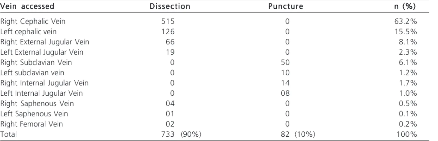
The first option was to access the right cephalic vein. When this displayed an inadequate diameter for placement of the catheter, we proceeded to dissection of the external jugular vein. The puncture was performed when we could not access by dissection. The puncture was first attempted in the subclavian vein and, as a second option, the internal jugular vein. Table 1 shows the access routes and techniques employed. No catheter was introduced when there was the presence of fever of unknown origin, any systemic infectious condition (bacteremia or septicemia) or signs of skin infection near the site for insertion.
We used intraoperative fluoroscopy to position the catheter tip in the superior vena cava, near the entrance to the right atrium. We did not perform routine venography or Doppler ultrasound of the great vessels, but in individuals who had symptoms of deep vein thrombosis.
Patients who had catheters implanted in the saphenous vein showed great vessels thrombosis or non-progression of the catheter due to mediastinal lymphadenopathy or infection in the upper cervical or subclavian regions.
Complications were classified into immediate / early (intraoperative and postoperative before catheter use) and late (those occurring after the use of the catheter).
RESULTS
RESULTS
RESULTS
RESULTS
RESULTS
Patients’ age ranged from 12 to 85 years, with an average of 50.8. Six hundred patients were female (75.7%) and 193 male (24.3%).
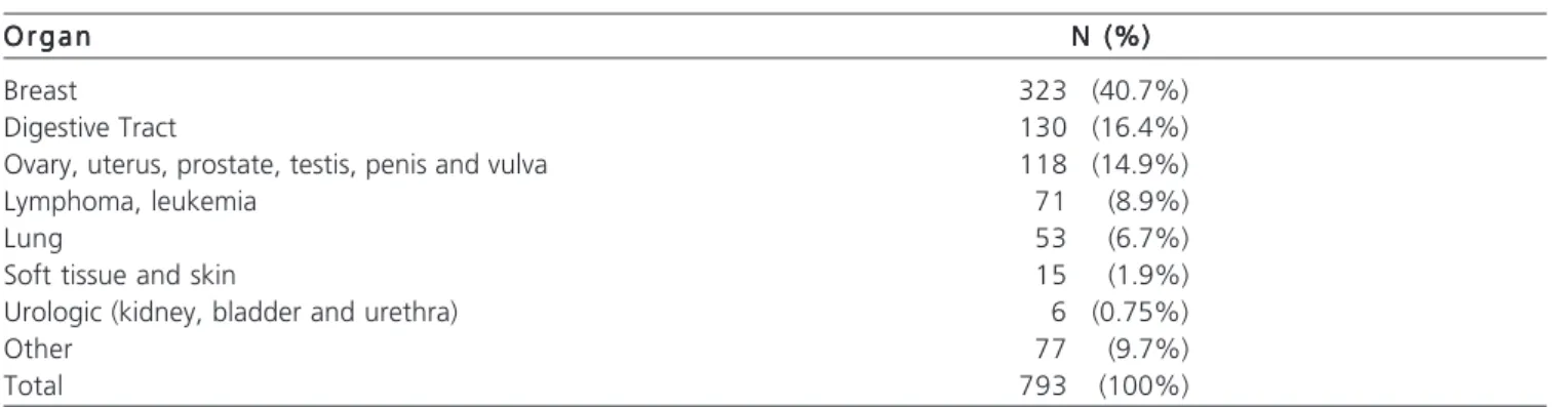
The main clinical indications for implantation of catheters were chemotherapy for treatment of solid tumors (87.5%) and hematologic diseases (12.5%). In 40.7% of patients, the tumor was located in the breast (Table 2). The procedure ranged from eight to 110 minutes, with an average of 17 minutes.
Of the 815 catheters, 82 (10%) had some type of complication. We removed 236 catheters, of which 201 (85%) were by elective indication due to the end of treatment and 35 (15%) resulted from complications that could not be controlled with clinical measures.
Among the early complications observed, there were six hematomas in the insertion site, five of which treated successfully by clinical measures and one, caused by puncture of the right carotid artery, treated conservatively; one small right pneumothorax which was also treated conservatively; one hemothorax and hemomediastinum resulting from puncture and insertion of the venous dilator in the left subclavian artery, the chest being drained; one bleeding, for which thromboprophylaxis was suspended and a pressure dressing applied for 48 hours; one case of bad positioning of the catheter, which was resolved by its replacement.
The other complications occurred at a later stage and involved 71 of the 815 catheters (8.7%). In four patients, more than one catheter complication was observed during follow-up: one patient had infection and venous thrombosis; one had infection and occlusion; one, infection and hematoma; and another, venous thrombosis and hematoma.
Infectious complications occurred in 35 patients (4.3%). Pseudomonas sp, Klebsiella pneumoniae, Salmonella sp and E. coli were the major etiological agents of catheter infections found in cultures and treated with proper antibiotics longer period of time, ranging from 14 to 21 days; the antibiogram showed sensitivity to ciprofloxacin, imipenem and meropenem. The catheter had to be removed in 16 cases due to prolonged fever or worsening of the patient’s clinical status. In 19 cases there was an improvement in the clinical picture, with preservation of the catheter (preservation rate of 54.3%). Wound infection occurred in ten patients, all treated with intravenous antibiotics, and in six cases there was catheter removal.
Non-infectious complications occurred in 26 cases. In six cases, there was obstruction of the catheter. In 20
Table 1 Table 1 Table 1 Table 1
Table 1 - Access routes used for the implantation of a central venous catheter.
Vein accessed Vein accessed Vein accessed Vein accessed
Vein accessed D i s s e c t i o nD i s s e c t i o nD i s s e c t i o nD i s s e c t i o nD i s s e c t i o n P u n c t u r eP u n c t u r eP u n c t u r eP u n c t u r eP u n c t u r e n (%)n (%)n (%)n (%)n (%)
Right Cephalic Vein 515 0 63.2%
Left cephalic vein 126 0 15.5%
Right External Jugular Vein 66 0 8.1%
Left External Jugular Vein 19 0 2.3%
Right Subclavian Vein 0 50 6.1%
Left subclavian vein 0 10 1.2%
Right Internal Jugular Vein 0 14 1.7%
Left Internal Jugular Vein 0 08 1.0%
Right Saphenous Vein 04 0 0.5%
Left Saphenous Vein 01 0 0.1%
Right Femoral Vein 02 0 0.2%
cases there was deep vein thrombosis associated with the catheter. In cases of obstruction, we trying to maintain the patency of the catheters, but in three cases patency was impossible and the catheters had to be removed, being replaced by another.
There were twenty cases of deep vein thrombosis: the majority affected the subclavian vein, followed by the internal jugular vein and there was only one case of superior vena cava thrombosis. By implementing the systemic anticoagulation with low molecular weight heparin and warfarin, it was possible to preserve ten of the 20 catheters (preservation rate of 50%). The catheters were removed in ten cases of deep vein thrombosis, which were associated with non-functioning catheters. None of these cases developed pulmonary thromboembolism. There was only one case of superior vena cava syndrome.
DISCUSSION
DISCUSSION
DISCUSSION
DISCUSSION
DISCUSSION
With the advent of chemotherapy, the search for safe and long lasting vascular accesses increased. Nowadays, the totally implantable central venous catheters are an excellent means of access to the bloodstream and are considered effective and associated with reduced incidence of complications. Kurul et al. 5 reported a
significant reduction in the rates of complications as the team gained experience with central venous accesses 2,3,5.
A previously controversial condition, the cost-effectiveness of implantable devices becomes evident within six months, related to lower rate of complications and need for maintenance care 6,7.
The preferred access route for the implementation of the majority of long-term catheters is the dissection of the right cephalic vein, successfully used in 63.2% of our cases. This vein is a safe route because it is a superficial one, its dissection requires minimal tissue handling and, enabling faster and effective control of possible complications.
For some the preferred route is the subclavian vein 8, which is more associated with complications such
as pneumothorax, hemothorax, arterial injury or compression of the catheter between the first rib and the clavicle. The surgical team is critical to the success of the procedure. The surgeon should be used to deal with different access routes, in order to provide the safest one, as well as be prepared for possible changes in the intraoperative conduct.
Early complications related to the procedure were properly diagnosed and treated. Such complications accounted for 1.3% of cases, hematoma at the insertion site being the most frequent, and were treated with surgical drainage. In this study there was no case of embolism by catheter fracture. Embolisms occur when there is a rupture of the catheter and a large fragment migrates to the vessel directly to the lung or heart, or when there is air in the system, infusion systems disconnections or empty solutions bottles, thereby characterizing gas embolisms, which are rare events 9. Due to the improvement of equipment and
insertion techniques, the complication rates are getting smaller. However, some recent studies show significant complication rates, with up to 2% incidence of pneumothorax, 14% of cardiac arrhythmia, 3% of arterial puncture, 3% of guide-wire bending and 3% of kinking of the introductory sheath 8,10,11. In this study, the incidence of
pneumothorax, arterial puncture, hemothorax and hemomediastinum and bad positioning was 0.12%, much lower than that shown in the literature; in addition, there was no case of cardiac arrhythmia.
Despite care, infection remains the main late complication, catheter-related bacteremia being the most frequent. The infection rate can reach 31%12. In this study,
it was observed that catheter infections occurred in 4.3% of patients. Infection arise from the contamination by micro-organisms of the site of infusion or of the catheter from skin colonization, contaminated material, malfunction of the air inlet filter and from the catheter connections 9. This
condition should be suspected whenever the patient has fever and wound erythema. The proper use of asepsis techniques during catheter manipulation reduces its occurrence 9,13. In our study, antibiotic prophylaxis was used
in all procedures.
Table 2 Table 2Table 2 Table 2
Table 2 - Location of tumors with indication of central venous catheter.
O r g a n O r g a nO r g a n O r g a n
O r g a n N (%)N (%)N (%)N (%)N (%)
Breast 323 (40.7%)
Digestive Tract 130 (16.4%)
Ovary, uterus, prostate, testis, penis and vulva 118 (14.9%)
Lymphoma, leukemia 71 (8.9%)
Lung 53 (6.7%)
Soft tissue and skin 15 (1.9%)
Urologic (kidney, bladder and urethra) 6 (0.75%)
Other 77 (9.7%)
Non-infectious late complications can be divided in catheter obstructions and deep vein thrombosis. The incidence of these two is far from negligible. In the literature, the rates vary between 7% and 50% 14-17. In our study there were 26 of
these complications (3.2%). Deep vein thrombosis (DVT) in the veins of the upper compartment of the body is usually secondary to central venous catheters and cancer-related hypercoagulable state 18. Regarding the catheter, there is:
chemical structure, diameter, number of lumens, position the catheter tip, insertion side, implantation technique, prior use of central venous access and catheter-related infections. Patients’ characteristics include: platelet count, presence and type of malignancy, chemotherapy protocol and hypercoagulable states 19. The implantation of central venous
access causes the endothelium to lose its integrity and leads to activation of procoagulant factors and platelets, thus forming thrombus 20. The use of anticoagulants after placement of a
totally implantable catheter for chemotherapy aiming to reduce thrombosis is controversial in the literature, with studies showing decreased thromboembolic events 20-23 and other not 24-26. In
this sample, there were 20 cases of deep vein thrombosis (2.4%), but removal of the catheter was necessary in ten patients (50%), due to their malfunctioning.
Deep vein thrombosis associated with central venous catheter is usually asymptomatic or presents with nonspecific symptoms 18,19. Symptomatic patients commonly
report discomfort in the shoulder or neck, and exhibit erythema, distal paraesthesia, congestion of the subcutaneous collateral veins and edema in the ipsilateral upper limb, the degree of venous obstruction being related to the signs and symptoms. In cases of obstruction of the superior vena cava, there is facial swelling, headache, visual changes, dizziness and breathlessness 18, simulating a
superior vena cava syndrome.
Obstructions are defined when there is an impossibility or difficulty of infusing substances or drawing blood, being classified as total or partial. As regards to the source of obstruction, they can be mechanical, when compression or bending occurs; thrombotic, through the formation of fibrin internally or external to the catheter due to vascular lesions or hypercoagulable states; and non-thrombotic, due to precipitation of substances infused through the catheter 9,18,26. In this article there were only six
cases of obstruction, and in three the catheter was removed and replaced in the same procedure.
Totally implantable catheters for chemotherapy are a safe means for the administration of substances, in view of the low number of complications observed in this study. An experienced surgeon, ahead of the team, with a good technique for catheter implantation and strict asepsis, and monitoring of patients throughout the treatment, reduces early complications and prevents late ones.
R E S U M O R E S U M O R E S U M O R E S U M O R E S U M O
Objetivo: Objetivo: Objetivo: Objetivo:
Objetivo: estudar retrospectivamente os resultados obtidos com a implantação de cateteres totalmente implantáveis em pacien-tes submetidos à quimioterapia. Métodos:Métodos:Métodos:Métodos:Métodos: foram colocados 815 cateteres totalmente implantáveis em 793 pacientes submetidos ao regime de quimioterapia preferencialmente utilizando-se a veia cefálica direita. Foram avaliadas as complicações precoces e tardias. Resultados: Resultados: Resultados: Resultados: Resultados: a análise retrospectiva mostrou duração média dos cateteres de 339 dias. Em 733 (90%) cateteres não se observou nenhuma complicação. Entre as complicações precoces observamos um pneumotórax, um mau posicionamento de cateter, uma punção arterial, um sangramento, um hemotórax e hemomediastino e seis hematomas na loja de implantação. Entre as complicações tardias, ocorreram 35 infecções relacionadas ao cateter, dez infecções no sítio cirúrgico, seis obstruções e 20 tromboses. Foram retirados 236 cateteres, 35 devido às complicações e 201 por final de tratamento. Conclusão:Conclusão:Conclusão:Conclusão:Conclusão: os cateteres totalmente implantáveis para quimioterapia são meios seguros para a administração de substâncias, em vista do baixo número de complicações observadas neste estudo.
Descritores: Descritores: Descritores: Descritores:
Descritores: Quimioterapia. Cateteres. Cateteres de demora. Cateteres venosos centrais. Infecção.
REFERENCES
REFERENCES
REFERENCES
REFERENCES
REFERENCES
1. Miranda RB, Lopes JRA, Cavalcante RN, Kafejian O. Perviedade e complicações no seguimento de cateteres venosos totalmente implantáveis para quimioterapia. J vasc. bras. 2008;7(4):316-20. 2. Hartkamp A, van Boxtel AJ, Zonnenberg BA, Witteveen PO. Totally
implantable venous access devices: evaluation of complications and a prospective comparative study of two different port systems. Neth J Med. 2000;57(6):215-23.
3. Biffi R, Pozzi S, Agazzi A, Pace U, Floridi A, Cenciarelli S, Peveri V, et al. Use of totally implantable central venous access ports for high-dose chemotherapy and peripheral blood stem cell transplantation: results of a monocentre series of 376 patients. Ann Oncol. 2004;15(2):296-300.
4. Nishinari K, Malavolta LC, Saes GF, Langer M, Carvalho Sobrinho A, Zerati AE, et al. Cateteres venosos totalmente implantáveis para quimioterapia: experiência em 415 pacientes. Acta oncol bras. 2003;23(2):432-40.
5. Kurul S, Saip P, Aydin T. Totally implantable venous-access ports: local problems and extravazation injury. Lancet Oncol. 2002;3(11):684-92.
6. Biffi R, de Braud F, Orsi F, Pozzi S, Mauri S, Goldhirsch A, Nolè F, et al. Totally implantable central venous access ports for long-term chemotherapy. A prospective study analyzing complications and costs of 333 devices with a minimum follow-up of 180 days. Ann Oncol. 1998;9(7):767-73.
8. Minassian VA, Sood AK, Lowe P, Sorosky JI, Al-Jurf AS, Buller RE. Longterm central venous access in gynecologic cancer patients. J Am Coll Surg. 2000;191(4):403-9.
9. Jesus VC, Secoli SR. Complicações acerca do cateter venoso central de inserção periférica (PICC). Ciênc cuid saúde. 2007;6(2):252-60. 10. Capaccioli L, Nistri M, Distante V, Rontini M, Manetti A, Stecco A. Insertion and management of long-term central venous devices: role of radiologic imaging techniques. Radiol Med. 1998;96(4):369-74.
11. Ballarini C, Intra M, Pisani Ceretti A, Cordovana A, Pagani M, Farina G, et al. Complications of subcutaneous infusion port in the general oncology population. Oncology. 1999;56(2):97-102. 12. Wolosker N, Yazbek G, Nishinari K, Malavolta LC, Munia MA,
Langer M, et al. Totally implantable venous catheters for chemotherapy: experience in 500 patients. Sao Paulo Med J. 2004;122(4):147-51.
13. Beckers MM, Ruven HJ, Seldenrijk CA, Prins MH, Biesma DH. Risk of thrombosis and infections of central venous catheters and totally implanted access ports in patients treated for cancer. Thromb Res. 2010;125(4):318-21.
14. Balestreri L, De Cicco M, Matovic M, Coran F, Morassut S. Central venous catheter-related thrombosis in clinically asymptomatic oncologic patients: a phlebographic study. Eur J Radiol. 1995;20(2):108-11.
15. Gould JR, Carloss HW, Skinner WL. Groshong catheter-associated subclavian venous thrombosis. Am J Med. 1993;95(4):419-23. 16. Pintor Holguín E, Sáez Noguero F, Piret Ceballos MV, González
Armengol, Ruiz Yagüe M, Patiño Barrios R, et al. Axillary-subclavian thrombosis: review of its etiology and features in recent years. An Med Interna. 1997;14(2):67-70.
17. De Cicco M, Matovic M, Balestreri L, Panarello G, Fantin D, Morassut S, et al. Central venous thrombosis: an early and frequent complication in cancer patients bearing long-term silastic catheter. A prospective study. Thromb Res. 1997;86(2):101-13.
18. Joffe HV, Goldhaber SZ. Upper-extremity deep vein thrombosis. Circulation. 2002;106(14):1874-80.
19. Verso M, Agnelli G. Venous thromboembolism associated with long-term use of central venous catheters in cancer patients. J Clin Oncol. 2003;21(19):3665-75.
20. Akl EA, Kamath G, Yosuico V, Kim SY, Barba M, Sperati F, et al. Thromboprophylaxis for patients with cancer and central venous catheters: a systematic review and a meta-analysis. Cancer. 2008;112(11):2483-92.
21. Kirkpatrick A, Rathbun S, Whitsett T, Raskob G. Prevention of central venous catheter-associated thrombosis: a meta-analysis. Am J Med. 2007;120(10):901.e1-13.
22. Young AM, Billingham LJ, Begum G, Kerr DJ, Hughes AI, Rea DW, et al. Warfarin thromboprophylaxis in cancer patients with central venous catheters (WARP): an opel-label randomised trial. Lancet. 2009;373(9663):567-74.
23. Verso M, Agnelli G, Bertoglio S, Di Somma FC, Paolett F, Ageno W, et al. Enoxaparin for the prevention of venous thromboembolism associated with central vein catheter: a double-blind, placebo-controlled, randomized study in cancer patients. J Clin Oncol. 2005;23(18):4057-62.
24. Petersen LJ. Anticoagulation therapy for prevention and treatment of venous thromboembolic events in cancer patients: a review of current guidelines. Cancer Treat Rev. 2009;35(8):754-64. 25. Niers TM, Di Nisio M, Klerk CP, Baarslag HJ, Büller HR, Biemond BJ.
Prevention of catheter-related venous thrombosis with nadroparin in patients receiving chemotherapy for hematologic malignancies: a randomized, placebo-controlled study. J Thromb Haemost. 2007;5(9):1878-82.
26. Couban S, Goodyear M, Burnell M, Dolan S, Wasi P, Barnes D, et al. Randomized placebo-controlled study of low-dose warfarin for the prevention of central venous catheter-associated thrombosis in patients with cancer. J Clin Oncol. 2005;23(18):4063-9.
Received on 30/06/2012
Accepted for publication 20/08/2012 Conflict of interest: none
Source of funding: none
How to cite this article: How to cite this article:How to cite this article: How to cite this article:How to cite this article:
Oliveira EB, Reis MA, Avelar TM, Vieira SC. Totally implantable central venous catheters for chemotherapy: experience with 793 patients. Rev Col Bras Cir. [periódico na Internet] 2013;40(3). Disponível em URL: http://www.scielo.br/rcbc
Address correspondence to: Address correspondence to:Address correspondence to: Address correspondence to:Address correspondence to: Mauricio Aguiar Reis