Araujo Neto SA et al. / Normal anatomy and variants of the CAT and HAS
Radiol Bras. 2016 Jan/Fev;49(1):49–52 49
0100-3984 © Colégio Brasileiro de Radiologia e Diagnóstico por Imagem
Iconographic Essay
Multidetector computed tomography angiography of the celiac
trunk and hepatic arterial system: normal anatomy and main
variants
*
Angiotomografia multidetectores do tronco celíaco e sistema arterial hepático: anatomia normal e suas principais variantes
Araujo-Neto SA, Mello-Júnior CF, Franca HA, Duarte CMA, Borges RF, Magalhães AGX. Multidetector computed tomography angiography of the celiac trunk and hepatic arterial system: normal anatomy and main variants. Radiol Bras. 2016 Jan/Fev;49(1):49–52.
Abstract
R e s u m o
Although digital angiography remains as the gold standard for imaging the celiac arterial trunk and hepatic arteries, multidetector computed tomography in association with digital images processing by software resources represents a useful tool particularly attractive for its non invasiveness. Knowledge of normal anatomy as well as of its variations is helpful in images interpretation and to address surgical planning on a case-by-case basis. The present essay illustrates several types of anatomical variations of celiac trunk, hepatic artery and its main branches, by means of digitally reconstructed computed tomography images, correlating their prevalence in the population with surgical implications.
Keywords: Anatomy; Celiac trunk; Hepatic artery; Multidetector computed tomography.
Embora a angiografia digital permaneça como padrão ouro no estudo do tronco celíaco e sistema arterial hepático, o exame por tomo-grafia multidetectores associada às ferramentas informáticas de reconstrução de imagens digitais tem representado uma alternativa útil, principalmente por serem métodos não invasivos. O conhecimento detalhado tanto da anatomia normal quanto das variações anatô-micas ajuda na interpretação de exames radiológicos e na adequação do planejamento cirúrgico para cada paciente. Este texto ilustra uma série de variações anatômicas do tronco celíaco e sistema arterial hepático, por meio de imagens tomográficas com reconstruções digitais, correlacionando as prevalências populacionais e implicações cirúrgicas.
Unitermos: Anatomia; Tronco celíaco; Artéria hepática; Tomografia computadorizada multidetectores.
* Study developed at Universidade Federal da Paraíba (UFPB), João Pessoa, PB, Brazil.
1. PdD, Associate Professor II of Medical Radiology at Universidade Federal da Paraíba (UFPB), João Pessoa, PB, Brazil.
2. PhD, Associate Professor IV of Medical Radiology at Universidade Federal da Paraíba (UFPB), João Pessoa, PB, Brazil.
3. Graduate Students of Medicine at Universidade Federal da Paraíba (UFPB), João Pessoa, PB, Brazil.
Mailing Address: Dr. Severino Aires Araujo Neto. Avenida Sapé, 1780, ap. 2201, Bairro Manaíra. João Pessoa, PB, Brazil, 58038-382. E-mail: severinoaires@hotmail. com.
Received May 12, 2014. Accepted after revision November 6, 2014.
modern image reconstruction programs, this imaging method becomes an additional option for the detailed inves-tigation of arteries with the significant advantage of its noninvasiveness(5). Multidetector CT angiography allows the visualization of small caliber short arteries by means of maximum intensity projection (MIP) and three-dimensional volume rendering (VR) techniques.
The present essay is aimed at describing the normal anatomy and commonly found CAT and HAS variations.
The images shown in the present essay were collected from the personal files of the authors and acquired in a Bril-liance 64-channel multidetector CT apparatus (Philips Medi-cal System; Best, The Netherlands).
The scan protocol, with small sporadic variations, con-sisted in axial sections, slice thickness of 1 mm, pitch 0.8. The contrast agent Ultravist (Bayer) was utilized, at a con-centration of 769 mg/mL, intravenously injected by means of an injection pump at a rate of 5 mL/s, with bolus tracking time delay. A standard field of view (250 mm) was utilized. The images reconstruction thickness was 2 mm. An Extended Brilliance Work Space workstation was utilized with the software Philips Brilliance for computed tomography (Philips Medical Systems; Best, The Netherlands).
Severino Aires Araujo Neto1, Carlos Fernando de Mello Júnior2, Henrique Almeida Franca3, Cláudia Martina Araújo Duarte3, Rafael Farias Borges3, Ana Guardiana Ximenes de Magalhães3
http://dx.doi.org/10.1590/0100-3984.2014.0041 INTRODUCTION
Developments in surgical techniques such as upper ab-dominal videolaparoscopic surgeries, liver transplantation and gastrectomies, besides invasive and noninvasive proce-dures in the abdomen, require of the health professional a wide knowledge about the anatomy of the celiac arterial trunk (CAT), hepatic arterial system (HAS) and their main varia-tions(1–4).
Araujo Neto SA et al. / Normal anatomy and variants of the CAT and HAS
Radiol Bras. 2016 Jan/Fev;49(1):49–52 50
In order to define the arterial pattern, analyses were performed in the axial plane with reconstruction techniques in the coronal and sagittal planes in multiplanar reconstruc-tions (MPR), as well as three-dimensional reconstrucreconstruc-tions with the MIP and VR techniques. The normal patter and the main CAT and HAS variations were demonstrated.
CELIAC ARTERIAL TRUNK AND HEPATIC
ARTERIAL SYSTEM: NORMAL ANATOMY
AND VARIATIONS
CAT variations are not infrequent(1). Song et al. have studied 5,002 cases of CAT and reported the occurence of variations in 10.9% of cases(5). However, as concomitant CAT and HAS variations are considered, the rate increases to 55% of patients(3).
The normal celiac trunk – 89.1% of cases(5) – is de-scribed as the trifurcation originating the left gastric artery, splenic artery and common hepatic artery(3,5) (Figure 1). Normally, the left gastric artery is the first branch of the CAT and runs cranially toward the smaller curvature of the stom-ach were it undergoes anastomosis with the right gastric artery; the splenic artery is the branch of the trunk with largest cali-ber and runs tortuously toward the spleen; the common he-patic artery runs to the right where it divides into gas-troduodenal artery inferiorly, and hepatic artery propria su-periorly(2,4).
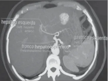
The most common CAT variations are the following: hepatosplenic trunk, representing about 3% of cases, where the common hepatic artery and the splenic artery originate from a single trunk, and the left gastric artery is located above this trunk, either in the aorta or in other artery of the upper abdomen (Figures 2 and 3)(6); splenogastric trunk (4%), where the left gastric artery originates from the splenic ar-tery, forming a common trunk (Figure 4); hepatogastric
Figure 2. Computed tomography, coronal section with VR, demonstrating a hepatosplenic trunk. The arrow indicates the left gastric artery emerging from the aorta, above the hepatosplenic trunk.
A B
Figure 4. Contrast-enhanced axial computed tomography showing a splenogastic trunk (consisting of splenic artery and left gastric artery – indicated by the arrow). In this case, the common hepatic artery is a branch from the aorta.
Figure 3. Contrast-enhanced axial computed tomography. The image demon-strates a case of hepatosplenic trunk with left gastic artery relocation, so in this case it emerges from the left hepatic artery.
(left hepatic artery)
(hepatosplenic trunk)
(left gastric artery)
(splenic artery)
(common hepatic artery)
Figure 1. Contrast-enhanced axial CT demonstrating a normal CAT. The celiac artery trunk represents an arterial triad consisting of the left gastric artery, com-mon hepatic artery and splenic artery indicated by the arrows.
(left gastric artery) (common hepatic artery)
(splenic artery)
Araujo Neto SA et al. / Normal anatomy and variants of the CAT and HAS
Radiol Bras. 2016 Jan/Fev;49(1):49–52 51
trunk (1%), with the left gastric artery and common hepatic artery originating from a single trunk (Figure 5). The ab-sence of the CAT is rarely described in the literature (0.1%)(5).
As regards the HAS, it is described as normal in cases where the common hepatic artery originates the hepatic tery propria after the emergence of the gastroduodenal ar-tery; and the hepatic artery propria, on its turn, divides into right and left hepatic arteries within the hepatoduodenal liga-ment, at few centimeters from the liver surface.
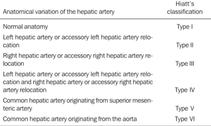
According to Koops et al., the frequencies of the nor-mal HAS pattern as per the Hiatt’s classification (Table 1), are contained in the interval 59–79.1% (type I) (Figure 6).
Table 1—Anatomical variations of the hepatic artery according to Hiatt’s classi-fication.
Anatomical variation of the hepatic artery
Normal anatomy
Left hepatic artery or accessory left hepatic artery relo-cation
Right hepatic artery or accessory right hepatic artery re-location
Left hepatic artery or accessory left hepatic artery relo-cation and right hepatic artery or accessory right hepatic artery relocation
Common hepatic artery originating from superior mesen-teric artery
Common hepatic artery originating from the aorta
Hiatt’s classification
Type I
Type II
Type III
Type IV
Type V Type VI
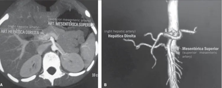
Amongst the most described variations, the following fre-quencies were found: 3–17% (type II), relocation of the left hepatic artery; 7–18% (type III), relocation of the right patic artery (Figure 7); and 1.5–5% (type V), common he-patic artery originating from the superior mesenteric artery. Also, according to Hiatt, it is possible to find non-classified variations with a frequency of 1–4.1%(7).
Normally, cases of Hiatt’s type III are the most preva-lent and play a relevant role in procedures involving the liver, as after originating from the superior mesenteric artery, the right hepatic artery runs posteriorly to the portal vein, which might confuse the surgeon, since in the normal pattern (type I) such artery is located anteriorly to the portal vein, within the hepatoduodenal ligament. Thus, one of the reasons for understanding those variations is avoiding iatrogenic events(8).
In Hiatt’s type II – left hepatic artery relocation –, pro-cedures such as gastrectomy should be cautiously performed, considering that in most of such cases, the left hepatic ar-tery emerges from the left hepatic arar-tery; thus, in case of Figure 5. Contrast-enhanced sagittal computed tomography. A: The image shows a case of hepatogastric trunk. The arrows indicate the arteries composing this trunk (common hepatic artery and left gastric artery). B: The image shows that the splenic artery, in this case, emerges from a common trunk with the superior mesenteric artery.
(common hepatic artery)
(left gastric artery)
(splenic artery)
(superior mesenteric artery)
Figure 6. Contrast-enhanced axial computed tomography. The image presents a case of normal pattern of the HAS, with the hepatic artery propria originating from the common hepatic artery, after the emergnce of the gastroduodenal artery; and right and left hepatic arteries emerging from the hepatic artery propria (Hiatt’s type I).
(left hepatic artery)
(right hepatic artery)
(gastroduodenal artery)
(splenic artery)
(hepatic artery propria)
(common hepatic artery)
Araujo Neto SA et al. / Normal anatomy and variants of the CAT and HAS
Radiol Bras. 2016 Jan/Fev;49(1):49–52 52
section of the left gastric artery, a possible ischemia of the whole functional left hepatic lobe might occur.
Understanding the pattern of variation of the hepatic arteries becomes imprescindible for the development of the liver transplant(9).
With the introduction of laparoscopic surgeries with reduction of the surgical field view, it is necessary to under-stand the pattern of variations of the upper abdomen(10).
The arterial patterns are relevant in the planning of the whole surgical and radiological procedure involving the upper abdomen(5).
Considering the relevance of the mentioned variations, the authors suggest that radiologists should investigate the arterial pattern and inform them in reports of surgeries and invasive examinations of the upper abdomen.
CONCLUSION
Considering that the vascularization of a great part of the gastrointestinal system occurs from CAT and HAS branches, the knowledge about anatomical variations and respective frequencies is of paramount relevance in the plan-ning of upper abdomen surgeries to avoid procedural errors and medical iatrogenic events.
REFERENCES
1. Iezzi R, Cotroneo AR, Giancristofaro D, et al. Multidetector-row CT angiographic imaging of the celiac trunk: anatomy and normal variants. Surg Radiol Anat. 2008;30:303–10.
2. Özbülbül NI. CT angiography of the celiac trunk: anatomy, vari-ants and pathologic findings. Diagn Interv Radiol. 2011;17:150–7. 3. Ugurel MS, Battal B, Bozlar U, et al. Anatomical variations of he-patic arterial system, coeliac trunk and renal arteries: an analysis with multidetector CT angiography. Br J Radiol. 2010;83:661–7. 4. Wang MJ, Cheng Z, Wang R, et al. Unusual course of the common hepatic artery originating from the celiac trunk. Surg Radiol Anat. 2010;32:883–5.
5. Song SY, Chung JW, Yin YH, et al. Celiac axis and common he-patic artery variations in 5002 patients: systematic analysis with spiral CT and DSA. Radiology. 2010;255:278–88.
6. Prakash, Rajini T, Mokhasi V, et al. Coeliac trunk and its branches: anatomical variations and clinical implications. Singap Med J. 2012;53:329–31.
7. Koops A, Wojciechowski B, Broering DC, et al. Anatomic varia-tions of the hepatic arteries in 604 selective celiac and superior me-senteric angiographies. Surg Radiol Anat. 2004;26:239–44. 8. Hiatt J, Gabbay J, Busuttil R. Surgical anatomy of the hepatic
arter-ies in 1000 cases. Ann Surg. 1994;220:50–2.
9. Todo S, Makowaka L, Tzakis AG, et al. Hepatic artery in liver transplatation. Transplant Proc. 1987;19(1 Pt 3):2406–11. 10. Scott-Conner CE, Hall TJ. Variant arterial anatomy in laparoscopic
cholecystectomy. Am J Surg. 1992;163:590–2.
Figure 7. The image presents a case of right hepatic artery relocation, where it emerges from the superior mesenteric artery (Hiatt’s type III). Contrast-enhanced axial computed tomography (A) and contrast-enhanced computed tomography with VR reconstruction (B) showing right hepatic artery relocation.
A B
(right hepatic artery)
(superior mesenteric artery)
(right hepatic artery)