O
r i g i n a la
rt i c l e3 0 9 Arq Bras Oftalmol. 2017;80(5):309-12 http://dx.doi.org/10.5935/0004-2749.20170075
ABSTRACT
Purpose: To evaluate the corneal biomechanical features and central corneal
thickness in ankylosing spondylitis patients and to evaluate correlations of these parameters with disease activity.
Methods: The study included 51 patients diagnosed with ankylosing spondylitis (mean age, 40.80 ± 13.15 years; range, 18-72 years) and 34 age- and sex-matched healthy controls (mean age, 42.00 ± 12.32 years; range, 18-60 years). All underwent a complete ophthalmological and physical examination, including visual acuity testing and biomicroscopic anterior and posterior segment examinations. Corneal hysteresis, corneal resistance factor, Goldmann-correlated intraocular pressure, and corneal compensated intraocular pressure were evaluated with an ocular response analyzer, and the central corneal thickness was measured with Sirius® corneal to-mography. The Bath Ankylosing Spondylitis Disease Activity Index, Functional Index, and Metrology Index scores were recorded.
Results: In the ankylosing spondylitis patients, the mean disease duration was 7.73 ± 6.05 (range, 1-30) years. There was no statistically significant difference between the patients and controls in the corneal biomechanical features. The Goldmann-correlated intraocular pressure and corneal compensated intraocular pressure both showed positive correlations with age (p=0.003 and p=0.001, res pectively). There was a negative correlation between corneal hysteresis and disease duration (p=0.002), and between central corneal thickness and the Bath Ankylosing Spondylitis Metrology Index score (p=0.003).
Conclusion: This study demonstrated a significant negative correlation between corneal hysteresis and disease duration in ankylosing spondylitis patients. Fur-thermore, the central corneal thickness value decreased with an increase in Bath Ankylosing Spondylitis Metrology Index score, which may result in an underestimate of intraocular pressure readings and thus an inaccurate risk assessment of glaucoma.
Keywords: Spondylitis, ankylosing; Corneal biomechanics; Corneal tomography; Severity of illness index
RESUMO
Objetivo: Avaliar as características biomecânicas da córnea e espessura central da córnea em pacientes com espondilite anquilosante e analisar a correlação destes parâmetros no grupo de estudo com a atividade da doença.
Métodos: Foram incluídos no estudo 51 pacientes com diagnóstico de espondilite anquilosante e 34 controles saudáveis com idade e sexo. Todos os sujeitos foram sub-metidos a um exame oftalmológico e físico completo, incluindo exames de acuidade visual, exames de segmento anterior e posterior biomicroscópicos. Foram avaliados o coeficiente de resistência da córnea, a pressão intraocular correlacionada com Goldmann e a pressão intraocular compensada da córnea com o analisador de res-posta ocular, a espessura corneana central com a tomografia corneana pelo Sirius®. Se o índice de atividade da doença de espondilite anquilosante de banho, o índice funcional de espondilite anquilosante de banho, o índice de metrologia de espondilite anquilosante de banho.
Resultados: Foram incluídos no estudo 51 pacientes com idade média de 40,80 ± 13,15 (intervalo: 18-72) anos e 34 casos de controle com idade média de 42,00 ± 12,32 (intervalo: 18-60) anos. No grupo espondilite anquilosante a duração média da doença foi de 7,73 ± 6,05 (1,00-30,00) anos. Não houve diferença estatisticamente significante entre dois grupos quanto às características biomecânicas da córnea. Na análise de correlação, no grupo de estudo; pressão intraocular correlacionada com Goldmann e pressão intraocular compensada da córnea estavam positivamente correlacionados com a idade (p=0,003, p=0,001, respectivamente). Houve uma correlação negativa entre a duração da doença e CH (p=0,002), e entre índice de metrologia de espondilite anquilosante de banho e espessura corneana central (p=0,003).
Conclusão: Este estudo demonstrou correlação negativa significativa entre a du-ração da doença e a histerese corneal em pacientes com espondilite anquilosante. Além disso, com um aumento na pontuação de índice de metrologia de espondilite anquilosante de banho, o valor de espessura corneana central também estava di-minuindo o que pode causar uma diminuição nas leituras de pressão intraocular artificialmente e resultar em avaliação de risco imprecisa de glaucoma.
Descritores: Espondilite anquilosante; Biomecânica da córnea; Tomografia da córnea; Ín dice de gravidade de doença
Corneal biomechanical features in patients with ankylosing spondylitis
Características biomecânicas da córnea entre pacientes com espondilite anquilosante
Kubra Serefoglu CabuK1, emine iSil ÜStÜn2, KurSat atalay1, ahmet Kirgiz1, ruKiye aydin3
Submitted for publication: September 21, 2016 Accepted for publication: May 15, 2017
1 Ophthalmology Department, Bagcilar Training and Research Hospital, Bagcilar, Istanbul, Turkey. 2 Physical Medicine and Rehabilitation Department, Bagcilar Training and Research Hospital, Bagcilar,
Istanbul, Turkey.
3 Ophthalmology Department, Medipol University, Bagcilar, Istanbul, Turkey.
Funding: No specific financial support was available for this study.
Disclosure of potential conflicts of interest: None of the authors have any potential conflict of interest to disclose.
Corresponding author: Kubra Serefoglu Cabuk. Ophthalmology Department. Bagcilar Training and
Research Hospital. Merkez Mahallesi, PK:34200, Bagcilar, İstanbul - E-mail: [email protected] Approved by the following research ethics committee: Bagcilar Training and Research Hospital
(#2015/401). INTRODUCTION
Ankylosing spondylitis (AS) is a chronic, progressive, inlammatory disease of the sacroiliac joints and the axial skeleton, which can also afect extra-articular parts of the body, such as the intestines, urinary system, and eyes(1). In general, its pathogenesis arises from envi-ronmental factors that trigger an abnormal immunological response in predisposed individuals, causing inlammation.
To help manage patients with AS, clinical scoring systems that deine the disease activity and its efects on the quality of life have been developed. The Bath Ankylosing Spondylitis Disease Activity Index (BASDAI) relects the entire spectrum of disease, with higher
BASDAI scores indicating greater disease activity(2). The Bath Ankylosing Spondylitis Functional Index (BASFI) is an approach to deining and monitoring the functional ability of patients with AS(3). The Bath Ankylosing Spondylitis Metrology Index (BASMI) is a metrological index that is used to measure spinal mobility, with higher BASMI scores indicating greater limitation of movement due to the AS(4).
Co r n e a lb i o m e C h a n i C a lf e at u r e s i npat i e n t sw i t ha n k y l o s i n gs p o n d y l i t i s
3 1 0 Arq Bras Oftalmol. 2017;80(5):309-12
cause many complications, such as synechia, cataract, glaucoma, and macular edema, with the possible consequence of visual loss.
Glaucoma is a major cause of blindness and is associated with progressive damage to the optic nerve. Increased intraocular pressure (IOP) is the most important risk factor for glaucoma and also the main target of therapy aimed at controlling the disease(7). It is widely accepted that central corneal thickness is a predictive factor for the risk of glaucoma progression(8). IOP tends to be underestimated in thin corneas; as a result, patients with this condition may not receive treatment(9,10). Recent evidence shows that using biomechanical pro-perties, such as corneal hysteresis, to adjust IOP may be less biased by corneal thickness and better associated with glaucoma status(11).
The biomechanical properties of the cornea may change during the course of some chronic inlammatory diseases, such as rheuma-toid arthritis or systemic lupus erythematosus(12-14). Although a de-crease in central corneal thickness (CCT) was found in AS patients in recent studies(15,16), changes in corneal biomechanical properties have not previously been studied in detail. The aim of this study was to evaluate corneal biomechanical features and CCT in AS patients and to evaluate correlations of these indings with disease activity.
METHODS
In total, 51 patients diagnosed with AS who attended the Physio-therapy and Rehabilitation Department of Bagcilar Education and Research Hospital, Istanbul, Turkey, between January 2015 and July 2015 were included in the study. The mean age was 40.80 ± 13.15 years (range, 18-72 years); there were 36 men and 15 women. The diagnosis of AS was made according to the modiied New York crite-ria(17), and the disease duration was deined as the time since onset of AS-speciic symptoms(18). Physical and locomotor system exami-nations were performed for all patients, and their medical history and medications were recorded. Routine laboratory tests, including for HLA-B27, were performed and the patients’ BASDAI, BASFI, and BASMI scores were recorded.
The control group included 34 age- and sex-matched healthy controls free from any history of rheumatic or ocular diseases. The mean age was 42.00 ± 12.32 years (range, 18-60 years); there were 21 men and 13 women. Anyone with a history of intraocular surgery, corneal disease, glaucoma, contact lens wear, refractive errors >1 diopter, uveitis, or any other ocular disease, or who was using syste-mic steroids, was excluded from the study. The study was approved by the local ethics committee (Ethics number: 2015-401). Informed consent was obtained from all participants.
All participants underwent a complete ophthalmological exami-nation, including visual acuity testing, assessment of the intraocular pressure (IOP) with an ocular response analyzer (ORA®, Reichert Inc., Depew, NY), tomography, and biomicroscopic anterior and posterior segment examinations. The ORA reports two IOPs: Goldmann-corre-lated IOP (IOPg) and corneal compensated IOP (IOPcc)(19); it was also used to evaluate corneal hysteresis (CH) and corneal resistance factor (CRF). CH, which is an indicator of corneal viscosity, was measured as the diference between two bidirectional (inward and outward) applanation pressure measurements recorded by the ORA(20). The CRF is considered to be an indicator of the overall resistance of the cornea and is mainly associated with the cornea’s elastic properties(20). The mean values of four measurements, for which all the signals had a waveform score >6.0, were used in the analysis(20,21). Single eye of each patient having higher waveform score is included in the study.
The central corneal thickness was measured using a Sirius® topo-graphy system (Costruzione Strumenti Oftalmici, Florence, Italy), which consists of a combination of two rotating Scheimplug cam-eras and a Placido disk. All measurements were made by the same trained examiner following the manufacturer’s guideline.
The statistical analyses of the results were performed using SPSS Statistics, version 21 (IBM Corp., Armonk, NY). Results are presented as mean ± SD for continuous variables and as proportions (%) for
cate-gorical variables. Student’s t-test or the chi-square test was used for comparisons between the patient and control groups. Correlations were calculated using Pearson correlation analysis. Statistical signii-cance was set as p<0.05.
RESULTS
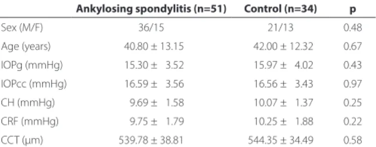
The demographic characteristics and corneal biomechanical features of study participants are summarized in table 1. In the AS group, the mean disease duration was 7.73 ± 6.05 years (range, 1-30 years). The mean values (ranges) of the BASFI, BASDAI, and BASMI scores were 1.46 ± 1.34 (0-5.10), 2.15 ± 1.27 (0.10-4.60), and 2.33 ± 1.56 (0.40-7.20), respectively. HLA B27 was positive in 41 (80%) of the AS group. Correlation analysis revealed positive correlations of IOPg and IOPcc with age in the AS group (r=0.30, p=0.03 and r=0.36, p=0.01, respectively), and negative correlations between CH and disease duration (r=-0.32, p=0.02) and between CCT BASMI (r=-0.31, p=0.03) (Table 2). Correlation analysis was applied to the control group to assess how corneal biomechanical features were associated with age. This showed a positive correlation between IOPcc and age (r=0.45, p=0.005) (Table 3).
DISCUSSION
In this study, we compared corneal biomechanical features and CCT between patients with AS and healthy controls. This did not reveal any diferences between the groups in IOPg, IOPcc, CH, CRF, or CCT, but IOPcc showed a positive correlation with age in both the AS and the control group. In patients with AS, there were negative correlations between CH and the disease duration and between CCT and the patients’ BASMI scores(22).
Ocular involvement is one of the most common extra-articular findings in AS patients. Although the pathogenesis of eye disease in AS has yet to be clearly identified, it has been suggested that environmental triggers and the activation of both innate and adaptive immunological systems may be implicated in genetically suscep-tible individuals(23,24). Acute anterior uveitis has been reported in about 30%-40% of individuals with AS and is strongly associated with HLA-B27 positivity(25). Furthermore, it has been observed that AS patients with uveitis had worse BASDAI and BASFI scores(26). Be-cause the effects of uveitis may be the topic of a future study, we did not include two patients with active uveitis and six patients with a previous history of uveitis in this study.
Data about corneal changes in AS patients are limited. A recent study evaluated the corneal parameters of 57 patients with AS by Scheimplug imaging and compared the results with 57 healthy control cases, observing that the mean CCT and corneal volume were reduced signiicantly in AS patients along with the Schirmer test
Table 1. The demographic features and corneal biomechanical features of study participants
Ankylosing spondylitis (n=51) Control (n=34) p
Sex (M/F) 36/15 21/13 0.48
Age (years) 040.80 ± 13.15 042.00 ± 12.32 0.67 IOPg (mmHg) 015.30 ± 03.52 015.97 ± 04.02 0.43
IOPcc (mmHg) 016.59 ± 03.56 016.56 ± 03.43 0.97
CH (mmHg) 009.69 ± 01.58 010.07 ± 01.37 0.25 CRF (mmHg) 009.75 ± 01.79 010.25 ± 01.88 0.22
CCT (µm) 539.78 ± 38.81 544.35 ± 34.49 0.58
Ca b u k kS, e t a l.
3 1 1 Arq Bras Oftalmol. 2017;80(5):309-12 Table 2. The results of the correlation analysis for the AS patients
IOPg (mmHg) IOPcc (mmHg) CH CRF CCT (µm)
Age
p -0.03 -0.01 -0.28 -0.67 -0.69
r -0.30 -0.36 -0.15 -0.06 -0.06
Disease duration
p -0.77 0.16 -0.02 -0.12 -0.11
r -0.04 -0.20 -0.32 -0.22 -0.23
BASFI
p -0.70 -0.40 -0.34 -0.58 -0.66
r -0.05 -0.12 -0.14 -0.08 -0.06
BASDAI
p -0.56 -0.29 -0.29 -0.61 -0.30
r -0.08 -0.15 -0.15 -0.07 -0.15
BASMI
p -0.77 -0.74 -0.24 -0.28 -0.03
r -0.04 -0.05 -0.17 -0.15 -0.31
HLA B27 positivity
p -0.33 -0.10 -0.14 -0.59 -0.71
r -0.14 -0.23 -0.21 -0.08 -0.05
BASFI= Bath Ankylosing Spondylitis Functional Index; BASDAI= Bath Ankylosing Spondylitis Disease Activity Index; BASMI= Bath Ankylosing Spondylitis Metrology Index; IOPg= Goldmann-correlated intraocular pressure; IOPcc= corneal compensated intraocular pressure; CH= corneal hysteresis; CRF= corneal resistance factor; CCT= central corneal thickness. The correlation coeicients (r) and p values were determined with Pearson correlation analysis, r= Perason’s correlation coeicient.
scores (disease duration, 60.0 months; range, 25.5–156.0 months; BASDAI, 3.68 ± 2.66)(15). Another study included 68 AS patients and 61 age-matched controls and reported that CCT was signiicantly decreased in the AS group (disease duration, 5.42 years; range, 1-25 years)(16). In our study, we also observed a decrease in CCT values in the AS group although the diference between the two groups was not statistically signiicant.
In the present study, we observed a positive correlation between IOPcc and age in both the AS and the control groups. Similarly, another recent study reported a signiicant association of both IOPg and IOPcc with older age in a large cohort(27). This association of increased IOP with older age should be kept in mind for both healthy individuals and patients with AS, especially during patient screening.
CH relects the ability of the corneal stromal tissue to absorb and disperse energy(11). In this study, CH decreased with longer durations of AS. Although the exact pathogenesis is not clearly understood, this decrease in CH may be associated with structural changes in proteoglycans and glycosaminoglycans, that preserve the elasticity and rigidity of the corneal stroma(28). The mean disease duration for the patients in the present study was 7.73 ± 6.05 years; these results suggest that, in later periods of the disease, CH would change
further, resulting in signiicant diferences with the healthy controls. Glaucoma is a chronic progressive disease that increases with age(29). Decreased CH has been deined a risk factor for all types of glaucoma and for progressive glaucomatous optic neuropathy(30). Comprehen-sive studies are warranted to determine whether decreased CH is associated with glaucoma in the later stages of AS.
BASFI, BASDAI, and BASMI are the three main determinants of function, disease activity, and metrology in AS. In this study, only CCT was negatively correlated with BASMI scores, with no other associa-tions found between corneal biomechanical features and the clinical activity index scores. The negative association between CCT and BASMI scores may be related to the indings of previous studies that also reported decreased CCT in AS patients. Clearly, if more severe AS cases are involved in a study, more diminished CCT values would be expected, which would result in a signiicant diference between AS patients and healthy controls. CCT is especially important when evaluating IOP, and a decreased CCT value may be associated with a lower IOP readings. When evaluating patients at an advanced stage of AS, clinicians should be aware of this condition and the increased risk of glaucoma being overlooked(31).
The main limitation of this study was the low number of patients. Determining the disease duration as “from the beginning of the irst AS-related symptoms” could potentially have resulted in inaccuracy. For the patients in this study, the disease durations were generally not long and their disease severity scores were not very high, which may also have inluenced the results.
In conclusion, we showed a signiicant negative correlation between CH and disease duration in AS patients. CCT decreased with higher BASMI scores, which could result in an underestimate of IOP readings and thus in an inaccurate risk assessment for glaucoma.
REFERENCES
1. Sieper J, Braun J, Rudwaleit M, Boonen A, Zink A. Ankylosing spondylitis: an overview. Ann Rheum Dis. 2002;61 3 Suppl 3):iii8-18.
Table 3. The results of correlation analysis performed for the control group
IOPg (mmHg) IOPcc (mmHg) CH CRF CCT (µm)
Age
p 0.07 0.005 -0.46 0.41 0.87
Coeicient 0.31 0.450 -0.13 0.14 0.03
Co r n e a lb i o m e C h a n i C a lf e at u r e s i npat i e n t sw i t ha n k y l o s i n gs p o n d y l i t i s
3 1 2 Arq Bras Oftalmol. 2017;80(5):309-12
2. Garrett S, Jenkinson T, Kennedy LG, Whitelock H, Gaisford P, Calin A. A new approach to deining disease status in ankylosing spondylitis: the Bath Ankylosing Spondylitis Disease Activity Index. J Rheumatol. 1994;21(12):2286-91.
3. Calin A, Garrett S, Whitelock H, Kennedy LG, O’Hea J, Mallorie P, et al. A new approach to deining functional ability in ankylosing spondylitis: the development of the Bath Ankylosing Spondylitis Functional Index. J Rheumatol. 1994;21(12):2281-5. 4. Jenkinson TR, Mallorie PA, Whitelock HC, Kennedy LG, Garrett SL, Calin A. Deining
spinal mobility in ankylosing spondylitis (AS). The Bath AS Metrology Index. J Rheuma-tol. 1994;21(9):1694-8.
5. Jakob E, Reuland MS, Mackensen F, Harsch N, Fleckenstein M, Lorenz HM, et al. Uveitis subtypes in a german interdisciplinary uveitis center-analysis of 1916 patients. J Rheu-matol, 2009;36(1):127-36.
6. Zeboulon N, Dougados M, Gossec L. Prevalence and characteristics of uveitis in the spondyloarthropathies: a systematic literature review. Ann Rheum Dis. 2008;67(7): 955-9.
7. Sommer A, Tielsch JM, Katz J, Quigley HA, Gottsch JD, Javitt J, et al. Relationship between intraocular pressure and primary open angle glaucoma among white and black Americans. The Baltimore Eye Survey. Arch Ophthalmol. 1991;109(8):1090-5. 8. Gordon MO, Beiser JA, Brandt JD, Heuer DK, Higginbotham EJ, Johnson CA, et al. The
Ocular Hypertension Treatment Study: baseline factors that predict the onset of pri-mary open-angle glaucoma. Arch Ophthalmol. 2002;120(6):714-20; discussion 829-30. 9. Ocular Hypertension Treatment Study Group.; European Glaucoma Prevention Study Group., Gordon MO, Torri V, Miglior S, Beiser JA, Floriani I, Miller JP, Gao F, Adamsons I, Poli D, D’Agostino RB, Kass MA. Validated prediction model for the development of primary open-angle glaucoma in individuals with ocular hypertension. Ophthalmo-logy. 2007;114(1):10-9.
10. Valbon BF, Guerra F, Silva RS, Canedo AL, Ambrósio Júnior R. Hipertensão ocular “mascarada” por edema de córnea após cirurgia de catarata. Rev Bras Oftalmol. 2009; 68(6):348-54.
11. Deol M, Taylor DA, Radclife NM. Corneal hysteresis and its relevance to glaucoma. Curr Opin Ophthalmol. 2015;26(2):96-102.
12. Yazici AT, Kara N, Yüksel K, Altinkaynak H, Baz O, Bozkurt E, et al. The biomechanical properties of the cornea in patients with systemic lupus erythematosus. Eye (Lond). 2011;25(8):1005-9.
13. Prata TS, Sousa AK, Garcia Filho CA, Doi LM, Paranhos A Jr. Assessment of corneal bio-mechanical properties and intraocular pressure in patients with rheumatoid arthritis. Can J Ophthalmol. 2009;44(5):602.
14. Gomes BA, Santhiago MR, Jorge PA, Kara-José N, Moraes HV Jr, Kara-Junior N. Corneal involvement in systemic inlammatory diseases. Eye Contact Lens. 2015;41(3):141-4.
15. Gunes A, Erkol Inal E, Tok L, Tok O. Assessment of corneal parameters with Scheimplug imaging in patients with ankylosing spondylitis. Semin Ophthalmol. 2017;32(3):276-80. 16. Ortak H, Inanır A, Demir S, Uysal A, Şahin Ş, Sağcan M, et al. Decreased central corneal
thickness in ankylosing spondylitis. Int Ophthalmol. 2014;34(2):263-8.
17. van der Linden S, Valkenburg HA, Cats A. Evaluation of diagnostic criteria for ankylosing spondylitis. A proposal for modiication of the New York criteria. Arthritis Rheum. 1984; 27(4):361-8.
18. Feldtkeller E, Erlendsson J. Deinition of disease duration in ankylosing spondylitis. Rheumatol Int. 2008;28(7):693-6.
19. Morita T, Shoji N, Kamiya K, Hagishima M, Fujimura F, Shimizu K. Intraocular pressure measured by dynamic contour tonometer and ocular response analyzer in normal tension glaucoma. Graefes Arch Clin Exp Ophthalmol. 2010;248(1):73-7.
20. Shah S, Laiquzzaman M, Cunlife I, Mantry S. The use of the Reichert ocular response analyser to establish the relationship between ocular hysteresis, corneal resistance factor and central corneal thickness in normal eyes. Cont Lens Anterior Eye. 2006; 29(5):257-62.
21. Mandalos A, Anastasopoulos E, Makris L, Dervenis N, Kilintzis V, Topouzis F. Inter-exa-miner reproducibility of Ocular Response Analyzer using the waveform score quality index in healthy subjects. J Glaucoma. 2013;22(2):152-5.
22. Lam AK, Chen D, Tse J. The usefulness of waveform score from the ocular response analyzer. Optom Vis Sci. 2010;87(3):195-9.
23. Gouveia EB, Elmann D, Morales MS. Ankylosing spondylitis and uveitis: overview. Rev Bras Reumatol. 2012;52(5):742-56.
24. Wendling D. Uveitis in seronegative arthritis. Curr Rheumatol Rep. 2012;14(5):402-8. 25. Robinson PC, Brown MA. Genetics of ankylosing spondylitis. Mol Immunol. 2014;
57(1):2-11.
26. Martin TM, Rosenbaum JT. An update on the genetics of HLA B27-associated acute anterior uveitis. Ocul Immunol Inlamm. 2011;19(2):108-14.
27. Chan MP, Grossi CM, Khawaja AP, Yip JL, Khaw KT, Patel PJ, Khaw PT, Moran JE, Vernan SA, Foster PJ; UK Biobank Eye and Vision Consortium. Associations with Intraocular Pressure in a Large Cohort: Results from the UK Biobank. Ophthalmology. 2016;123(4): 771-82.
28. Kotecha A. What biomechanical properties of the cornea are relevant for the clini-cian? Surv Ophthalmol. 2007;52(6 Suppl 2):S109-14.
29. Guedes G, Tsai JC, Loewen NA. Glaucoma and aging. Curr Aging Sci. 2011;4(2):110-7. 30. Chee RI, Silva FQ, Ehrlich JR, Radclife NM. Agreement of licker chronoscopy for struc-tural glaucomatous progression detection and factors associated with progression. Am J Ophthalmol. 2013;155(6):983-90.