1
Original Article
Echocardiographic Monitoring of Balloon Atrial
Septostomy
Carlos Henrique de Marchi, Moacir Fernandes de Godoy, Márcio Antônio dos Santos,
Airton Camacho Moscardini, Sírio Hassen Sobrinho, Ulisses Alexandre Croti
São José do Rio Preto, SP - Brazil
Faculdade de Medicina de São José do Rio Preto
Mailing address: Carlos Henrique de Marchi – Al. Dr. Ulysses da Silveira Guimarães, 273 - Cep 15061-723 - São José do Rio Preto, SP, Brazil – E-mail: [email protected]
Received for publication: 02/20/2004 Accepted for publication: 08/13/2004 English version by Stela Maris Costalonga
Objective
To assess balloon atrial septostomy monitored with echocar-diography.
Methods
From August 1997 to January 2004, 31 infants with conge-nital heart diseases indicated for balloon atrial septostomy under-went the procedure with exclusive echocardiographic monitoring. Success was defined as the obtainment of an atrial septal defect diameter ≥ 4 mm and ample mobility of its margins.
Results
The male sex predominated (83.9%). The median age was 5 days (1 – 150), and the median weight was 3,300 g (1,800 -7,500). Transposition of the great arteries occurred in 80.6% of the patients, tricuspid valve atresia in 12.9%, total anomalous pulmonary venous drainage in 3.2%, and pulmonary atresia with intact septum in 3.2%. The procedure was successful in all patients. The size of the atrial septal defect increased from 1.8 ± 0.8 mm to 5.8 ± 1.3 mm (P < 0.0001) and oxygen arterial saturation from 64.5 ± 18.9% to 85.1 ± 9.2% (P < 0.0001). The following complications occurred: 3 balloon rup-tures, one lesion of the right femoral vein, one supraventricular tachycardia, and one atrial flutter.
Conclusion
Balloon atrial septostomy monitored with echocardiography is a safe and effective procedure. It may be performed at bedside, avoiding transporting of the patient, identifies the catheter loca-tion, reduces the occurrence of severe complications, and asses-ses the immediate result of the procedure.
Key words
congenital heart disease; echocardiography; balloon atrial septostomy
The survival of children with certain congenital heart diseases depends on the presence of an adequate atrial septal defect. Balloon atrial septostomy is a palliative technique that enables blood cir-culation in those children by creating or widening an atrial septal defect, enabling a better blood mixture in the atria.
Surgical removal of part of the atrial septum to create commu-nication was described by Blalock and Hanlon 1 in 1950. A great
landmark in the treatment of transposition of the great arteries (TGA) and interventionist catheterization occurred when Rashkind and Miller 2 performed, in the hemodynamics laboratory, the first
atrial septostomy with a balloon catheter introduced through the femoral vein without requiring a thoracotomy.
Classically described for the treatment of transposition of the great arteries, balloon atrial septostomy has also proved to be effective in patients with tricuspid atresia, total anomalous pul-monary venous drainage, pulpul-monary atresia with intact ventricular septum, and hypoplastic left heart syndrome 3-6.
Although balloon atrial septostomy has provided satisfactory results, the procedure is not free from complications. Rashkind 7
reported the possible problems associated with the procedure, such as difficulty in the venous access for the introduction of 5.5-or 6.5-F catheters in neonates, perf5.5-oration of cardiac chambers, balloon rupture with embolism of rubber fragments, and lesions in the pulmonary vein, right and left atrioventricular valves, and infe-rior vena cava.
Perry et al 8 reported the use of echocardiography to help in
locating and placing venous or arterial catheters in children. Even in the hemodynamics laboratory, echocardiography could contribute to positioning the catheters and decreasing the time of radiation exposure, mainly in complex heart diseases. After editing the arti-cle, the authors added a note reporting the echocardiographic follow-up of 3 balloon atrial septostomies.
The use of echocardiography for monitoring balloon atrial sep-tostomy was initially reported by Matsunaga et al 9 in Japan, who
performed the procedure in 8 children, 6 of whom had d-transpo-sition of the great arteries, and 2 had total anomalous pulmonary venous drainage. They were followed by Allan et al 10 in England
and Perry et al 11 in the United States of America.
The possibility of performing the procedure more rapidly in neonatal intensive care units with the need for neither transporting high-risk neonates nor using radiation has been reported by Baker et al 12. The latter authors performed balloon atrial septostomy at
2
In Brazil, the performance of balloon atrial septostomy at bed-side under echocardiographic control has been reported by Serra et al 13. The procedure was performed in 9 newborn infants, 8 of
whom had transposition of the great arteries and one had pulmo-nary atresia with intact ventricular septum. The lack of complica-tions, wide atrial septal defect, and clinical improvement were results obtained in all cases. Mattos et al 14, in Brazil, performed
atrial septostomy in the intensive care unit in 3 neonates, 2 of whom had transposition of the great arteries and one had tricuspid atresia. The procedure was unsuccessful in only one 36-day-old patient, who might have had stiffness of the atrial septum.
This study aimed at assessing the efficacy and safety of balloon atrial septostomy monitored with echocardiography in a pediatric cardiological unit of the Hospital Universitário.
Methods
This study was approved by the committee on ethics in research of our institution according to resolution 196/96 of the CNS. The procedure was performed after the guardians of the children gave written informed consent.
From August 1997 to January 2004, 31 children with conge-nital diseases underwent balloon atrial septostomy monitored exclu-sively with echocardiography. Twenty-eight procedures were per-formed at our institution’s hospital, and 3 procedures were perfor-med at other hospitals in the city.
We selected children with the echocardiographic diagnosis of congenital heart disease associated with atrial septal defect or restrictive oval foramen of an insufficient size to allow adequate blood mixture and to assure circulation. Atrial septal defect or oval foramen was considered restrictive when their diameter on echocardiography was smaller than 4 mm 15.
Based on the clinical suspicion of congenital heart disease, a 2-dimensional transthoracic echocardiography with color flow map-ping was performed at bedside. The examinations performed at our hospital used an ATL device (Advanced Technology Laboratory, Bothell, WA), Apogee CX 200 model, with a 5-mHz mechanical transducer. The examinations performed at other hospitals used the Esaotebiomédica device (Florence, Italy), SIM 7000 CFM mo-del, with a 5-mHz mechanical transducer.
Standard echocardiographic windows and views were used, according to the recommendations of the American Society of Echocardiography 16,17, and sequential segmentary analysis was
performed 18.
Simple transposition of the great arteries was defined as the congenital heart disease with atrioventricular concordance, ven-triculoarterial discordance, and possible associations with patent ductus arteriosus, patent oval foramen, or ventricular septal defect < 3 mm, or both 19.
The cyanotic neonates treated after the year 2000 received prostaglandin E1 at the dosage of 0.01 to 0.1 µg/kg/min in con-tinuous intravenous infusion, even before diagnostic confirmation. All children were under continuous monitoring of oxygen arte-rial saturation through pulse oximetry obtained with a Dixtal monitor (Dixtal Biomédica Ind. e Com. Ltda, Manaus, AM, Brazil), DX 2405 model. For analysis purposes, the oximetry values considered were those obtained immediately before the procedure (Sat 1) and approximately 30 minutes after its end (Sat 2).
The procedure was performed at bedside in patients whose diagnostic echocardiography was sufficient to provide the necessary information. The procedure was performed at the hemodynamics laboratory when complementation with angiographic study was required, such as in patients diagnosed with total anomalous pul-monary venous drainage, in those with transposition of the great arteries, whose echocardiogram did not precisely define the pattern of coronary circulation, and in those who required a better asses-sment of the pulmonary branches.
The routine for performing balloon atrial septostomy was as follows: a) patient placed in the dorsal decubitus position in a warmed crib, if a neonate; b) sedation with oral chloral hydrate at the dosage of 50 mg/kg/dose, if the patient was on spontaneous ventilation, or intravenous midazolam at the dosage of 0.1 mg/ kg, if the patient was on mechanical ventilation; c) lower limbs fixed in abduction; d) asepsis and antisepsis of the right inguinal region; e) anesthetic infiltration of the right inguinal region with 2% lidocaine; f) preferential dissection of the right saphenous-femoral venous junction; the umbilical route was an alternative access; g) introduction of the 5-F or 6-F Rashkind catheter, whose models were the Rashkind balloon (USCI-CR Bard, Inc, Billerica, MA) and the Fogarty balloon catheter for atrial septostomy (Ed-wards-Baxter Healthcare); h) the procedure’s echocardiographic monitoring was initiated with the echocardiographer wearing glo-ves, positioned contralaterally to the hemodynamics professional; the echocardiographic transducer was disinfected with povidone-iodine solution and placed on the subcostal window to show the anatomical details of venous drainage and the atria; i) under echo-cardiographic monitoring, the atrial septostomy catheter was pas-sed through the inferior vena cava to the right atrium and oval foramen, then the left atrium, where it was inflated with saline solution, being then rapidly pulled back to the right atrium, and deflated; j) the following parameters were estimated with the aid of echocardiography: diameter of the inflated balloon, mobility of the margins, and diameter of the atrial septal defect after each procedure (the abbreviation ASD1 was used for atrial septal defect or oval foramen before the procedure, and ASD2 for the atrial septal defect resulting from the procedure); l) the initial procedure was performed with an inflated balloon diameter of 11 to 12 mm, which was progressively increased until the maximum limit of 15 to 16 mm.
The procedure was repeated until meeting the criterion of satisfaction, ie, the obtainment of a satisfactory atrial septal defect, defined as that whose diameter was ≥ 4 mm and whose margins had wide mobility 15. The echocardiographic examinations were
recorded on videotapes.
3
Results
Of the 31 infants undergoing echocardiographically assisted balloon atrial septostomy, 26 (83.9%) were of the male sex, and 5 (16.1%) were of the female sex.
Their mean age was 14.6±28.3 days, the median being 5 days (range: 1 to 150 days).
The mean weight was 3,328.8±947.1 g, and the median was 3,300 g (range, 1,800 to 7,500 g).
Transposition of the great arteries was the most frequent anatomi-cal diagnosis found in 25 (80.6%) infants, followed by tricuspid atresia in 4 (12.9%), total anomalous pulmonary venous drainage in one (3.2%), and pulmonary atresia with intact septum in one (3.2%).
Of the 25 patients with transposition of the great arteries, 16 were of the simple type (absence or ventricular septal defect or ventricular septal defect < 3 mm), and 9 had the association of ventricular septal defect ≥ 3 mm. Of the cases cited, one also had coarctation of the aorta, and 2 had pulmonary stenosis, one of the valvular type and another of the subvalvular type. The patient with pulmonary valve stenosis also had disconnected pulmonary arteries. Of the infants with tricuspid atresia, 3 had ventriculoarterial concordance with pulmonary stenosis, and the other had ventri-culoarterial discordance without pulmonary stenosis.
The case of total anomalous pulmonary venous drainage was of the supracardiac type.
Twenty (64.5%) infants were on prostaglandin E1 at the time of the procedure. The following 11 patients, who were treated before the year 2000, were not using that medication: one infant with total anomalous pulmonary venous drainage; 4 infants with tricuspid atresia; and 6 infants with transposition of the great arteries. Seventeen (54.8%) infants were on assisted ventilation with orotracheal intubation, and the remaining infants were on sponta-neous ventilation with oxygen supplementation.
Twenty-seven (87%) infants underwent the procedures at bed-side in the ICU, except for the following: the infant with the total anomalous pulmonary venous drainage; the infant with tricuspid atresia; the infant with transposition of the great arteries whose coronary anatomy remained doubtful on echocardiography; and the infant with transposition of the great arteries associated with pulmonary valve stenosis and nonconfluent pulmonary arteries. In those cases, balloon atrial septostomy was performed in the he-modynamics laboratory at the time of cardiac catheterization, but also under exclusive echocardiographic control. Of the proce-dures performed at bedside, 3 were performed at hospitals that did not belong to our institution, but by the same team of hemo-dynamics professionals and echocardiographers. That was due to the severity of the patients’ clinical findings, which did not allow the transfer of patients between hospitals.
The femoral access was used in 30 (96.8%) infants, and, in only one, the catheter was introduced through the umbilical route. Echocardiography monitored and guided the position of the balloon catheter from the inferior vena cava until the left atrium. Undesired trajectories, such as to the superior vena cava, right or left ventricles, and loop formation in the right atrium, were im-mediately identified and corrected. Figure 1 shows the steps of the echocardiographically monitored procedure.
Figure 2 depicts an atrial septal defect resulting from balloon atrial septostomy observed during the Jatene surgery 5 days after the procedure, evidencing the satisfactory result obtained with rupture of the membrane of the fossa ovalis.
Thirty infants were considered for the statistical analysis of the size of the atrial septal defect, oxygen arterial saturation, and number of tractions. Patient #1, who marked the beginning of
A A A A A BBBBB
C C C C C DDDDD LA
LA LA LA LA
OF OF OF OF OF
A D A D A D A D A D
RA RA RA RA RA
BC BC BC BC BC
LA LA LA LA LA
A D A DA D A D A D
RA RA RA RA RA
ASD ASDASD ASDASD
LA LA LA LA LA
Fig. 1 – Echocardiography monitored balloon atrial septostomy. A) coronal subcostal view showing a patent oval foramen. B) catheter (clear arrow) in the inferior vena cava and RA; C) inflated balloon in the LA; D) resultant ASD with mobility of its margins (solid arrow). RA- right atrium, LA- left atrium, OF- oval foramen, BC- balloon catheter, and ASD- atrial septal defect.
Fig. 2 – Aspect of the atrial septal defect 5 days after echocardiography monitored balloon atrial septostomy. B shows a pair of tweezers pulling the membrane of the torn oval fossa. RA- right atrium, ASD- atrial septal defect.
RA
RA
RA
RA
RA
RA
RA
RA
RA
RA
4
T T T T
Table I - Vable I - Vable I - Vable I - Vable I - Variables of echocardiography monitored balloon atrial septostomyariables of echocardiography monitored balloon atrial septostomyariables of echocardiography monitored balloon atrial septostomyariables of echocardiography monitored balloon atrial septostomyariables of echocardiography monitored balloon atrial septostomy
Case Place Ventilation PG Access ASD ASD Sat Sat NP Complications
E1 1 2 1 2
1 bed Intubated no femoral na na na na Na no 2 bed Intubated no femoral 1 6 30 70 3 no 3 bed Spontaneous no femoral 3 7 70 88 4 no 4 bed Intubated no femoral 1 5 76 93 6 no 5 bed* Intubated no femoral 3 8 80 85 6 no 6 bed Intubated no femoral 1 9 35 55 5 no 7 bed Spontaneous no femoral 1 5 70 85 4 NSSVT and balloon rupture 8 bed Spontaneous yes femoral 2 7 30 70 6 no 9 hemo Spontaneous no femoral 2 8 90 90 5 no 10 bed Intubated yes femoral 2 6 40 80 4 no 11 hemo Spontaneous yes femoral 1 5 70 90 6 no 12 bed Spontaneous no femoral 2 4 86 92 7 balloon rupture 13 bed Intubated yes femoral 4 5 85 90 5 atrial flutter 14 bed Spontaneous yes femoral 2 7 65 87 9 no 15 bed Spontaneous yes femoral 3 8 70 90 5 no 16 bed Intubated yes femoral 1 5 65 85 5 no 17 bed Intubated yes femoral 1 5 65 88 3 no 18 bed Intubated yes femoral 1 5 60 85 3 no 19 bed Spontaneous yes femoral 2 4 62 83 4 no 20 hemo Spontaneous yes femoral 2 7 70 95 3 no 21 bed Spontaneous yes femoral 2 7 60 90 5 no 22 bed Spontaneous no femoral 3 5 85 85 3 no 23 bed Intubated yes femoral 1 5 30 90 5 no 24 bed* Intubated yes umbilical 3 5 40 70 5 balloon rupture 25 bed Intubated yes femoral 1 6 36 75 4 no 26 bed* Spontaneous yes femoral 1 5 70 92 6 no 27 bed Intubated yes femoral 2 6 65 80 2 no 28 bed Intubated yes femoral 1 6 80 94 3 no 29 bed Intubated yes femoral 1 4 80 96 7 no 30 hemo Spontaneous no femoral 2 4 85 88 5 femoral vein laceration 31 bed Intubated yes femoral 1 6 85 93 3 no * procedures performed at hospitals not belonging to our institution; na- not available; PG- prostaglandin; ASD 1- initial atrial septal defect; ASD 2- final atrial septal defect; Sat 1- initial arterial oxygen saturation; Sat 2- final arterial oxygen saturation; NP- number of tractions of the balloon; NSSVT- nonsustained supraventricular tachycardia.
this study, was excluded for not having complete data. Table I shows the complete data of the patients.
Enlargement of the atrial septal defect size (fig. 3) to 4 mm or more was obtained in all patients, and it was the first satisfaction criterion for ending the procedure. The atrial septal defect size increased from 1.8 ± 0.8 mm to 5.8 ± 1.3 mm after the procedure (P < 0.0001).
Wide mobility of the margins of the atrial septal defect was obtained in all patients, and it was the second satisfaction criterion for ending the procedure.
Arterial oxygen saturation (fig. 4) increased from 64.5 ± 18.9% to 85.1 ± 9.2% (P < 0.0001) after the procedure. Only patients # 9 and 22 maintained the same saturation.
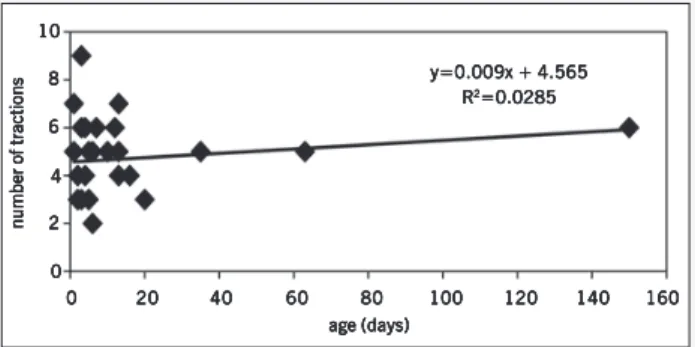
The number of tractions necessary for obtaining a satisfactory interatrial communication ranged from 2 to 9, with a median of 5. No correlation was observed between the patient’s age and the number of tractions (R2 = 0.0285) (fig. 5).
Complications related to the procedures occurred in 5 (16.1%) patients: 3 balloon ruptures, one supraventricular tachycardia, one atrial flutter, and a lesion in the right femoral vein. Patient #7 had supraventricular tachycardia and balloon rupture. All ca-ses of balloon rupture caused no embolic complications. The 2 cases of supraventricular arrhythmia were reverted only with manipulation of the catheter in the atrial cavities. Patient #30 had laceration of the right femoral vein during the introduction of the balloon catheter; after its ligature, the procedure was performed through the contralateral route, with no further damage to the right lower limb.
7 6 5 4 3 2 1 0
ASD 1 ASD 2
Fig. 3 – Enlargement of the size of the atrial septal defect after echocardiography monitored balloon atrial septostomy, ASD 1: initial atrial septal defect in mm, ASD 2: final atrial septal defect in mm. Means and standard deviation.
Discussion
5
90 85 80 75 70 65 60 55 50 45 40 35 30 25 20 15 10 5
0
Fig. 4 – Increase in arterial oxygen saturation after echocardiography monitored balloon atrial septostomy. Sat 1: initial saturation in%, Sat 2: final saturation in%. Means and standard deviation.
Sat 1 Sat 2
The older patient was the one with tricuspid atresia with ventriculoarterial concordance and pulmonary stenosis, who under-went echocardiography monitored balloon atrial septostomy at the age of 150 days. Despite the advanced age, a satisfactory interatrial communication of 8 mm was obtained after 6 tractions of the balloon. The child is on a 4-year follow-up, has completed the second stage of the Fontan operation (total cavopulmonary anastomosis), and is currently in good clinical condition.
Balloon atrial septostomy has been proved to be effective in infants below the age of 2 months 4 due to the lower resistance of
their atrial septum; Jamjureeruk et al 20 have already reported
successful procedures in infants up to the age of 6 months in a study with 47 infants with various diagnoses and ages ranging from 31 to 180 (mean, 83.5 ± 48.5) days.
Echocardiographic monitoring of balloon atrial septostomy allows the procedure to be performed in a more appropriate environment for the patient: at bedside in the ICU or even at medical services away from the referral center. Thus, it provides important advan-tages, such as avoiding the transportation of severely ill children with heart disease and reducing the time preceding the procedure. Recently, Martin et al 21 have assessed the advantages of
transporting the team of specialists of the Real Brompton Hospi-tal in London to the local neonaHospi-tal ICU, where the child with transposition of the great arteries was hospitalized to undergo echocardiographically monitored balloon atrial septostomy, thus avoiding the emergency transference of the child to the specialized hospital, with clinical, social, and organizational advantages of the specialized centers.
Even when cardiac catheterization was indicated, such as for infants with total anomalous pulmonary venous drainage and trans-position of the great arteries, for defining the anatomy of the coronary or pulmonary arteries, the use of echocardiography resul-ted in a shorter time of exposure to radiation, greater safety for positioning the balloon catheter, and immediate assessment of the result.
Immediate assessment of the result at every traction of the balloon catheter, showing the size of the interatrial communication obtained and the mobility of its margins, contributes by avoiding the interruption of the procedure after only stretching the interatrial communication or oval foramen without tearing the atrial septum. Baker et al 22, studied 43 children with transposition of the
great arteries undergoing balloon atrial septostomy by using only hemodynamic criteria, such as equalization of the interatrial pres-sures, lack of resistance to traction of the inflated balloon, enlar-gement of the interatrial communication through balloon measu-rement, and improvement in arterial oxygen saturation. Those authors found a small interatrial communication in the postmortem examination, and another interatrial communication was no longer detected in the hemodynamic study performed due to progressive cyanosis, despite an initially satisfactory result. The authors con-cluded that an initially adequate interatrial communication does not guarantee long-term satisfactory results.
Mullins et al 4 studied 12 children with total anomalous
pul-monary venous drainage undergoing balloon atrial septostomy without echocardiographic monitoring. Two of the children did not benefit from the procedure, and early surgical intervention was required when a small resultant interatrial communication was found, although the mean interatrial gradient was lower than one mmHg after septostomy in both. The lack of an interatrial pressure gradient even with a small interatrial communication may be consequent to a greater right atrial compliance. Balloon atrial septostomy in children with total anomalous pulmonary venous drainage is technically more difficult than in other cardiac defects, due to the small left atrial size and the difficulty in locating the catheter in the absence of usual hemodynamic criteria to identify the left atrium (presence of pulmonary veins, greater pres-sure, and greater oxygen saturation). Echocardiography plays an important role in such cases.
Both the enlargement of the atrial septal defect size and the elevation in the oxygen arterial saturation were statistically signi-ficant. Clinical and hemodynamic improvements have also been observed in all patients, mainly in the one with anomalous pulmonary venous drainage.
The possibility of identifying the catheter inside the heart and its relation with the cardiac structures defined on echocardiography makes the procedure undoubtedly safer, reducing the risk of per-forations or lesions in the atrioventricular valves. Echocardiography may have contributed to the small number of complications related to the procedure (16.1%) and the absence of severe events in our study. The supraventricular arrhythmias had a short duration and were reverted only with the manipulation of the catheter inside the atria. The lesion in the right femoral vein evolved satisfactorily with no complication in the lower limb. The cases of rupture of the balloon resulted from problems with the material and were not directly related to the technique of the procedure.
Currently, the economic factor is also a point of interest in
number of tractionsnumber of tractionsnumber of tractionsnumber of tractionsnumber of tractions
10 10 10 10 10 8 8 8 8 8 6 6 6 6 6 4 44 4 4 2 22 22 0 00 0 0
y=0.009x + 4.565 y=0.009x + 4.565y=0.009x + 4.565 y=0.009x + 4.565 y=0.009x + 4.565
R R R R R22222=0.0285=0.0285=0.0285=0.0285=0.0285
0 0 0 0
0 2020202020 4040404040 6060606060 8080808080 100100100100100 120120120120120 140140140140140 160160160160160 age (days)
age (days)age (days) age (days)age (days)
6
1. Blalock A, Hanlon CR. The surgical treatment of complete transposition of the aorta and the pulmonary artery. Surg Gynec Obstet 1950; 90: 1-15.
2. Rashkind WJ, Miller WW. Creation of atrial septal defect without thoracotomy. JAMA 1966; 196: 991-2.
3. Mattos SS, Rodrigues JV, Severi R et al. Management of tricuspid atresia in neona-tes. Report of three cases and review of literature. J Pediatr (Rio J) 1994;70:33-8. 4. Mullins CE, el-Said GM, Neches WH et al. Balloon atrial septostomy for total
ano-malous pulmonary venous return. Br Heart J 1973; 35: 752-7.
5. Shams A, Fowler RS, Trusler GA, Keith JD, Mustard WT. Pulmonary atresia with intact ventricular septum: report of 50 cases. Pediatrics 1971;47:370-7. 6. Barbero Marcial M, Tanamati C. Síndrome de hipoplasia do coração esquerdo. In:
Santana MVT, editor. Cardiopatias Congênitas no Recém-Nascido Diagnóstico e Tratamento. São Paulo: Atheneu; 2000; 123-32.
7. Rashkind WJ. The complications of balloon atrioseptostomy. J Pediatrics 1970; 76: 649-50.
8. Perry LW, Galioto FM Jr, Blair T, Shapiro SR, Ruckman RN, Scott LP. Two-dimen-sional echocardiography for catheter location and placement in infants and chil-dren. Pediatrics 1981; 67: 541-7.
9. Matsunaga S, Suzuki K, Ichinose E et al. Application of two dimensional echocar-diography for the intracardic manipulation: the evaluation of atrial septal move-ment before and after ballon atrial septostomy. J Cardiogr 1981; 11:217-24. 10. Allan LD, Leanage R, Wainwright R, Joseph MC, Tynan M. Ballon atrial
septosto-my under two dimensional echocardiographic control. Br Heart J 1982; 47: 41-3. 11. Perry LW, Ruckman RN, Galioto FMJr, Shapiro SR, Potter BM, Scott LP.
Echocar-diographically assisted ballon atrial septostomy. Pediatrics 1982; 70:403-8. 12. Baker EJ, Allan LD, Tynan MJ, Jones OD, Joseph MC, Deverall PB. Ballon atrial
septostomy in the neonatal intensive care unit. Br Heart J 1984; 51: 377-8.
References
13. Serra A, Chamiê F, Paupério H et al. Atriosseptostomia com balão através da eco-cardiografia bidimensional. Arq Bras Cardiol 1988; 50: 179-82.
14. Mattos SS, Severi R, Marques DL et al. Atriosseptostomia em unidade de terapia intensiva sob monitorização ecocardiográfica. Experiência inicial do Instituto do Coração de Pernambuco. Arq Bras Cardiol 1993; 61: 33-6.
15. Baylen BG, Grzeszczak M, Gleason ME et al. Role of balloon atrial septostomy before early arterial switch repair of transposition of the great arteries. J Am Coll Cardiol 1992; 19: 1025-31.
16. Henry WL, DeMaria A, Gramiak R et al. Report of the American Society of Echocar-diography Committee on nomenclature and standards in two-dimensional echo-cardiography. Circulation 1980; 62: 212-8.
17. Snider AR, Serwer GA, Ritter SB, editores. Echocardiography in pediatric heart disease. 2o ed. St. Louis (Missouri): Mosby-Year Book; 1997.
18. Lucchese FA, Becker AE, Macartney FJ et al. Classificação das cardiopatias con-gênitas. Arq Bras Cardiol 1980; 35: 427-34.
19. Liebman J, Culum L, Belloc NB. Natural history of transposition of the great arte-ries. Circulation 1969; 40: 237-62.
20. Jamjureeruk V, Sangtawesin C, Layangool T. Balloon atrial septostomy under two-dimensional echocardiography control: a new outlook. Pediatr Cardiol 1997; 18: 197-200.
21. Martin AC, Rigby ML, Penny DJ, Redington AN. Bedside balloon atrial septostomy on neonatal units. Arch Dis Child Fetal Neonatal Ed 2003; 88: F339-40. 22. Baker F, Baker L, Zoltun R, Zuberbuhler JR. Effectiveness of Rashkind procedure in
transposition of the great arteries in infants. Circulation 1971; 43 (suppl 1): 1-6. 23. Zellers TM, Dixon K, Moake L, Wright J, Ramaciotti C. Bedside balloon atrial
sep-tostomy is safe, efficacious, and cost-effective compared with sepsep-tostomy per-formed in the cardiac catheterization laboratory. Am J Cardiol 2002; 89: 613-5.
medicine. In this regard, Zellers et al 23 have also shown that the
echocardiographic monitoring of balloon atrial septostomy at bed-side is advantageous, with a cost of practically half of that performed at the hemodynamics laboratory.
In conclusion, the echocardiographic monitoring of balloon atrial septostomy allows the procedure to be performed at bedside, avoiding the transportation of severely ill patients, and enabling the adequate positioning of the balloon catheter and its relation with the cardiac structures without the use of ionizing radiation. It also allows the immediate assessment of the result of the
pro-cedure after each traction of the balloon catheter, which makes the procedure effective and safe.