1N e u rology Division, Hospital das Clínicas, São Paulo University, São Paulo SP, Brazil (HC/FMUSP);2Radiology Department, HC/FMUSP Received 9 March 2005, received in final form 24 August 2005. Accepted 5 September 2005.
Dra. Adriana Bastos Conforto - Neurology Department / Hospital das Clínicas / São Paulo University - Avenida Dr. Enéas de Carv a l h o Aguiar 255/5131 - 05409-011 São Paulo SP - Brasil. E-mail: [email protected], [email protected]
FACIAL SENSORY SYMPTOMS
IN MEDULLARY INFARCTS
Adriana Bastos Conforto
1, Fábio Iuji Yamamoto
1, Cláudia da Costa Leite
2,
Milberto Scaff
1, Suely Kazue Nagahashi Marie
1ABSTRACT -Objective:To investigate the correlation between facial sensory abnormalities and lesional topography in eight patients with lateral medullary infarcts (LMIs). Method:We reviewed eight sequen-tial cases of LMIs admitted to the Neurology Division of Hospital das Clínicas/ São Paulo University between J u l y, 2001 and August, 2002 except for one patient who had admitted in 1996 and was still followed in 2002. All patients were submitted to conventional brain MRI including axial T1-, T2-weighted and Fluid attenuated inversion-re c o v e ry (FLAIR) sequences. MRIs were evaluated blindly to clinical features to deter-mine extension of the infarct to presumed topographies of the ventral trigeminothalamic (VTT), lateral spinothalamic, spinal trigeminal tracts and spinal trigeminal nucleus. Results:S e n s o ry symptoms or signs w e re ipsilateral to the bulbar infarct in 3 patients, contralateral in 4 and bilateral in 1. In all of our cases with exclusive contralateral facial sensory symptoms, infarcts had medial extensions that included the VTT t o p o g r a p h y. In cases with exclusive ipsilateral facial sensory abnormalities, infarcts affected lateral and posterior bulbar portions, with slight or no medial extension. The only patient who presented bilateral facial symptoms had an infarct that covered both medial and lateral, in addition to the posterior re g i o n of the medulla. Conclusion:Our results show a correlation between medial extension of LMIs and pres-ence of contralateral facial sensory symptoms.
KEY WORDS: ventral trigeminothalamic tract, spinal trigeminal nucleus, ischemic stroke, Wallenberg syn-drome.
Sintomas sensitivos na face em infartos medulares
RESUMO -Objetivo:Investigar a correlação entre alterações de sensibilidade na face e topografia lesional em oito pacientes com infartos bulbares laterais (IBLs). Método:Revisamos oito casos seqüenciais de IBLs admitidos na Divisão de Clínica Neurológica do Hospital das Clínicas da Universidade de São Paulo entre julho de 2001 e agosto de 2002, exceto por um caso que havia sido admitido em 1996 e estava sendo segui-do em 2002. Tosegui-dos os pacientes foram submetisegui-dos a ressonância magnética de encéfalo, incluinsegui-do seqüên-cias pesadas em T1, T2 e FLAIR. As ressonânseqüên-cias foram avaliadas por investigadores que não tiveram aces-so às características clínicas. Avaliaram-se as extensões dos infartos em relação às topografias presumidas dos tratos trigeminotalâmico ventral (TTV), espinotalâmico lateral, trigeminal espinhal e do núcleo trigemi-nal espinhal. Resultados:Sintomas ou sinais sensitivos foram ipsilaterais ao infarto bulbar em 3 pacientes, contralaterais em 4 e bilaterais em 1. Em todos os casos de comprometimento exclusivo da sensibilidade da hemiface contralateral, os infartos tiveram extensões mediais que incluíram a topografia do TTV. Em casos de anormalidades sensitivas faciais exclusivamente ipsilaterais, os infartos afetaram as porções late-ral e posterior do bulbo, com pouca ou nenhuma extensão medial. O único paciente que apresentou sin-tomas faciais bilaterais tinha um infarto comprometendo as porções medial e lateral, além da região pos-terior do bulbo. Conclusão:Nossos resultados mostram uma correlação entre extensão medial de IBLs e presença de sintomas sensitivos faciais contralaterais.
PA L AV R A S - C H AVE: trato trigeminotalâmico ventral, núcleo trigeminal espinhal, síndrome de Wa l l e n b e rg.
Wa l l e n b e rg ’s syndrome (WS) is usually caused by infarction of the lateral portion of the medul-la, more often caused by vertebral art e ry (VA) dis-e a s dis-e1 - 3. In classical WS, pain and temperature
sen-sation loss on the face is ipsilateral to the lesion
in the medulla. However, contralateral and bilat-eral sensory abnormalities may also occur4-7.
n e rve or ventral trigeminothalamic tract (VTT) are located in the posterolateral medulla1 , 4. The VTT
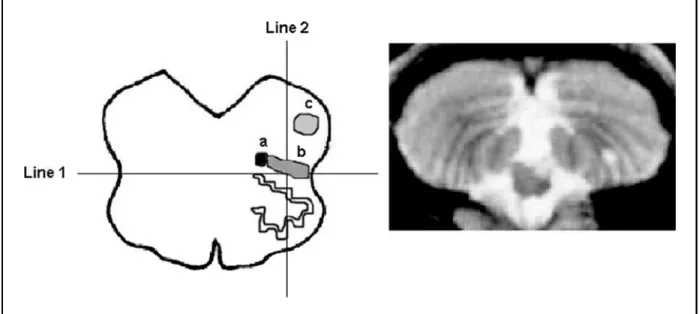
is positioned adjacent to the medial lemniscus or medial to the lateral spinothalamic tract (LST) in the dorsomedial corner of the inferior olive (Fig 1)4. DSN/T lesions are associated with decrease in
pain and temperature sensation on the ipsilater-al face, while injuries to the VTT crossing fibers p roduce diminished sensation on the contralater-al face4 , 8. Thus, it would be expected that infarc t s
extending medially and anteriorly in the dorsolat-eral medulla would cause pain and temperature s e n s o ryloss on the contralateral face, opposite to the side of the lesion.
Some studies re p o rted lateral medullary infarc t s (LMIs) to be more medially located in patients with contralateral facial pain/temperature sensory loss than in those with ipsilateral facial sensory abnor-m a l i t i e s4 , 9but others did not confirm these
find-i n g s8. We have investigated the correlation
bet-ween facial sensory abnormalities and involvement of the VTT topography in eight patients with me-dullary infarcts (MIs).
METHOD
T h e re were 5 men and 3 women ranging in age fro m 40 to 64 years (one less than 45 years), admitted to the N e u rology Division of Hospital das Clínicas/ São Paulo University between July, 2001 and August, 2002 except for one patient who had been admitted in 1996 and was still been followed in 2002. Vascular risk factors
w e re arterial hypertension (n=4), diabetes mellitus (n=2), smoking (n=6) and Chagas’ disease (n=1)1 0. All of the
patients were evaluated by the Neurology Division staff . Side and type of sensory findings on the face, arm and/or leg, as well as other neurological symptoms and signs w e re reviewed. Investigation included biochemical and s e rological testing, electro c a rdiogram, chest radiogra-p h y, echocardiogram, cervical Doradiogra-pradiogra-pler ultrasound, crani-al computed tomography (CT), magnetic resonance imag-ing (MRI) and magnetic resonance angiography (MRA). Digital subtraction angiography (DSA) and transcranial Doppler were perf o rmed in 4 and 2 patients, re s p e c t i v e-l y. Patients e-less than 45 years were submitted to exhaus-tive haematologic, immunologic and cere b rospinal flu-id analysis. Criteria of the Lausanne Stroke Registry were used to define presumed causes of infarc t i o n1 1.
All patients were submitted to conventional brain MRI including axial T1-, T2-weighted and Fluid attenu-ated inversion-re c o v e ry (FLAIR) sequences from a 1.5-T MR unit (GE Signa System, General Electric). Diffusion-weighted images (DWI) were performed in 3 patients. The investigator evaluated MRI lesional topography based on transverse anatomical templates of the medul-la oblongata4(Fig 1, left) was blind to patient history
and neurological examination. Two lines were drawn on the transverse MRI section demonstrating the MI. Line 1 divided each hemi-medulla in anterior and pos-terior regions. Line 2 bisected Line 1, dividing the hemi-medulla in medial and lateral regions. Medullary sec-tions were also classified as rostral (massive bulging of the re s t i f o rm body), middle (bulging of the inferior olive) and caudal (relatively round shape without bulging of the lateral surface)3.
RESULTS
T h e re were 4 right and 4 left MIs. Characteristics of the 8 cases are shown in the Table. All of the patients had sensory abnormalities: 7 patients had pain and/ or temperature hypesthesia and 4 patients had facial paresthesias. Deep sensation was normal in all of them. Sensory symptoms or signs were ipsilateral to the bulbar infarct in 3 patients (37.5%), contralateral in 4 (50%) and bilat-eral in 1 (12.5%).
In all of the patients with facial sensory abnor-malities ipsilateral to the lesion (Cases 1-3), MIs included lateral and posterior medullary regions.
In Cases 1 and 2, infarcts had also slight medial extensions. In all of the patients with contralater-al facicontralater-al symptoms or signs (Cases 4-7), infarcts had g reater medial extension than in Cases 1-3 (Fig 2, Table). In Case 8, the patient had bilateral facial symptoms and the lesion also extended medially in the medulla (Fig 1, right).
The middle portion of the medulla was aff e c t-ed in all infarcts. In cases 1, 3 and 8, there was also involvement of the caudal medulla (Fig 2). Two of these patients had exclusive ipsilateral facial symp-toms and the other patient, bilateral sympsymp-toms. Unilateral cerebellar infarcts, ipsilateral to the bul-bar lesion, were present in Cases 2-5 and bilater-al infarcts, in Case 8. Glossopharyngebilater-al and vagus n e rve involvement was present in 6 of these cas-es (85.7%); miosis and ptosis occurred in 5 (71.4%); limb and/or gait ataxia were found in 3 (42.8%). VA athero s c l e rosis was the presumed mecha-nism of infarction in all of the 3 patients with ipsi-lateral facial sensory symptoms. Athero s c l e ro s i s was the presumed mechanism in 1 of the patients with contralateral symptoms (Case 4). In the oth-er 4 patients, mechanisms woth-ere undetoth-ermined but VA or posterior inferior cerebellar art e ry (PICA) dissections were considered probable candidate s t roke mechanisms in 3 patients (Cases 5, 7 and 8).
DISCUSSION
Our findings suggest that contralateral facial s e n s o ryabnormalities are related to medial exten-sion of the infarct and lack of contralateral symp-toms, to absence of medial involvement in agre e-ment with other re p o rt s4 , 9. Case 8 presented a tru
n-cal sensory level. Such a finding has been pre v i-ously re p o rted in LMIs4. Case 3 had arm pare s t h
e-sias ipsilaterally to the LMI. This pattern of senso-ry abnormality has also been described9.
Based on current neuroanatomical inform a t i o n and meticulous neurological evaluation, Curr i e r and colleagues1 2associated sensory symptoms
con-tralateral to the LMI to ventral and dorsoventral syndromes. More than twenty years later, Matsu-moto and colleagues4c o rrelated imaging findings
and neurological symptoms, concluding that patients with LMIs and contralateral facial symp-toms had lesions more medially located than those with ipsilateral sensory defects. In a patient with bilateral facial symptoms, lesions encopassed both DSN/T and VTT projections. In four patients with m e d u l l a ry infarcts confirmed by MRI, two had
lateral , one contralateral and one, bilateral sen-s o ry abnormalitiesen-s. On the other hand, Chia and colleagues did not find an association between LMI location and side of facial sensory abnormal-i t abnormal-i e s4 , 8. They re p o rted 53.8% of ipsilateral and
46,2% of contralateral facial sensory loss in 13 patients. Likewise, in a series of 130 patients with p u re LMIs, no significant correlations were found between horizontal patterns of infarction and ipsi-lateral or contraipsi-lateral sensory symptoms6. Ipsila-teral, contralateral and bilateral facial symptoms or signs were described respectively in 26%, 25% and 8% of the patients. Comparisons were made between 5 groups of lesions. In 3 of these gro u p s (“typical”, “large” and “ventral”; n=59) lesions ex-tended medially and there f o re, potentially involv-ed the VTT while in the other 2 groups (“lateral” and “dorsal”, n=9), VTT topography was spare d . Specific comparisons between facial sensory symp-toms and infarcts involving or not the VTT topog-raphy were not perf o rmed. However, if the data f rom the first 3 groups had been pooled and com-p a red with combined results from the last 2 g roups, facial symptoms in lesions potentially
enco-passing VTT topography would be contralateral or bilateral in 42/59 (71.2%) patients and ipsilat-eral in 17/59 (28.8%), while in lesions sparing the V T T, contralateral or bilateral symptoms would occur in 1/9 (11.1%) and ipsilateral symptoms, in 8/9 patients (88.9%).
D i ff e rent criteria for classification of lesional topography may partially explain discrepancies in the literature. However, diff e rences in mechanisms of stroke and anatomical PICA and VA variations may also contribute to conflicting results in diff e r-ent series of patir-ents. The lateral medulla is more often supplied by perforating branches from the VA, and less frequently through the medial PICA branch or the proximal basilar art e ry3 , 9 , 1 3. It has
been suggested that the speed of vascular lesion development as well as collateral blood flow have a major role in defining imaging and clinical char-acteristics in MIs3. In the present series, VA
ather-o s c l e rather-osis was the prather-obable cause in 2 patients with ipsilateral facial involvement and the possible mechanism in the third case. Among 5 cases with contralateral or bilateral sensory loss, VA or PICA
Table. Neurological symptoms/signs and side of medullary infarctions and location of lesions ro s t ro c a u d a l l y / m e d i a l - l a t e r a l l y (n=8).
Case Side of Side of Sensory Sensory Lateral Medial Anterior Posterior Caudal Middle Rostral sensory bulbar symptom/ symptom/
symptom/ infarction sign sign (limbs) sign (face) (face)
1 R R P Absent + ± – + + + –
2 R R HP L arm and + ± – + – + –
leg P and HP
3 L L P/HP L arm P + – – + + + –
4 L R HP L arm and + + – + – + –
leg P and HP
5 L R P/HP R arm HP + + ± + – + –
6 R L HP R arm and + + – + – + +
leg HP
7 R L HP R arm HP; – + – + – + –
8 R/L L P/HP left arm and + + – + + + –
leg P + HP; right leg P right trunk and leg HP with T8 sensory level
dissections were considered likely stroke mecha-nisms in 3 patients.
Sensory symptoms and signs have been found to be the most common neurological manifesta-tion in LMIs6 , 1 4. Our results confirm the corre l a t i o n
between medial extension of the infarct involving VTT topography, and side of facial sensory symp-toms, as previously described4. Simultaneous
com-parisons between side of sensory facial symptoms, topography of medullary lesions and pre s u m e d causes of stroke according to complete neuro v a s-cular investigation should be perf o rmed in a larg-er slarg-eries of patients in ordlarg-er to detlarg-ermine whethlarg-er characteristics of facial sensory symptoms and signs can also be related to LMI mechanisms.
REFERENCES
1. Roig C, Barraquer- B o rdas L. History of Wa l l e n b e rg’s syndrome. Rev Neurol 1996;24:96-100.
2. Sacco RL, Freddo L, Bello JA, Odel JG, Onesti ST, Mohr JP. Wa l l e n b e rg ’ s lateral medullary syndrome: clinical-magnetic resonance imaging cor-relations. Arch Neurol 1993;50:609-614.
3. Kim JS, Lee JH, Suh DC, Lee MC. Spectrum of lateral medullary syn-d rome: correlation between clinical finsyn-dings ansyn-d magnetic re s o n a n c e imaging in 33 subjects. Stroke 1994;25:1405-1410.
4. Matsumoto S, Okuda B, Imai T, Kameyama M. A sensory level on the t runk in lower lateral brainstem lesions. Neurology 1988;38:1515-1519. 5. Bogousslavsky J, Regli F, Maeder P, Meuli R, Nader J. The etiology of posterior circulation infarcts: a prospective study using magnetic res-onance imaging and magnetic resres-onance angiography. Neuro l o g y 1993;43:1528-1533.
6. Kim JS. Pure lateral medullary infarction: clinical-radiological corre l a-tion of 130 acute, consecutive patients. Brain 2003;126:1864-1872. 7. Kim JS, Lee JH, Choong CG. Patterns of lateral medullary infarc t i o n :
vascular lesion-magnetic resonance imaging correlation of 34 cases. Stroke 1998;29:645-652.
8. Chia LG, Shen WC. Wa l l e n b e rg’s lateral medullary syndrome with loss of pain and temperature sensation on the contralateral face: clin-ical, MRI and electrophysiological studies. J Neurol 1993;240:462-467 . 9. Vuilleumier P, Bogousslavsky J, Regli F. Infarction of the lower brain-stem: clinical, aetiological and MRI- topographical correlations. Brain 1995;118:1013-1025.
10. C a rod-Artal FJ, Va rgas A P, Melo M, Horan TA. American trypanoso-miasis (Chagas’ disease): an unrecognised cause of stroke. J Neuro l Neurosurg Psychiatry 2003;74:516-518.
11. Bogousslavsky J, Van Melle G, Regli F. The Lausanne Stroke Registry: analysis of 1,000 consecutive patients with first stroke. Stro k e 1988;19:1083-1092.
12. Currier RD, Giles CL, De Jong RN. Some comments on Wa l l e n b e rg ’ s lateral medullary syndrome. Neurology 1961;11:778-791.
13. A m a renco P, Roullet E, Hommel M, Chaine P, Marteau R. Infarction in the territory of the medial branch of the posterior inferior cere b e l l a r artery. J Neurol Neurosurg Psychiatry 1990;53:731-735.