9 9 9 9 9
Hospital Angelina Caron – Campina Grande do Sul, PR
Mailing address: Niraj Mehta – Rua Almirante Tamandaré, 503/52 – 80050-230 Curitiba, PR – Brazil - E-mail: [email protected]
Objective - To identify useful clinical characteristics for selecting patients eligible for mapping and ablation of atrial fibrillation.
Methods - We studied 9 patients with atrial fibrillati-on, without structural heart disease, associated with: 1) antiarrhythmic drugs, 2) symptoms of low cardiac output, and 3) intention to treat. Seven patients had paroxysmal atrial fibrillation and 2 had recurrent atrial fibrillation.
Results - In the 6 patients who underwent mapping (all had paroxysmal atrial fibrillation), catheter ablation was successfully carried out in superior pulmonary veins in 5 patients (the first 3 in the left superior pulmonary vein and the last 2 in the right superior pulmonary vein). One patient experienced a recurrence of atrial fibrillation after 10 days. We observed that patients who had short episodes of atrial fibrillation on 24-hour Holter monitoring before the procedure were those in whom mapping the focus of ta-chycardia was possible. Tata-chycardia was successfully suppressed in 4 of 6 patients. The cause of failure was due to the impossibility of maintaining sinus rhythm long enough for efficient mapping.
Conclusion - Patients experiencing short episodes of atrial fibrillation during 24-hour Holter monitoring were the most eligible for mapping and ablation, with a final success rate of 66%, versus the global success rate of 44%. Patients with persistent atrial fibrillation were not good candidates for focal ablation.
Key words: atrial fibrillation, radiofrequency catheter ablation, mapping
Arq Bras Cardiol, volume 78 (nº 1), 9-16, 2002
N i r a j M eh t a , M a r i a Zi l d a n y Pi n h ei r o Tá vo r a , N o r i a k i Ta k esch i t a , Ed i l b er t o Fi g u ei r ed o , Ri ca r d o M . L o u r en ço , H él i o Ger m i n i a n i , D a l t o n Pr éco m a
Curitiba, PR - Brazil
Useful Clinical Features for the Selection of Ideal Patients
with Atrial Fibrillation for Mapping and Catheter Ablation
Atrial fibrillation is the most common supraventricular arrhythmia observed in clinical practice, corresponding to 35% of hospital admissions due to arrhythmias 1. This type of arrhythmia leads to changes in the patient’s quality of life and is responsible for significant morbidity and mortality 2,3. Its treatment involves long-term use of antiarrhythmic drugs with only partial control of tachyarrhythmia crises 4-7.
A major revolution has occurred in the treatment of arrhythmias in this last decade with the arrival of ablation of supraventricular tachyarrhythmia using radiofrequency energy 8. Patients who depended on antiarrhythmic drugs, which are not always effective, were totally cured after radiofrequency ablation 9,10. If this technique is used effec-tively in patients with atrial fibrillation, it may benefit them, even if it may be used in only a portion of patients.
Currently, 2 technical approaches exist for curative catheter ablation of atrial fibrillation: the creation of lines of block simulating the surgery of Cox et al 11, and the search for ectopic foci responsible for atrial fibrillation 12.
Several physicians have tested the possibility of creating lines of block to simulate the Cox surgery, and although initially successful when performed by Swartz et al 13, this technique has proven difficult, complicated, and slow, leading to complications in 31% of patients. Apart from the significant incidence of complications, the 79% success rate achieved by Swartz et al has not been repro-duced by others, even though they used advanced tech-nology mapping (Carto system) 14. Possibly, better designed catheters for a more complete and faster creation of lines of block are necessary so that this technique can be feasible and applicable in most electrophysiology study centers.
1 0 1 0 1 0 1 0 1 0
tachycardia caused by nodal reentry have a higher inciden-ce of atrial fibrillation 16, and the frequency of crises can be reduced after elimination of tachycardia 17.
Recently, we have observed that in some patients a focus of very rapidly occurring atrial tachycardia can lead to atrial fibrillation. Ablation of atrial tachycardia may lead to definite elimination of atrial fibrillation 12.
Because patients who present with focal atrial fibrilla-tion clearly benefit from mapping and ablafibrilla-tion techniques, selecting ideal candidates is very important. Currently, we do not know the actual incidence of focal atrial tachycardia as the cause of atrial fibrillation. Likewise, we do not yet know how to select ideal candidates. Another remaining question is whether or not this technique can be widely used in all electrophysiological study centers.
This article reports our experience in a limited number of patients with paroxysmal or recurrent atrial fibrillation, who underwent attempts at mapping and focal ablation, taking into account clinical features that may be useful for the selection of the ideal candidate for mapping and ablation.
Methods
From March 1999 to January 2000, 9 male patients with paroxysmal or recurrent atrial fibrillation were referred for electrophysiological study and radiofrequency ablation. The criteria for selecting patients was the presence of paroxysmal or sustained atrial fibrillation, without structural heart disease, related to the following clinical data: 1) refractoriness to conventional antiarrhythmic medications, including amiodarone, 2) presence of symptoms of low output or any other disabling symptom, and 3) intention to treat. All patients underwent 24-hour Holter monitoring and echocardiography. We considered paroxysmal atrial fibrillation as fibrillation that can be spontaneously reverted at least once, and recurrent atrial fibrillation as fibrillation that required electrical cardioversion to be reverted. Clinical features of patients are shown in table I. No patient had structural heart disease on ECG, and all had normal sized left atrium (<4cm). In patients with recurrent atrial fibrillation, the period of sustained arrhythmia was less than 6 months in 1 patient and less than 2 years in the other. Initially, all patients but the first (who had rare episodes of atrial fibrillation and whose arrhythmia type was irregular atrial tachycardia) received anticoagulation with dicumarol or warfarin for 3 weeks before the procedure. One week before the procedure, oral anticoagulation medication was discontinued and was replaced by subcutaneous heparin 20mg, up to the day of the electrophysiological study. Antiarrhythmic drugs except for amiodarone were disconti-nued for a particular period of time. Amiodarone was discontinued for at least a week prior to the procedure.
Electrophysiological procedures were performed in a hemodynamic room with a 16-channel-cardiac-polygraph (TEB®). Initially, patients with recurrent atrial fibrillation underwent electrical cardioversion. Patients whose
cardioversion was successful and those with sinus rhythm underwent femoral vein puncture to position the diagnostic catheters. In patients that did not have spontaneous atrial extrasystoles, drug stimulation techniques (isoproterenol infusion and adenosine) and electrical stimulation were used to induce atrial fibrillation or atrial extrasystoles. We did not use other provocative maneuvers. A classical transseptal puncture was made, using the Brockenborough technique, and then a 7F ablation catheter was manipulated with an electrode distance of 2mm (Cordis-Webster®) through the left atrium. The mapping technique used was the detection of earlier electrical activity during atrial extrasystoles and the recording of double potentials (pulmonary vein potential followed by atrial electrogram). To perform mapping, fluoroscopy was done in the anteroposterior projection, and in some patients the right anterior oblique projection associated with a cranial projection of 15 degrees was used. In the sites of precocious electrical activity, a 15-W radiofrequency current was delivered for up to 15s each application. Application was discontinued if the patient complained of pain. Applications were performed in only 1 pulmonary vein. After the procedure, patients were followed-up and put on 24-hour Holter monitoring (perfor-med up to 15 days after the procedure). Procedural suc-cess was defined as the definite interruption of tachycar-dia and the inability to induce it again. Clinical success was the absence of symptoms in a 4.8-month follow-up. Twenty-four-hour Holter monitoring was performed up to 15 days after the procedure.
Antiarrhythmic drugs were discontinued in patients who were successfully treated based on laboratory fin-dings. We administered antiarrhythmic drugs to those patients who did not experience successful ablation. All patients were administered warfarin for up to 3 months after the procedure.
Results
We observed a global success rate in this study of 44%. However, the final success rate (without recurrence) in the subgroup of patients with paroxysmal nonsustained atrial fibrillation on 24-hour Holter monitoring prior to the procedure was 66% (4 of 6 patients) (table II). Causes of failure are described in table III. We did not observe complications following the procedure.
1 1 1 1 1 1 1 1 1 1
of mapping, sustained atrial fibrillation occurred, which was not reverted, not even after the use of 5 shocks of 360J. In another patient (number 4), radiofrequency was not applied because the patient did not have ectopy, making it impossi-ble for mapping to be performed.
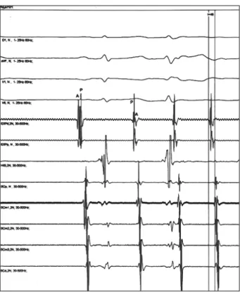
Two of the 3 patients in whom proper mapping was achieved and radiofrequency was applied had sponta-neous induction of atrial fibrillation provoked by ectopic beats (fig. 1). In the sites selected for ablation, earlier electrical activity occurred in the left superior pulmonary vein in 4 patients and in the right superior pulmonary vein in 2 patients (table IV). Laboratory success was achieved in 5 patients: 4 are free of preexcitation, atrioventricular reentry tachycardia, and we could not induce atrial fibrillation in patient number eight. Laboratory success was not achieved in patient number 9 due to rare atrial ectopy, which made it, impossible to institute precise mapping. Radiofrequency was applied at sites with precocious electrograms and in the presence of double potentials (pulmonary vein potential and atrial electro-gram). Three successful electrograms with these features were observed (figs. 2, 3, 4 and 5). In 2 cases, ablation was performed inside the pulmonary vein (fig. 6), and in the Table I – Clinical characteristics of patients referred for electrophysiological study
Pts Age (years) Sex Clinical Presentation Electrophysiological Study indication Previous Holter
1 44 M Paroxysmal AF Refractoriness to drugs AF (NSAT-20)
2 68 M Paroxysmal AF Low output AF (NSAT-565)
3 82 M Paroxysmal AF Low output AF (NSAT-16)
4 48 M Paroxysmal AF Refractoriness to drugs AF (NSAT-33)
5 51 M Recurrent AF Refractoriness to drugs AF(afterECV,NSAT)
6 38 M Paroxysmal AF Intention to treat AF (NSAT-2084).
7 51 M Recurrent AF Refractoriness to drugs AF
8 35 M Paroxysmal AF Refractoriness to drugs AF (NSAT-18)
9 30 M Paroxysmal AF Intention to treat Rare atrial ectopy
M- male; AF- atrial fibrillation; ECV- electrical cardioversion; NSAT- nonsustained atrial tachycardia. The number that follows “NSAT” corresponds to the number of nonsustained atrial tachycardia on 24-hour Holter monitoring.
Table II – Success rate of ablation in patients with short-duration atrial fibrillation (nonsustained atrial tachycardia) on 24-hour Holter monitoring prior to the procedure compared with the total group of patients
Mapped patients Laboratory success rate Recurrence Final success rate
NSAT to 24-hour Holter monitoring 100% (6 of 6 patients) 83% (5 of 6 patients) 16% (1 of 6 patients) 66% (4 de 6 pacientes) (6 patients)
All patients (9 patients) 88% (8 of 9 patients) 55% (5 of 9 patients) 11% (1 of 9 patients) 44% (4 of 9 patients)
NSAT- nonsustained atrial tachycardia.
Table III - Causes of failure
Pts Transeptal puncture Causes of unsuccessful mapping
4 Yes Insufficient atrial ectopy 5 Yes Insufficient mapping duration (degeneration
to AF nonrespondent to cardioversion) 7 No (AF nonresponsive to cardioversion)
Mapping not performed 9 Yes- Insufficient atrial ectopy
(application unsuccessful)
AF- atrial fibrillation.
Table IV – Radiofrequency application sites and results in mapped patients
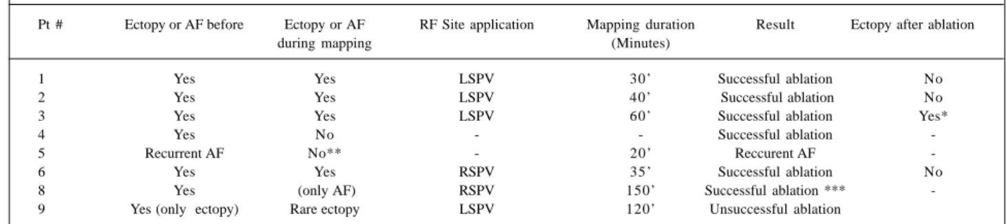
Pt # Ectopy or AF before Ectopy or AF RF Site application Mapping duration Result Ectopy after ablation
during mapping (Minutes)
1 Yes Yes LSPV 30’ Successful ablation No
2 Yes Yes LSPV 40’ Successful ablation No
3 Yes Yes LSPV 60’ Successful ablation Yes*
4 Yes No - - Successful ablation
-5 Recurrent AF No** - 20’ Reccurent AF
-6 Yes Yes RSPV 35’ Successful ablation No
8 Yes (only AF) RSPV 150’ Successful ablation ***
-9 Yes (only ectopy) Rare ectopy LSPV 120’ Unsuccessful ablation
1 2 1 2 1 2 1 2 1 2
other 3 ablation was applied in the pulmonary vein ostia with low precocity related to the P wave (fig. 3). When ablation was applied inside the pulmonary vein, precocity related to the P wave (118ms and 70ms) was more evident when compared with ablation applied to the pulmonary vein ostia (28ms, 48ms, and 36ms).
Clinical success (absence of symptoms) was obtained in 4 patients (numbers 1, 2, 3, and 6), in an average 4.8-month follow-up. On 24-hour Holter monitoring, we also observed the elimination of nonsustained atrial tachycardia (numbers 1, 2, 6). One patient who achieved laboratory success (noninduction of atrial fibrillation) experienced recurrence of atrial fibrillation 10 days after the procedure (number 8).
In all patients who achieved laboratory success (number 1, 2, 3, 6, and 8), antiarrhythmic drugs were discon-tinued so that clinical results and possible recurrences could be observed. In patients who did not achieve laboratory success (number 4, 5, 7, and 9) or in whom clinical recurrences occurred (number 8), antiarrhythmic drugs were reintroduced.
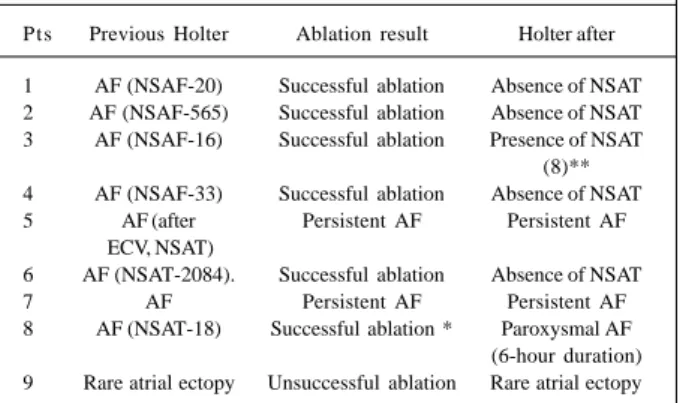
Twenty-four hour Holter monitoring preablation and postablation (table V) reveals an interesting finding. The presence of frequent episodes of nonsustained atrial tachycardia on 24-hour Holter preablation was associated with a greater rate of success.
Discussion
Catheter ablation of atrial fibrillation is currently one of the greatest challenges in electrophysiology. This arrhy-thmia is difficult to control with antiarrhythmic drugs and has the potential of becoming permanent as the number and duration of episodes increase. The possibility of cure, even if only in a small number of patients, is extremely rewarding, Fig. 1 – Records of 24-hour Holter monitoring with several electrocardiographic
tracings. Several episodes of short-standing atrial fibrillation were observed (in fact, they were nonsustained atrial tachycardia). They were mainly seen in the last 2 tracings of the first column.
Fig. 2 – Record of surface derivations I, II, V1, and V6 and of intracavity electrograms of the right atrium (ADPL, ADL, ADH, IE4), His bundle (HIS), and left atrium (SCP, SCM, SCD) of spontaneous induction of atrial fibrillation initiated by atrial extrasystoles. It was observed that the fifth QRS is preceded by an atrial extrasystole (ectopic P wave observed in DII), followed by a rapid sequence of atrial extrasystoles with induction of atrial fibrillation on the surface electrocardiogram and maintenance of intracavity extrasystole activation.
Table V – Findings of 24-hour Holter monitoring before and after ablation of patients referred for electrophysiological study
Pts Previous Holter Ablation result Holter after
1 AF (NSAF-20) Successful ablation Absence of NSAT 2 AF (NSAF-565) Successful ablation Absence of NSAT 3 AF (NSAF-16) Successful ablation Presence of NSAT
(8)** 4 AF (NSAF-33) Successful ablation Absence of NSAT 5 AF (after Persistent AF Persistent AF
ECV, NSAT)
6 AF (NSAT-2084). Successful ablation Absence of NSAT
7 AF Persistent AF Persistent AF
8 AF (NSAT-18) Successful ablation * Paroxysmal AF (6-hour duration) 9 Rare atrial ectopy Unsuccessful ablation Rare atrial ectopy
1 3 1 3 1 3 1 3 1 3
Fig. 3 – Record of surface derivations I, II, V
1, and V6 and of intracavity electrograms of
catheter ablation by double distal electrodes (EXD) and by the double of the proximal electrode (EXP), of the left atrium (SCD, SCM2, SCM3, SCP) and of the right atrium (ADA) of the application site of successful radiofrequency. In the first intracavity electrogram of the catheter ablation (EXD), a 118ms precocious ectopic beat was observed in relation to the surface P wave.
Fig. 4 – Record of surface derivations I, toVF, V1, and V6 and of intracavity electro-grams of the ablation catheter by the double distal electrode (EXD) and by the dou-ble proximal electrode (EXP), of His bundle (HIS), and of the left atrium (SCP, SCm1, SCm, SCm3, SCD) of the application site of successful radiofrequency. In the first in-tracavity electrogram of the ablation catheter (EXD), we can observe that the first beat is sinusal (with the pulmonary vein potential following the atrial electrogram). In the following beats, we can observe fusion and inversion of electrograms (atrium potential follows the potential of pulmonary vein).
Fig. 5 – Record of surface derivations I, VL, VF, and V1 and of intracavity electrograms of catheter ablation by the distal electrode pair (EXP), of the left atrium (SCD, SCM, SCP) and of the His bundle (HIS) of the successful application site of radiofrequency. In this electrogram, we observe that the first beat is an atrial ectopy (with a left atrium potential following the electrogram of a 36ms precocious pulmonary vein in relation to the distal coronary sinus electrogram-SCD). In the following sinusal beats, we observed electrogram inversion (pulmonary vein potential follows left atrium potential).
due to the significant morbidity and mortality associated with this frequent disease.
Criteria for selecting patients for mapping and cathe-ter ablation have not been appropriately defined. Because this ablation technique has only just recently begun to be used for atrial fibrillation, we do not know exactly which pa-tients benefit most from the procedure. Although this cura-tive technique is highly desirable for all patients, the procedure is complex, involves risks, and the reproducibility success rate has not been totally proved. For this reason, it is important to identify the patient with the greatest chance of a cure with the ablation procedure.
Atrial fibrillation focal ablation was first described by Haïssaguerre et al 18, and its use is considered a great achievement because it was believed that atrial fibrillation could never be suppressed by a focal application. In another study with 9 patients carried out by the same group of researchers, the most relevant clinical data reported for identifying patients eligible for the ablation technique is the finding of atrial fibrillation associated with episodes of irre-gular monomorphic tachycardia or atrial extrasystoles without structural heart disease 12.
1 4 1 4 1 4 1 4 1 4
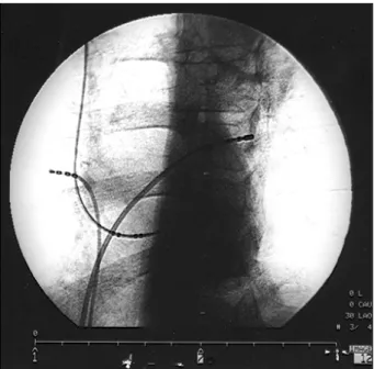
Fig. 6 – A left anterior oblique cine frame at a 30° position of the ablation catheter at the successful application site. We observed that the ablation catheter is inside the superior left pulmonary vein placed through a transeptal puncture.
by Arentz et al, 20 in 232 patients with paroxysmal atrial fibrillation to on 24-hour Holter monitoring who underwent ablation, only 23 patients with atrial tachycardia or frequent monomorphic atrial extrasystoles were selected. Of those, 10 had frequent paroxysmal atrial fibrillation (more than 2 episodes per week) and were resistant to more than 1 antiarrhythmic drug. In this subgroup, it was possible to identify a focal source in only 3 patients who successfully underwent ablation.
These extremely rigorous criteria for attempts at mapping and ablation were quickly changed. Interesting observations arose from studies of chronic atrial fibrillation, where the attempt at eliminating arrhythmia was based on the performance of linear lesions simulating Cox surgery. In these studies, the organization of atrial activity either shortened or prevented episodes of atrial fibrillation, suggesting focal activity with atrial extrasystoles, which are the main triggering mechanisms of atrial fibrillation episo-des before ablation. These atrial ectopies were identified in at least 40% of patients and had not been obvious before the procedure due to long-lasting atrial fibrillation. We also observed that the pulmonary veins are the main source of these ectopic foci 20-22. It is still unknown whether in these patients with chronic atrial fibrillation ablation of these triggering foci prior to linear ablation is enough to suppress arrhythmia 23. These impressive findings raised expecta-tions that those patients with long- lasting atrial fibrillation could also be eligible for mapping and ablation. This belief encouraged the inclusion of an increasing number of patients with long-standing paroxysmal fibrillation or recurrent atrial fibrillation. Lau et al 24 selected for mapping and ablation not only patients with paroxysmal atrial fibrillation but also patients with chronic internal
defibrilla-tion. In this study, 20% of patients with chronic atrial fibrillation had spontaneous atrial fibrillation reentry, which was used as a guide for mapping and ablation. Todd et al 25 demonstrated that focal atrial arrhythmias commonly occur after electrical cardioversion in patients with recurrent atrial fibrillation. In this study, 39% of patients (14 of 36) had focal atrial tachycardias that were mapped. This finding was very important because it increased confidence in the possibility that patients with chronic atrial fibrillation could be included for mapping and ablation. Takahashi et al 26 performed successful ablation in 3 of 6 patients with recurrent atrial fibrillation. Mapping was not successful in 2 patients (1 due to the absence of ectopy and 1 due to the presence of multiple foci).
Structural heart disease is another factor that excludes patients for mapping. Structural heart disease implies greater electrical disarrangement and fewer chances of focal ablation. However, in clinical studies, data are controversial. In the study by Todd et al 25, the absence of structural heart disease was the only independent predictor of a focal source of atrial fibrillation (12 of 24 patients without structural disease had focal atrial tachycardia). Although the presence of structural heart disease seems to clearly hinder successful ablation, Vergara et al 27, when analyzing clinical predictors of successful atrial fibrillation ablation, observed that structural heart disease was not associated with fewer chances for ablation. In this study, a greater number of atrial fibrillation foci in the same patient and the presence of left atrial flutter were associated with a greater incidence of recurrence, reflecting a greater extension of atrial disease.
The presence of structural heart disease is no longer a contraindication for mapping and ablation of focal atrial fibrillation in a series of patients studied by the group of French researchers, previously mentioned 28. In the last 10 months of the study, this group 29 used as a criterion for inclusion of patients for mapping and focal ablation of resistant atrial fibrillation. This fast and progressive evolution as an indica-tion for mapping and ablaindica-tion in patients for identifying discre-et foci as the source of atrial fibrillation caused difficulty for the clinical cardiologist in the selection and referral of patients for electrophysiological study.
In our limited series of patients, we observed that mapping of pulmonary veins to search for atrial tachycardia foci that cause atrial fibrillation is feasible, although the selection of the ideal patient for the procedure is of utmost importance to achieve the best results.
1 5 1 5 1 5 1 5 1 5
patients with recurrent atrial fibrillation, because they had sinus rhythm with frequent atrial extrasystoles of the same morphology.
Causes of failure in our series were mainly rare atrial ectopy and atrial fibrillation resistant to electrical cardio-version. In 1 patient (number 9), atrial ectopy was very rare during the procedure, hindering precise mapping. In another case (number 4), the rarity of ectopic episodes hin-dered mapping, not allowing the selection of the focus for application of radiofrequency. In patient number 8, we were able to perform mapping and ablation of a focus in the right superior pulmonary vein, despite infrequent ectopy during mapping. We considered noninducibility of atrial fibrillation as a laboratory success. This patient, however, had recurrence of atrial fibrillation 10 days after the proce-dure. In late follow-up, this patient had significant clinical improvement in symptoms without the use of antiarrhy-thmic medication.
We were not able to perform focal mapping in the 2 patients (number 5 and 7) with recurrent atrial fibrillation. In one, conversion of atrial fibrillation to sinus rhythm (number 7) was possible. He was a former smoker of 60 cigarettes a day and had a dilated anteroposterior thoracic diameter, in which, even cardioversion techniques with 2 simultaneous charges were unsuccessful. In the other patient, atrial fibrillation was converted, but episodes of nonsustained atrial tachycardia degenerated to atrial fibrillation in a short period of time and were refractory to cardioversion. Internal defibrillation may be a useful technique for converting atrial fibrillation in these patients.
Atrial fibrillation ablation is a complex procedure, because it involves a series of premises: 1) the patient needs to be in sinus rhythm with extrasystoles frequent enough to enable mapping of pulmonary veins, 2) transseptal puncture is necessary, 3) evolvement to atrial fibrillation during the procedure makes it more difficult because it requires repea-ted electrical cardioversion, 4) the number of radiofrequen-cy applications must be limited so that pulmonary vein stenosis can be avoided, 5) mapping needs to be precise so that, ideally, only 1 pulmonary vein is selected for the application of radiofrequency.
We observed that patients with recurrent atrial fibrilla-tion were not ideal candidates for mapping of pulmonary ve-ins due to the difficulties in cardioversion and in the mainte-nance of sinus rhythm for mapping of atrial extrasystoles. If these patients wanted to undergo the procedure, we sug-gested that after successful electrical cardioversion, 24-hour Holter monitoring be performed. Patients who experi-enced frequent atrial ectopy were considered eligible. Ho-wever, a greater chance existed in these patients that the procedure would be more difficult.
In the subgroup of patients with short episodes of ap-parent atrial fibrillation on 24-hour Holter monitoring (6 patients), the success rate was 66% (4 of 6 patients). These episodes of atrial fibrillation were actually episodes of
non-sustained atrial tachycardia, allowing mapping of the focal source in the pulmonary veins. We believe that these patients are ideal candidates for mapping and ablation. Two patients (number 2 and 6) experienced more than 500 episodes of nonsustained atrial tachycardia simulating atrial fibrillation on 24-hour monitoring prior to the procedure. In these patients, mapping was made easier by the high density of the arrhythmia, resulting in success in both cases.
We did not observe complications due to the procedu-re. The main apprehension in applying radiofrequency in the interior or around the pulmonary veins is the risk of pulmonary vein stenosis, which may lead to pulmonary hy-pertension. This is the most common complication resul-ting from linear ablation (extensive application with the in-tent to form continuous lines) in the left atrium around pul-monary veins 30. However, pulmonary vein stenosis has been described in the focal ablation of atrial fibrillation 31,32. In these studies 31, ablation was performed in more than 1 pulmonary vein with multiple procedures, or electrical isolation of the pulmonary vein was attempted 32. In our series, we performed only 1 procedure in all patients, and we did not make radiofrequency applications in more than 1 pulmonary vein. We took special care during the radiofre-quency application (applications lasted only 15s, and pain was used as a criterion for interrupting the application). Perhaps the absence of complications in our series is due to this set of factors.
Currently, indications for mapping and attempting ablation of atrial fibrillation are not yet definitely establi-shed. This is a field of intensive research and constant chan-ge. The results of our study, even though preliminary, seem to indicate that mapping and attempting ablation of atrial fibrillation are feasible and may be attempted in all patients with paroxysmal atrial fibrillation, without structural heart disease and with short episodes of nonsustained atrial fi-brillation on 24-hour Holter monitoring.
Our series of patients is limited and perhaps a larger group of patients might enable better selection of candidates for mapping. Systematic application of a greater number of provocative maneuvers to induce atrial ectopy would proba-bly allow more efficient mapping in some patients. Maneu-vers used in this study were: isoproterenol infusion, atrial sti-mulation, and adenosine infusion. We did not use the Valsa-va maneuver or others mentioned in the literature, such as ca-rotid massage and volume infusion, digoxin or verapamil. Of these, only the Valsalva maneuver has made significant alterations in the frequency of ectopies 28.
1 6 1 6 1 6 1 6 1 6
1. Bialy D , Lehman MH, Schumacher DN, et al. Hospitalization for arrhythmias in the United States: Importance of atrial fibrillation. J Am Coll Cardiol 1992: 19-41. 2. Prystowsky EN, Benson DW Jr, Fuster V, et al. Management of patients with
atrial fibrillation: a statement for healthcare professionals from the Committee on Electrocardiography and Electrophysiology, American Heart Association. Circulation 1996; 93: 1262-77.
3. Atrial fibrillation investigators. Risk factors for stroke and efficacy of anti-thrombotic therapy in atrial fibrillation: analysis of a pooled data from five randomized controlled trials. Arch Intern Med 1994: 154: 1449-57. 4. Coplen SE, Antman EM, Berlin JA, Hewitt P, Chalmers TC. Efficacy and safety of
quinidine therapy for maintenance of sinus rhythm after cardioversion. A meta-analysis of randomized control trials. Circulation 1990; 82: 1106-16. 5. Juul Moller S, Edvardsson N, Rehnqvist Ahlberg N. Sotalol versus quinidine
for the maintenance of sinus rhythm after direct current conversion of atrial fibrillation. Circulation 1990; 82: 1932-9.
6. Crijins HJ, Van Gelder IC, Van Gilst WH, Hillege H, Gosselink ATM, Lie KI. Serial antiarrhythmic drug treatment to maintain sinus rythm after electrical cardioversion for chronic atrial fibrillation or atrial flutter. Am J Cardiol 1991; 68: 335-41. 7. Gosselink ATM, Crijins HJ, Van Gelder IC, Hillege H, Wiesfeld AC, Lie KI.
Low-dose amiodarone for maintenance of sinus rhythm after cardioversion of atrial fibrillation or flutter. JAMA 1992; 267: 3289-93.
8. Jackman WM, Wang X, Friday KJ, et al. Catheter ablation of acessory atrio-ventricular pathways (Wolff-Parkinson-White syndrome) by radiofrequency current. N Engl J Med 1991; 324: 1605-11.
9. Calkins H, Souza J, El-Atassi R, et al. Diagnosis and cure of the Wolff-Parkinson-White syndrome or paroxysmal supraventricular tachycardia during a single electrophysiologic test. N Engl J Med 1991; 324: 1612-7.
10. Lesh MD, Hare GFV, Schamp DJ, et al. Curative percutaneous catheter ablation using radiofrequency energy for acessory pathways in all locations: Results in 100 consecutive patients. J Am Coll Cardiol 1992; 19: 1203-9.
11. Cox JL, Boineau JP, Schuessler RB, et al. Successful surgical treatment of atrial fibrillation. Review and clinical update. JAMA 1991; 266: 1976-80. 12. Jaïs P, Haïssaguere M, Shah DC, et al. A focal source of atrial fibrillation treated
by discrete radiofrequency ablation. Circulation 1997; 95: 572-6.
13. Swartz JF, Pellersels G, Silvers J, Patten L, Cervantez D. A catheter-based curative approach to atrial fibrillation in humans. Circulation 1994; 90(pt 2): I-335. 14. Pappone C, Oreto G, Lamberti F, et al. Catheter ablation of paroxysmal atrial
fibrillation using a 3D mapping system. Circulation 1999; 100: 1203-8. 15. Waspe L, Brodman R, Kim SG. Susceptibility to atrial fibrillation and ventricular
tachyarrhythmias in the Wolff-Parkinson-White syndrome: role of the accessory pathway. Am Heart J 1986; 112: 1141-52.
References
16. Hamer ME, Wilkinson WE, Clair WK, Page RL, McCarthy EA, Pritchett ELC. Incidence of symptomatic atrial fibrillation in patients with paroxysmal supraventricular tachycardia. J Am Coll Cardiol 1995; 25: 984-8.
17. Delise P, Gianfranchi L, Paparella N, et al. Clinical usefulness of slow pathway ablation in patients with both atrioventricular node reentrant tachycardia and atrial fibrillation. Eur Heart J 1996; 17(suppl): 271.
18. Haïssaguerre M, Marcus FI, Fischer B, et al. Radiofrequency catheter ablation in unusual mechanisms of atrial fibrillation: Report of three cases. J Cardiovasc Electrophysiology 1994; 5: 743-51.
19. Haïssaguerre M, Jaïs P, Shah DC, et al. Spontaneous initiation of atrial fibrillation by ectopic beats originating in the pulmonary veins. N Engl J Med 1998; 339: 659-66. 20. Arentz T, Ott P, Stockinger J, Blum T, Rosenthal JV, Kalusche D. Radiofrequency
catheter ablation of focal atrial fibrillation. PACE 1998; 21(II): 963. 21. Konings KTS, Kirschhof CJHJ, Smeets JRLM, Wellens HJJ, Penn O, Alessie MA.
High-density mapping of electrically induced atrial fibrillation in humans. Circulation 1994; 89: 1665-80.
22. Ortiz J, Niwano S, Abe H, Rudy Y, Jonhson NJ, Waldo AL. Mapping the conversion fo atrial flutter to atrial fibrillation and atial fibrillation to atrial flutter: Insights into mechanism. Circ Res 1994; 74: 882-94.
23. Haïssaguerre M, Shah DC, Jaïs P, Clémenty J. Role of catheter ablation for atrial fibrillation. Curr Opinion Cardiology 1997; 12: 18-23.
24. Lau CP, Tse HF, Ayers GM: Radiofrequency ablation of focal atrial fibrillation as guided by site of spontaneous reinitiation of atrial fibrillation after defibrillation. PACE 1998; 21(II): 922.
25. Todd DM, Hobbs WJC, Fynn SP, Armstrong K, Garratt CJ. How common is focal a-trial fibrillation in patients undergoing cardioversion? PACE 1999; 22(II): 792. 26. Takahashi A, Jaïs P, Shah D, et al. Successful focal ablation of ectopy reinitiating
persistent atrial fibrillation. PACE 1999; 22(II): 905.
27. Vergara I, Martin D, Bahnson T, Sorrentino RA, Greenfield RA, Wharton JM. Clinical predictors of successful focal atrial fibrillation ablation. PACE 1999; 22(II): 772.
28. Shah DC, Haïssaguerre M, Jaïs P, Takahashi A, Hocini M, Clémenty J. Provocati-ve manoeuProvocati-vers for inducing pulmonary Provocati-vein ectopy. PACE 1999; 22(II): 738. 29. Haïssaguerre M, Jaïs P, Shah DC, et al. Plurifocal sources of atrial fibrillation
initiation from the pulmonary veins. PACE 1999; 22(II): 707.
30. Robbins IM, Colvin EV, Doyle TP, et al. Pulmonary vein stenosis after catheter ablation of atrial fibrillation. Circulation 1998; 98: 1769-75.
31. Jaïs P, Shah D, Haïssaguerre M, et al. Pulmonary vein patency after radiofrequen-cy ablation. PACE 1999; 22: 738.