INTRODUCTION
The Brazilian Society of Hepatology has sponsored in October 18th, 2014 at São Paulo, the irst meeting concerning the management of autoimmune liver diseases (ALD), in order to draw evidence-based rec-ommendations concerning the diagnosis and treatment of autoimmune hepatitis (AIH), primary sclerosing cholangitis (PSC), primary biliary cirrhosis (PBC) and their overlap syndromes. The governing board of the Brazilian Society of Hepatology elected seven investiga-tors with recognized expertise and/or publications in the ield to organize the scientiic agenda. They have selected the topics to be reviewed, including the diag-nosis and treatment of AIH, PSC, PBC and overlap syndromes; the management of speciic complications of cholestasis, such as pruritus, fatigue and
hyperco-BRAZILIAN SOCIETY OF HEPATOLOGY
RECOMMENDATIONS FOR THE DIAGNOSIS
AND MANAGEMENT OF AUTOIMMUNE
DISEASES OF THE LIVER
Paulo Lisboa
BITTENCOURT
1, Eduardo Luiz Rachid
CANÇADO
2, Cláudia Alves
COUTO
3,
Cynthia
LEVY
4, Gilda
PORTA
2, Antônio Eduardo Benedito
SILVA
5,
Debora Raquel Benedita
TERRABUIO
2and Members of the Pannel of the 1st Consensus of
the Brazilian Society of Hepatology on the Diagnosis and Management of Autoimmune Diseases
of the Liver*
ABSTRACT - In order to draw evidence-based recommendations concerning the management of autoimmune diseases of the liver, the Brazilian Society of Hepatology has sponsored a single-topic meeting in October 18th, 2014 at São Paulo. An organizing committee comprised of seven investigators was previously elected by the Governing Board to organize the scientiic agenda as well as to select twenty panelists to make a systematic review of the literature and to present topics related to the diagnosis and treatment of autoimmune hepatitis, primary sclerosing cholangitis, primary biliary cirrhosis and their overlap syndromes. After the meeting, all panelists gathered together for the discussion of the topics and the elaboration of those recommendations. The text was subsequently submitted for suggestions and approval of all members of the Brazilian Society of Hepatology through its homepage. The present paper is the inal version of the reviewed manuscript organized in topics, followed by the recommendations of the Brazilian Society of Hepatology.
HEADINGS - Autoimmune hepatitis. Primary sclerosing cholangitis. Primary biliary cirrhosis. Diagnosis. Treatment.
Declared conflict of interest of all authors: none
1 Hospital Português, Salvador, BA, Brasil; 2 Faculdade de Medicina da Universidade de São Paulo, SP, Brasil; 3 Faculdade de Medicina da
Uni-versidade Federal de Minas Gerais, MG, Brasil; 4 University of Miami, USA; 5 Faculdade de Medicina da Universidade Federal de São Paulo, SP,
Brasil; 6 Hospital Israelita Albert Einstein, SP, Brasil; 7 Centro de Ciências da Saúde da Universidade Federal do Espírito Santo, Vitória, ES, Brasil; 8 Hospital Universitário da Faculdade de Medicina da Universidade Federal de Espírito Santo, ES, Brasil; Faculdade de Medicina da Universidade Federal de
Per-nambuco, PE, Brasil; 9 Departamento de Gastroenterologia da Universidade Federal de Santa Catarina, SC, Brasil; 10 Universidade Estadual de Campinas, SP, Brasil.
* Roberto José de Carvalho Filho5, Dalton Marques Chaves2, Irene Kazue Miura2, Liana Codes1, Luciana Costa Faria3, Andreia Silva Evangelista6, Alberto Queiroz Farias2,
Luciana Lofêgo Gonçalves7, Michele Harriz2, Edmundo Pessoa A Lopes Neto8, Gustavo Oliveira Luz2, Patrícia Oliveira5, Elze Maria Gomes de Oliveira5, Janaina Luz
Narciso Schiavon9, Tiago Seva-Pereira10, Edison Roberto Parise5.
Correspondence: Paulo Lisboa Bittencourt. Rua Prof. Clementino Fraga, 220, 1901 - CEP: 40170050 - Salvador, Bahia, Brasil. Email: [email protected]
- Class I: conditions for which there is evidence and/ or general agreement that a given diagnostic evalua-tion, procedure or treatment is beneicial, useful, and effective.
- Class II: conditions for which there is conflicting evidence and/or a divergence of opinion about the usefulness/eficacy of a diagnostic evaluation, proce-dure, or treatment.
- Class IIa: weight of evidence/opinion is in favor of usefulness/eficacy.
- Class IIb: usefulness/eficacy is less well established by evidence/opinion.
- Class III conditions for which there is evidence and/ or general agreement that a diagnostic evaluation, procedure/treatment is not useful/effective and in some cases may be harmful.
PART I: AUTOIMMUNE HEPATITIS
Clinical manifestations
Autoimmune hepatitis (AIH) is a chronic inlammatory liver disease characterized by hipergamaglobulimemia and reactivity autoantibodies, particularly for anti-smooth muscle (SMA), antinuclear (ANA), anti-liver kidney microsome type 1 (anti-LKM1), anti-soluble liver antigen (anti-SLA) and anti-liver cytosol type 1 (anti-LC1) antibodies. AIH primarily affects women and typically responds to immu-nosuppressive therapy with clinical, biochemical and histo-logical remission. Frequently, the diagnosis of AIH is made when in the presence of inespeic clinical symptoms, like fatigue, nausea, upper abdominal pain, rash, arthralgia and oligomenorrhea. Jaundice can be present in half of subjects and nearly 1/3 of patients can present with signs of advanced liver disease, such as ascites, splenomegaly, variceal bleeding and hepatic encephalopathy.
The disease can resemble an acute hepatitis in approxi-mately 30% of cases with symptoms of jaundice, choluria, fecal acholia, and eventually, pruritus, especially in young girls. Fulminant hepatic failure (FHF) can also be a manifes-tation of severe AIH, and the disease should be considered in the differential diagnosis of FHF. Nearly 15%-20% of the patients can be asymptomatic at the time of diagnosis that may be elicited due to the biochemical abnormalities, detected during check-up evaluations, or in patients with other extrahepatic autoimmune disorders, like thyroiditis, arthritis or diabetes mellitus(3,4).
The disease is usually classiied in two types based on the autoantibody profile. Autoimmune hepatitis type 1 (AIH-1) is deined by the presence of SMA and/or ANA, whereas AIH type 2 (AIH-2) is characterized by reactivity for anti-LKM1 and/or anti-LC1. Diagnostic criteria and treatment options do not differ between both groups, but patients with AIH-2 may have distinct clinical features at onset such as younger age, higher frequency of acute liver failure, lower gammaglobulin levels and an increased inci-dence of IgA deiciency(3,4).
A comparative study of 115 Brazilian and 161
North-Americans patients with AIH-1 showed that Brazilian patients had earlier disease onset, lower frequency of concurrent immune diseases, higher serum aspartate ami-notransferase (AST) and gammaglobulin levels, greater occurrence of smooth muscle antibodies (SMA), and lower frequency of antinuclear antibodies (ANA), when compared to their North-American counterparts. Besides, HLA-DR13 occurred more commonly in Brazilian patients, when compared to North-American subjects, who had more often HLA-DR3 and -DR4(5).
Data from a Brazilian cohort from the São Paulo Uni-versity showed that patients with AIH (n=268) had more frequently at disease onset clinical manifestation resembling acute hepatitis (56%)(6).Signs and symptoms of advanced chronic liver disease (CLD) were present in 25% of them and 10% were asymptomatic at diagnosis. Differences in clinical presentation between studies may be in part due to distinct genetic background but also to heterogeneity between parameters employed by the authors to evaluate types of clinical presentation. When the disease begins in the child-hood, patients more frequently have a acute clinical course and a higher frequency of cirrhosis at diagnosis (60%-80%)(7).
Brazilian patients with AIH-1 with less than 18 years, when compared to older subjects had higher frequency of acute disease, SMA reactivity and HLA-DR13 as well as a lower frequency of concurrent extrahepatic autoimmune diseases. Patients older than 18 years also showed similar clinical features of their North American conterparts regard-ing the autoantibody proile and the frequency of concurrent extrahepatic autoimmune(5).
A variant of AIH, called autoimmune sclerosing chol-angitis (ASC), characterized by the concomitance of col-angiograic indings of PSC and AIH in the same patient, has been frequently described in children(8). Even though this variant has been also reported in adults whether it is a different entity of part of AIH spectrum is not known. It would be better characterized elsewher in this manuscript as part of the spectrum of AIH and PSC overlap syndrome.
Autoimmune hepatitis can cause FHF in 2% to 16% of patients. Those frequencies may be underestimated as patients could also be erroneously classiied as cryptogenic FHF due to the fact that most of them do not have the full-blown AIH phenotype. The frequency of FHF cause with unknown etiology varies from 16%-47%, reaching 57% of the cases in Japan(9).
Recommendations
- In the majority of cases, patients with AIH have un-recognized CLD with acute hepatitis-like symptoms, but signs and symptoms of advanced CLD may also be present. Less frequently, the disease can be present without symptoms or with FHF (Class IIa).
- AIH primarily affects women in a 4:1 ratio, mostly in 5 to 25 years of age, but it can occur in all age and races (Class IIa).
- Autoimmune extrahepatic disorders are frequently seen in patients with AIH. Autoimmune thyroiditis and rheumatoid arthritis are the most common en-countered diseases. (Class I)
Diagnosis
The diagnosis of AIH is based on clinical, biochemical, serological and histological features and requires the exclu-sion of other causes of liver disease, such as viral hepatitis, hemochromatosis, Alpha-1 antitrypsin deficiency, drug-induced liver diseases, Wilson’s disease, non-alcoholic fatty liver disease and alcohol(11,12).
Aminotransferase levels can be more than 10 times the up-per limit of normal (ULN), the alkaline phosphatase (ALP) levels in general are less than three times the ULN, while the gamaglutamiltranspeptidase (GGT) levels are quite variable. Hypergammaglobulinemia, with a predominance of the IgG fraction, is the hallmark of the disease(12,13).
Determination of circulating autoantibodies (AA) are key to the diagnosis and characterization of ALD. Most frequently, AA are non-organ speciic and its expression can vary during the course of the disease, may also be present in other infectious liver diseases, rheumatological diseases, or even absent in 10% of cases(11,14). ANA, ASMA, anti-LKM1 are the classical AA used to diagnose and classify AIH. They are tested by indirect immunoluorescence (IFI) using tissue sections of kidney, liver and stomach of rodents as a substrate(15). Titers of AA in general do not correlate with severity of disease and its clinical course. Titers of SMA, but not ANA, are useful markers of disease activity and can be used to monitor response to treatment(3,16).
According to international criteria, AA titers are con-sidered positive, when present at a dilution of at least 1:40 in adults, while titers of at least 1:20 for SMA and ANA or titers of at least 1:10 for anti-LKM1 are considered positive in children(17). Homogeneous and speckled are the most frequent patterns of ANA in AIH patients(11,13,15,18). Centro-meric, nucleolar and nuclear dots cannot be considered as markers of AIH.
SMA reactivity, with or without ANA, characterizes AIH-1. Both can turn to be negative after treatment(33,16). The immunoluorescent staining of SMA is detected in the arte-rial walls of rodent kidney, liver and stomach. In the kidney, SMA can have three patterns: SMA-V (vessels), SMA-VG (vessels and glomeruli) and SMA-VGT (vessels, glomeruli and tubules). SMA-VG and AMA-VGT patterns are more speciic of AIH, but SMA-V pattern is frequently found in viral hepatitis(19,20). The speciicity for antigens present in the
microilaments (F-actin), characterizing anti-actin antibody (AAA) is speciic, but not pathognomonic, of AIH and is well correlated with the presence of SMA-VGT pattern in IIF(21).
Lack of IFI standardization limits its use on routine ba-sis(13,15) Usually, patients with reactivity for SMA are younger than patients with reactivity for ANA. In this regard, patients with AIH-1 and SMA also exhibit reactivity for AAA and are shown to carry HLA-DR13 and -DR3 genotypes in South America and HLA-DR3 and -DR4 in Europe and North-America.
Autoimmune hepatitis type 2 is characterized primarily by reactivity to anti-LKM1(21,22). It reacts with CYP2D6 antigens, but can be positive in up to 5% of hepatitis C patients(23).As previously pointed out, patients with AIH-2 are younger, have lower levels of gamaglobulin, carry more frequently HLA-DR7 and/or -DQ2 genotypes and are more prone to have FHF at presentation, when compared to their counterparts with AIH-1(24,25).
Other AA can be tested to better characterize AIH patients without those aforementioned conventional antibodies. Anti-soluble liver pancreas (Anti-SLA/LP), that the molecular target is as Sep(O-phosphoserine) tRNA:Sec(selenocysteine)tRNA synthase (SEPSECS) is highly speciic for AIH but has low sensitivity(26,27).It is mostly detected in asociation with ANA and SMA. Its presence identiies patients with a more severe disease and a worse outcome. It is also associated with HLA-DR3(28,29).Anti-LC1 is the second marker of AIH-2, detected in 24% to 32% of patients with anti-LKM1 and rarely as the only marker of AIH-2. It occurs mainly in young patients with an aggressive disease course with rapid progression to cirrhosis(30,31). The perinuclear anti-neutrophil cytoplasm (pANCA) antibody and anti-asialoglycoprotein receptor antibody (ASGPR) are less frequently used in clinical practice since they are not speciic for the diagnosis of AIH(29,32).
Liver biopsy should be performed, whenever possible, to conirm the diagnosis of AIH, to exclude other liver diseases as well as to guide therapy. It can be valuable also to stablish prognosis, as 30%-50% of the patients with AIH have cir-rhosis at disease onset(11,12). Typical, but not patognomonic, histologic features of AIH include the presence of interface hepatitis, moderate to severe lymphoplasmocytic portal inil-trate with rosetting of hepatocytes, varying degrees of lobular inlammation, bridging necrosis and sometimes multiacinar collapse. Granulomas are exceptional, as well as, severe bile duct lesions. Liver biopsy is considered a prerequisite for the diagnosis of AIH and is also helpful for assessment of portal inlammation and ibrosis staging. Noninvasive tests have gained currently widespread use in the quantiication of ibrosis in patients with hepatitis C and nonalcoholic steatohepatitis. However, they have not been validated for assessment of ibrosis in AIH and it could be hypothesized that their use in this setting would be hampered by the in-lammatory activity seen in this disease.
Autoimmune hepatitis frequently has an acute presenta-tion, sometimes FHF, resembling acute hepatitis or drug-induced liver disease. Patients with severe acute hepatitis or FHF may have autoantibodies in low-titer or absence of conventional autoantibodies. They may also have normal gammaglobulin or IgG levels and atypical histological ind-ings, including zone III centrolobular necrosis, perivenul-lar or central inlammation with lymphocytes and plasma cells, multiacinar collapse and lymphoid aggregates(9,35,36). Diagnostic scoring systems can aid in the diagnosis of these peculiar types of AIH, but it should be kept in mind that they have been standardized in cohorts of subjects with classical AIH. Sometimes, when liver biopsy is unfeasible, a short trial of corticosteroids is warranted in those dificult cases in order to stablish or refute the diagnosis of AIH based on the treatment response.
TABLE 1. International Autoimmune Hepatitis Study Group scoring system for the diagnosis of autoimmune hepatitis
Parameters Score
Female sex +2
ALP /ALT ratio (xULN)
<1,5 +2
1.5-3.0 0
> 3.0 -2
Globulins, Gammaglobulins or IgG (xULN)
>2.0 +3
1.5-2.0 +2
1.0-15 +1
<1.0 0
Autoantibodies (IIF titers in rodent tissues) Adults: ANA,SMA and anti-LKM1
>1/80 +3
1/80 +2
1/40 +1
<1/40 0
Children: ANA, SMA, anti-LKM1 >1/20 +3
Children: ANA, anti-LKM1:1/10-1/20 +2
Children: SMA 1/20 +2
Children: SMA 1/10 +1
Viral markers
Anti-HAV IgM, AgHBs or antiHBc IgM positive -3
Anti-HCV e RNA do HCV positivos -3
Anti-HAV IgM, AgHBs, anti-HBc IgM or anti-HCV
negative +3
Recent use of hepatotoxic drugs positive/negative -4/+1
Alcohol intake: <25g/day +2
>60g/day -2
Other autoimmune disease in the patient or irst degree
family member +2
Histology: Interface hepatitis +3
Rosettes +1
Severe lymphoplasmocytic iniltrate Iniltrado
inlamatório +1
None of histological indings above -5
Biliary changes suggestive of PBC or PSC -3
Other histological features suggestive of other etiology -3 Other autoantibodies in subjects without ANA, SMA and anti-LKM1
Anti-SLA/anti-LP, anti-LC1, anti-liver speciic protein, anti-asialoglycoprotein receptor: positive/negative +2/0 HLA-DR13 and -DR3 for AIH-1 or HLA-DR7 and
-DR3 for AIH-2 +1
Treatment response
Complete +2
Relapse during or after withdrawal of therapy after a
complete response +3
Deinite diagnosis: before treatment >15
Deinite diagnosis: after treatment >17
Probable diagnosis: before treatment 10-15
Probable diagnosis: after treatment 12-17
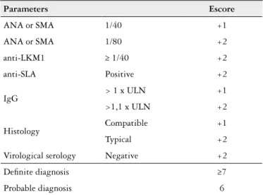
TABLE 2. Simpliied criteria for the diagnosis of AIH
Parameters Escore
ANA or SMA 1/40 +1
ANA or SMA 1/80 +2
anti-LKM1 ≥ 1/40 +2
anti-SLA Positive +2
IgG > 1 x ULN +1
>1,1 x ULN +2
Histology Compatible +1
Typical +2
Virological serology Negative +2
Deinite diagnosis ≥7
Probable diagnosis 6
Adapted from(33); AIH: autoimmune hepatitis; ULN: upper limit of normal; ANA: antinuclear
Recomendations
- The diagnosis of AIH should be performed in patients with elevated aminotransferases and gammaglobulin levels, reactivity for SMA, ANA, LKM1, anti-LC1 and anti-SLA and typical histological indings, after the exclusion of other liver disease, particularly viral hepatites and Wilson’s disease (Class I)
- The revised IAIHSG scoring system and the simpliied AIH criteria can be used for the diagnosis of AIH, but the former performs better in the diagnostic evaluation of atypical cases (Class IIa).
- Liver biopsy, whenever possible, should be perfomed in patients with AIH for histological diagnosis and prognostic assessement. However, it may not be entirely necessary in patients with classical full-blown disease (Class IIa).
- SMA, ANA, anti-LKM1 and anti-LC1 should be screened by indirect imunoluorescence using rodent tissues, while anti-SLA reactivity should be assessed by ELISA or immunoblotting (Class I).
Management and treatment of AIH
Treatment of AIH should begin preferably with dual drug therapy with azathioprine and corticosteroids, either prednisone or prednisolone in daily doses, respectively, of 50 mg and 30 mg. The American Association for the Study of Liver Diseases (AASLD) guidelines recommend weekly tapering of prednisone, but most centers in Brazil, probably due to the severity of AIH in our country, prefer to gradually reduce the dose of prednisone at monthly in-tervals(11). The treatment protocol at the University of São Paulo advocate reduction of prednisone after one month from 30 mg to 20 mg per day, if aminotransferase levels decrease, maintaining the dosage of azathioprine in 50 mg per day. After the second month, if aminotransferases keep falling, prednisone is decreased to achieve levels of 10 mg per day by 6 months. On the contrary, azathioprine is gradually increased up to 2 mg/kg/day (75-150mg/day), particularly if there is no change or an increase in either AST or ALT or requirement for increased dosages of cor-ticosteroids. Futher adjustments may be needed according to tolerance and staging of AIH and it is entirely acceptable to keep doses of prednisone and azathioprine around 15 mg/day and 150 mg/day, respectively, to achieve and keep aminotransferases in the normal range(37). Monotherapy with prednisone is rarely employed due to the adverse side effects of high doses of corticosteroids, in the absence of allergy to azathioprine or drug intolerance. Whenever re-quired, corticosteroid monotherapy is initially introduced with prednisone 60mg/day with dose reductions to 40 mg/ day and then 30 mg/day every two weeks depending on the levels of AST and/or ALT. After the third month of therapy, the drug is reduced to maintenance doses of 20 mg/ day. In the presence of normal aminotransferases, further reductions to 10-15 mg/day of prednisone monotherapy may be attempted, but remission is rarely maintained with those doses of corticosteroids.
In order to prevent or reduce corticosteroid side effects, budesonide has been initially evaluated in a pilot study from the Mayo Clinic without satisfactory results(38). However, one subsequent multicentre randomized controlled trials (RCT), comparing azathioprine and budesonide vs. azathioprine and prednisone, disclosed higher rates of remission and less side effects in the group of budesonide treated patients. However, overall rates of remission in this RCT, 60% in the budesonide-treated patients vs. 39% in the prednisone-treated subjects, were much lower, when compared to previously reported treatment outcomes. It should also be stressed that prednisone was rapidly tapered in this RCT, in accordance with AASLD guidelines, whereas stardand doses of 6-9 mg/ day of budesonide were maintained throughout the study(39). Budesonide is contraindicated in patients with cirrhosis and also in subjects with portal hypertension due to an increased risk of portal vein thrombosis. It is also not advisable to em-ploy the drug in AIH subjects with concurrent extrahepatic autoimmune disorders, that may beneit from prednisone treatment. Therefore, up to now it remains controversial whether budesonide should be preferred over prednisone in the irst-line treatment of AIH due to uncertainty regarding and in adittion higher cost.
Measurement of azathioprine metabolytes may be useful in certain clinical settings to adjust its dosage and to look for patient’s drug adherence. Two metabolytes can be measured: 6-thioguanine and 6-metilmercaptopurine. Therapeutic ef-fects of azathyoprine are ascribed to 6-thioguanine as well as dose-related myelotoxicity, whereas hepatotoxicity of the drug is related to 6-metilmarcaptopurine. In this regard, levels of 6-thioguanine and 6-metilmercaptopurine should be maintained, respectively, in the range of 235-450 pmol/8 x 10(8) red blood cells and less than 5.700 pmol/8 x 10(8) red blood cells(38), to avoid suboptimal treatment responses as well as myelotoxicity and hepatotoxicity. There is no role for the measurement of those metabolytes in stable patients easily controlled with standard doses of azathioprine (75-100mg/day). However, their monitoring could be useful in those patients with poor treatment responses requiring higher doses of the drug. When both drug metabolytes are above the aforementioned levels, an increase in azathioprine dosage would be inapropriate, leading only to increased drug toxicity without beneical therapeutical effects. However, in the presence of lower 6-thioguanine and higher than desired levels of 6-mercaptopurine, adittion of allopurinol in doses of 100mg/day coupled with a 25% to 50% reduction in total daily dosage of azathioprine, was shown to swich metabolic drug pathways toward 6-thioguanine and increased drug eficacy and safety proile. It should be emphasized that this approach was better validated in patients with inlammatory bowel disease (IBD) but not in subjects with AIH. Close monitoring of drug metabolytes is mandatory during this treatment strategy(40).
restoration of albumin and bilirrubin leves and INR to normal values. Therapy is guided toward complete treatment response, deined by normalization of either AST and ALT as well as IgG, which requires at least 6 months of immu-nossupression(10). Treatment is usually required for at least 24 months. Liver biopsy to assess histological remission is required before evaluation of treatment in patients with clini-cal or biochemiclini-cal remission. Timing for liver biopsy is not standardized. Most centers recommend histological evalu-ation 18 months after biochemical remission. The presence of circulating SMA, and particularly AAA, but not other AA, have been associated with a higher risk for histological activity of AIH, which may guide the appropriate timing of liver biopsy in this setting(16).
In the presence of histological remisson, with none of minimal portal inflammatory activity disclosed at liver biopsy, treatment withdrawal should be carefully evaluated and discussed with the patient, because relapse rates can be observed in up to 70%-80% of the subjects, requiring rein-troduction of higher doses of immunossupression or leading to disease dacompensation in those subjects with cirrhosis and poor liver function. Relapse usually occurs insidiously in the irst six months of treatment withdrawal and should be regularly monitored with periodic measurement of AST and ALT(4,41).It should also be taken into account, the AA proile, since anti-SLA reactivity is associated with higher relapse rates(26). It is uncertain whether the treatment outcomes after relapse would be worse or better when compared to previous response to therapy(4).
There are three management options after stablishment of clinical, biochemical and histological remission of AIH: 1) to withdraw treatment with the aim of achieving spontane-ous long-term remission; 2) to continue the same treatment schedule to maintain remisson, 3) to change the immunos-supressive regimen either maintaining azathioprine after the weaning of the corticosteroids or substituting both drugs to less toxic alternatives. In this regard, one pilot study has suggested that cloroquine would be a reasonable option(44).
The IAIHSG has deined relapse in the presence of a two-fold increase in aminotransferases after withdrawal of treatment(12). This usually occurs insidiously in the irst three months of follow-up. Slight increases of aminotransferases may be transient with spontaneous return to the normal limits thereafter. Thus, it is not stablished when and how treatment should be reinstituted after relapse. High doses of immunossupression may not be entirely required depending on the absence of symptoms and the degree of biochemical abnormalities.
There is no formal requirement in adjustment of AIH treatment during pregnancy and lactation, despite some reports on teratogenic effects, lymphopenia and tymic atro-phy of newborns of women taking azathioprine(45). Due to the fact that azathioprine is considered a class D drug for pregnancy by the FDA, some experts prefer to maintain only corticosteroids during pregnancy. In agreement, the policy of the University of São Paulo is to avoid azathioprine and to maintain prednisone 15-20 mg/day until delivery(46).
Due to the high levels of strogen encountered throughout pregnancy and the consequent switch of Th1 to Th2 immune responses, reduction of disease activity is generally observed during pregnancy with frequent AIH recrudescence during puerperium.
Either vaginal or cesarian delivery can be accomplished depending on the clinical conditions of the mother and the child. Vaginal delivery would be more appropriate in most subjects, particularly those without cirrhosis and portal hypertension. In their counterparts with large esophageal varices, not previously erradicated by banding, cesarian section would be more advised due to the risk of variceal hemorrhage during labor(47).
According to medical litterature, treatment response with azathioprine and prednisone with clinical, biochemical and histological remission is observed in 70%-80% after 3 years of therapy. Recent data, employing revised criteria encompassing complete normalization of liver enzymes as the main endpoint, revealed much lower rates of treatment response, as low as 35% in 5 years in the experience of the University of São Paulo(6). Autoimmune hepatitis refractory or with no response to treatment was reported in 7%-9% of the cases, whereas incomplete response was shown in around 10%-13% of treated subjects. In these patients, other treatment regimens have been evaluated, including calcineurin inhibitors, either cyclosporin or tacrolimus, mycophenolate mofetil, ursode-oxicholic acid (UDCA), anti-tumor necrosis factor (TNF) alpha agents as well as rituximabe. Some of those agents were also employed in the 10%-15% of subjects, who turn to be intolerant to either azathioprine or corticosteroids, due to the development of side effects(11,13,48-52).
There is no evidence-based rational for the use of the aforementioned drugs, currently employed after LT, to treat AIH. Most studies evaluating their use were uncontrolled and non-randomized. There are several reports, comprising more than 100 patients, evaluating cyclosporin use in patients either with refractory AIH or without response or intoler-ance to azathioprine and/or prednisone. Improvement of liver enzymers was observed in 93% of the cases. Only 7% of those treated subjects were refractory or intolerant to cyclo-sporin(11,13,48-52). One study reported signiicant improvement in AST and/or ALT with good drug tolerance in 19 (nine treatment naïve) adult patients with AIH followed by 26 weeks. Cyclosporin was used aiming to achieve trough levels between 100-300 ng/ml(53). Tacrolimus, on the other hand, have been employed for treatment of AIH since 1995 with improvement in liver enzymes reported in most of the treated patients(13,48-52). Both drugs share similar adverse events, but while diabetes, neurotoxicity, nefrotoxicity, diarrhea, pruritus and alopecia are more frequent with tacrolimus, arterial hypertension, dyslipidemia, hyrsutism and gengival hypertrophy are more commonly seen in cyclosporin-treated patients(48). In adittion, cyclosporin is generally preferred over tacrolimus due to availability of more data favoring its use in AIH.
with AIH non-responders to conventional treatment. In most of the studies, doses of mycophenolate mofetil ranged from 0,5 to 3g/day (average 2g/day). Treatment responses appeared to be better in patients with AIH intolerant to azathyoprine, when compared to their counterparts with refractory disease. Different from azathiyoprine, metabolism of mycopheno-late is not remycopheno-lated to 6-thioguanine metiltransferase. In one study evaluating mycophenolate mofetil in treatment-naïve patients with AIH, 88% of the subjects had biochemical remission in three months. Partial response, corticosteroid withdrawal were observed, respectively, in 12% and 58% of the patients(54).Drug-related side effects, particularly nauseas, diarrhea and abdominal pain, were noted in 3% to 33% of the patients(4,48,50-52,54). Despite its safety proile and eficacy, it can not be recommend as irst-line therapy due to the paucity of data regarding its use and its higher cost in comparison to azathioprine.
Initial reports with the use of anti-CD20 antibody, rituximabe, in the treatment of AIH and concurrent extra-hepatic autoimmune diseases such as idiopathic thrombo-cytopenic purpura, cryoglobulinemic glomerulonephritis and autoimmune hemolytic anemia were encouraging. One study included six patients with AIH either intolerant (n=3) or refractory (n=3) to conventional treatment. The treatment schedule proposed was 1g of rituximabe at once and 14 days thereafter with maintenance of azathioprine and gradual weaning of corticosteroids for 72 months. All patients had marked biochemical and/or histological improvement. The drug was well tolerated without sig-niicant side effects(55).
Anti-TNF alpha agents, such as inliximabe, etanercept and adalimumabe are commonly used for treatment of rheumatoid arthritis, psoriasis and IBD. Weiler-Normann et al(56) have recently reported the use of inliximabe in 11 patients with refractory AIH. They have employed the drug, given as an intravenous infusion of 5mg/kg/dose, at days one, 14 and 42 of therapy and repeated thereafter every four to six weeks, depending on the treatment response. Improve-ment in liver enzymes was seen in all patients. Biochemical and histological remission were achieved, respectively, in 8 patients and in all of the ive subjects submitted to follow-up liver biopsy. These results were promising in this dificult-to-treat patients, but it caution is advised with the use of these agents, due to their enhanced risk to induce viral and bacterial infections, particularly in subjects with cirrhosis(56).
Ursodeoxicholic acid (UDCA) is a choleretic and immu-nomodulatory drug that was also evaluated in the treatment of AIH. One Japanese study have reported clinical, biochemi-cal and histologibiochemi-cal remission with the use of 600mg/day of UDCA for treatment of patients with a less-aggressive form of AIH(57).Another American study failed to conirm the aforementioned results(58).On the other hand, UDCA were reported to be beneicial when added to conventional treatment with azathyoprine and prednisone in those patients with abnormal ALT and GGT, leading to normalization of these liver enzymes in 67% of the AIH treated subjects(6).
Features of AIH in children
Children with platelet and leukocyte counts, respectively, above 50.000/mm(3) and 3.000/mm(3) should be treated with dual therapy with prednisone 1,5-2 mg/kg/day to a maximum dose of 60mg/day and azathyoprine 1-2 mg/kg/day. Treatment protocol of the University of São Paulo advocate tapering every 4-6 weeks the dose of prednisone, with a 50% dose reduction in the irst subsequent consultation with gradual decreases thereafter to achieve maintenance dosages of 2,5 to 5 mg/day. Azathyoprine dosage can be adjusted according to treatment response and the development of side effects, such as leukopenia and trombocytopenia.
In children with platelet and leukocyte counts, respec-tively, below 50.000/mm(3) and 3.000/mm(3), monotherapy with prednisone is preferable in doses of 1,5-2 mg/kg/day with gradual tapering, as described above, until normalization of aminotransferases, gammaglobulins or IgG(59).
The use of budesonide for AIH in childhood is unsettled. The is only one RCT with a small sample size comparing budesonide vs. prednisone for treatement of AIH. In this RCT, rates of were similar in subjects treated with either budesonide or prednisone. However, side effects were less often observed in the budesonide treated group of children. Due to the limited experience with the use of budesonide, it can not be recommneded for treatment of AIH in children(60). There is also scarcity of data regarding the employment of other immu-nossupressors in pediatric AIH. Most published studies were uncontrolled and not randomized. In this regard, cyclosporin was associated with clinical and biochemical remission when used in children with AIH without cirrhosis or decompensated liver disease(61).Unfavorable side effects were observed, includ-ing nefrotoxicity, hysurtism, tumors, dislipidemia and arterial hypertension. The eficacy and safety proile of tacrolimus was not adequately evaluated(62). Mycophenolate mofetil, on the other hand, may be used in subjects with intolerance to azathioprine in association with prednisone(63).
Anti-CD20 antibodies (rituximabe) may be employed to rescue children with AIH refractory to conventional treat-ment. Remission can be achieved in this dificult to treat patients under rituximabe, but experience with this drug in this setting is very limited(64).
Criteria for evaluation of treatment response in AIH in children should include disappearence of symptoms, nor-malization of liver enzymes, gammaglobulins and IgG levels and no or minimal portal inlammation on liver biopsy(65). Children should be treated for at least 24 months. Liver bi-opsy to evaluate histological remission, is mandatory after treatment withdrawal in patients with AIH-1. Some experts suggest maintenance of immunossupression in patients with AIH-2 even in the presence of histological remission due to their higher rate of relapse after interruption of treatment. Recommendations
those drugs (Class I). In childhood AIH, dual therapy with prednisone 1,5-2 mg/kg/day (up to 60 mg/daily) and azathioprine 1-2 mg/kg/day is also recommended (Class I).
- Despite the lack of data to guide drug adjustments dur-ing immunossupressive therapy of AIH, it is suggested to taper the dose of prednisone at monthly intervals and to progressively increase the dose of azathioprine to achieve biochemical remission with as minimal side effects as possible with a median mantainance dose of prednisone and azathioprine, respectively, of 7,5-15 mg/day and 75-150 mg/day, not exceeding doses of azathioprine greater than 2mg/kg/day. Maintenance doses of those immunossupreisve drugs in children are usually 2,5-5mg/day for prednisone and up to 2mg/kg/ day for azathioprine (Class IIb)
- It is suggested to begin monotherapy with prednisone in AIH adult patients with contraindications to azathio-prine therapy. Treatment should begin with prednisone 60mg/day with subsequent tapering to 40mg/day and then 30 mg/day every two weeks. The corticosteroid dose should be decreased more gradually afterwards to maintenance levels not higher than 20 mg/day. In children, doses of corticosteroids should be tapered to achieve biochemical remission with minimal side effects.
- Despite one RCT demonstrating advantages of budesonide over prednisone in the treatment of AIH, the use of budesonide as irst-line therapy of AIH in adults, as well as in children cannot up to now be recommended (Class IIb).
- Clinical, biochemical and histological remission of AIH should be regarded as the primary end-point of treatment (Class I). In order to achieve this primary end-point, treatment should be maintained for at least 24 months. Liver biopsy should be performed at least 18 months after biochemical remission in order to assess histological remission (Class I).
- In patients with clinical, biochemical and histological remission, treatment withdrawal may be tried, after discussion of the beneits and risks with the patient. Close monitoring of AIH patients weaned off imunos-supression is mandatory. Alternatively monotherapy with azathioprine in doses up to 2mg/kg/day may be instituted as maintenance treatment indefinitevely (Class IIa)
- In AIH patients with intolerance to azathioprine or suboptimal responses to dual conventional therapy, measurement of azathioprine metabolytes can be useful to perform drug adjustments as well as to add alupurinol to swich drug metabolism to 6-thyoguanine, which is more safe and effective, when compared to azathioprine. Alternatively mycophenolate mofetil can be used in substitution for azathioprine (Class IIb).
- Either cyclosporin or tacrolimus may be used in AIH patients without response to conventinal treatment, but cyclosporin is usually prefered due to a larger experience with the use of this drug in refractory AIH (Class IIa).
PART II: PRIMARY SCLEROSING CHOLANGITIS
Diagnosis
Primary sclerosing cholangitis is a chronic cholestatic liver disease of unknown cause characterized by diffuse inlammation, ibrosis and stenosis of the intrahepatic and/ or extrahepatic biliary tree(66). The disease is considered to be immune-mediated but its etiopathogenesis is largely un-recognized. Genetic predisposition to PSC is complex, but is primarily linked to the major histocompatibility complex on chromosome 6(67). It occurs more commonly in males, with a male to female ratio of 2:1, with a mean age at diagnosis around 40 years(68).
The clinical presentation of PSC is variable. Nowadays, most patients are entirely asymptomatic at diagnosis, which is usually carried out due to investigation of abnormal liver enzymes, particularly ALP and GGT, typically in a male adult patient with IBD. Other clinical features include pru-ritus, right upper quadrant pain, fatigue, weight loss as well as fever and chills associated with bacterial cholangitis(69).
The clinical course of PSC may be complicated by the development of dominant stenosis in the biliary tree, cholan-giocarcinoma (CC), gallbladder cancer, and colorectal cancer as well as decompensation of CLD including those mani-festations of portal hypertension and end-stage liver failure. Symptoms attributable to chronic cholestasis such as fatigue, spontaneous fractures due to osteoporosis and pruritus may predominate in some subjects. Most of the patients with PSC either die due to liver failure, CC and colorectal neoplasia.
Primary sclerosing cholangitis usually affects the entire biliary tree. Aproximately 20% of the patients may have in-volvement restricted to the intrahepatic bile ducts and 5% of them only involvement of interlobular and septal bile ducts, which characterizes small-duct PSC, a condition with better prognosis deined by liver biopsy in a patient with IBD and a normal cholangiogram(70).
There is a strong association of PSC with IBD. Either ulcerative cholitis (UC) or Crohn’s disease (CD) are observed in 70% to 80% of the patients with PSC, but most of the cases of IBD in subjects with PSC are due to UC (80%). Indeterminate colitis and CD are responsible each for 10% of the remaining cases(71).
Other autoimmune or immune-mediated disorders have been also associated with PSC, including AIH, celiac disease, rheumatoid arthritis, Sjögren syndrome, glomerulonephritis, systemic lupus erithematosus, autoimmune hemolytic anemia and idiopathic thrombocytopenic purpura. It is not clear however, whether those conditons are true associations or heve been merely encountered in PSC patients by chance(72).
Cholangitis due to IgG4 is the hepatobiliary manifesta-tion of multisystemic IgG4 disease, a ibroinlammatory disorder with variable clinical features, maily affecting the biliary ducts and the pancreas, leading to autoimmune pan-creatitis and pancreatic pseudotumor. The disease occurs predominantly in older men, is frequently associated with lymphadenopathy, and responds well to steroid therapy. They often present more abruptly with painless obstructive jaundice, whereas obstructive jaundice is rarely present in PSC patients. It can be mistaken for pancreatic or bile duct cancer, as well as primary or secondary sclerosing cholangitis. Clinical manifestations are apparent in the pancreas, bile duct, gallbladder, salivary gland, retroperitoneum, kidney, lung and prostate, in which tissue ibrosis with obliterative phlebitis is pathologically induced.
Serum IgG4 levels and immunostaining for anti-IgG4 antibody in tissue specimens are useful in making the diag-nosis. IgG4-related sclerosing cholangitis is not associated with IBD. In many cases, stenosis is located in the lower part of the common bile duct, but thickening of the common bile duct wall is sometimes detected even in the segment in which abnormalities are not clearly observed upon cholan-giography(73,74).
The diagnosis of PSC is usually stablished in subjects with clinical and laboratory features of cholestasis with typical cholangiographic indings of the disease either at MRCP or ERC, including the presence of strictures alternating with dilatations or sacculations of intrahepatic and/or extrahe-patic bile ducts(75).
Abnormal levels of ALP and GGT are usually found in PSC subjects. An increase of AST and/or ALT levels, less than 2 to 3 times the upper limit of normal, is frequently seen in most patients, but normal AST/ALT levels can also be found. In this regard, ALT levels greater than 5 times the upper limit of normal should raise the suspition of AIH and PSC overlap syndrome. Hyperbilirubinemia is uncommon at disease onset and slightly higher IgG levels can be observed in 60% of the cases(76).
Autoantibodies may be present, particularly in low-titers. They are not important of the diagnosis of PSC as well as for the diagnosis of AIH and PSC overlap syndrome. Atypical perinuclear antineutrophil cytoplasmatic antiboby (pANCA) is often present, but it not speciic for either PSC or IBD(75).
Secondary causes of sclerosing cholangitis should be ruled out, particulary those associated with previous surgery, intra-arterial chemotherapy, recurrent bacterial cholangitis, intrahepatic lithiasis or common bile duct stones. Other dis-orders that may resemble PSC include IgG4 disease, portal biliopathy and HIV-associated colangiopathy(77).
In subjects with clear-cut cholangiographic criteria for sclerosing cholangitis, the presence of intrahepatic lithi-asis or choledocolithilithi-asis is not suficient to exclude PSC. In this respect, it should be emphasized that sludge and biliary stones can also aggravate the clinical course of the disease due to biliary stasis. Other clues for the diagnosis of PSC are important in this setting, including its clinical
and cholangiographic features as well as the presence of concurrent IBD.
Patients with clinical, laboratory and histological features of PSC with a normal cholangiogram may harbor small-duct PSC. Differential diagnosis of this condition from other causes of intrahepatic cholestasis may be challenging. Concurrent IBD as well as exclusion of secondary causes of sclerosing cholangitis usually favor the diagnosis of small-duct PSC(75).
Histological indings can support the diagnosis of PSC, but they are usually inespeciic in the early stages of the disease. Periductular obliterative ibrosis, also known as per-icholangitis or onionskin lesion is a characteristic feature of PSC or small-duct PSC, but can be rarely seen also in cases of secondary sclerosing cholangitis(78).
Liver biopsy is always needed to stablish the diagnosis of small-duct PSC, but is usually not indicated in subjects with full-blown PSC indings at MRCP or ERC, unless there is a clinical suspicion of AIH and PSC overlap syndrome(75).
In subjects with end-stage liver disease, cholangiograms with distortion and or rarefaction of biliary tree branches are usually seen, raising the suspicion for PSC. The absence of IBD as well as typical indings of PSC, particularly in the extrahepatic bile ducts may help to exclude the disease in this setting.
Recommendations
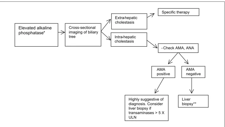
- Patients with cholestasis of unknown cause, particu-larly in the absence of antimithocondrial antibody (AMA) should be submitted to MRCP to rule out PSC (Class Ia).
- Liver biopsy should be considered in those subjects with normal MRCP under suspicion of small-duct PSC. Histology is not required for diagnosis of patients with large-duct PSC by MRCP. However, it may de needed to assess the presence of PSC with features of AIH, in those subjects with disproportionally higher aminotransferases levels more than 5 times the upper limit of normal (Class Ib).
- Colonoscopy is recommended for patients with PSC irrespective of the presence of symptoms. Multiple biopsies are recommend even if the endoscopic ap-pearance of the colonic mucosa is normal (Class Ia).
- Patients with the diagnosis of concurrent IBD should be submitted to colonoscopic screening for colorectal neoplasia (Class Ib).
Pharmacological treatment of PSC
and cytokine-induced cytotoxicity against hepatocytes and colangiocytes; and 4) immunomodulatory and anti-inlammatory effects(80). It is important to emphasize that the relative contribution of each of these mechanisms for the anticholestatic effect of UDCA is unknown. In the context of the PSC, UDCA has been evaluated at low doses (10 to 15 mg/kg/day), intermediate doses (17 to 23 mg/kg/day) and high doses (25 to 30 mg/kg/day), in several clinical studies, which have been compiled in three meta-analyzes.(81-83). Be-sides having different doses and designs and high risk of bias, these studies used different response criteria and, in general, included large numbers of patients with advanced disease, making it dificult to get deinitive conclusions about the eficacy and safety of UDCA in PSC. However, despite the abovementioned methodology limitations and until new evidence is generated in additional studies, we can conclude that: a) low-dose UDCA may result in clini-cal and biochemiclini-cal improvement, but without increase in survival; b) high doses can have a negative impact on the evolution of the disease, even in patients with early disease; c) intermediate doses can induce biochemical and histologi-cal response, without drug-related serious adverse events, but with uncertain impact on survival.
PSC carriers under treatment with UDCA should be periodically monitored with clinical examination and rou-tine liver tests, with two main objectives: to assess response to therapy and to identify potential disease progression. Recently, three studies have identiied clinical improvement in patients with PSC that showed a signiicant reduction in serum levels of ALP, deined as normalization of ALP or reduction to levels below 1.5 time the ULN at any time during follow-up or reduction ≥ 40% after one year of treatment with UDCA(84-86). Although these criteria need prospective validation, they may be useful for prognostic purposes. However, it is important to emphasize that late responses can occur (even after two years of therapy) and that the suspension of UDCA may lead to significant clinical and laboratory worsening(79,87). Thus there is no evidence that UDCA should be stopped in the absence of signiicant biochemical response, except in cases of sus-pected UDCA-related disease progression. Worsening of pruritus, fatigue and/or deterioration of hepatic synthesis tests, progressive elevation of serum ALP, and develop-ment, increase or rupture of esophagogastric varices are signs of disease progression.
Currently, there are no pharmacological alternatives for the speciic treatment of the PSC. Antiibrotic agents (colchicine, penicillamine, silymarin, etc.), antimicrobial (vancomycin, minocycline, metronidazole), immunobiologi-cal agents (inliximab, etanercept) and immunosuppres-sants (prednisone, prednisolone, budesonide, azathioprine, methotrexate, tacrolimus, cyclosporin, mycophenolate) have not been effective and/or safe for the treatment of PSC, and are not recommended(75). Examples of prom-ising drugs being evaluated in ongoing clinical trials: 24-norursodeoxycholic acid (an UDCA homologous; trial identiied as NCT01755507 on the ClinicalTrials.gov
website), docosahexaenoic acid (fatty acid omega-3 type; NCT00325013), obeticholic acid (farnesoid X receptor agonist; NCT02177136), BTT1023 (antibody anti-VAP-1; NCT02239211) and simtuzumab (humanized monoclonal antibody against the enzyme lysyl oxidase-like 2 [LOXL2]; NCT01672853).
Bezaibrate has been successfully used as a pharmaco-logical alternative in the PSC in a few cases reported in the literature, so that there is insuficient evidence to recommend its use(88,89).
Corticosteroids and other immunosuppressive agents are not recommended for the treatment of PSC, except in cases of PSC with features resembling AIH, the so called “AIH/ PSC overlap syndrome”(68) In this context, the combined regimen with prednisone and azathioprine is indicated, typically in association with UDCA. Immunosuppression (corticosteroids alone or in combination with azathioprine) is also recommended for the treatment of PSC associated to IgG4(68). In this case, the treatment has a minimum of three months of duration and can require maintenance therapy if there is recurrence or incomplete answer.
The most robust randomized controlled trials(90,91) and two meta-analyzes concluded that there is no signiicant impact of the use of UDCA on the incidence of CC(81,82). There is no evidence that the use of UDCA reduces the risk of development of gallbladder cancer in patients with PSC. As for colorectal cancer (CRC), the real impact of the use of UDCA remains unclear, with two studies suggesting a protective effect(92,93), four studies noting no impact(94-97) and one study indicating increased risk of CRC in UDCA users(98). Recent meta-analysis found no association between the use of UDCA and the risk of CRC or dysplasia in adults with PSC and IBD(99). Relecting the controversy, while the American guidelines do not recommend the use of UDCA as a prophylaxis agent against CRC(75), the European guidelines suggest its use for high-risk patients (those with extensive colitis, positive family history or previous CRC), although it recognizes that there is limited evidence(68).
Recommendations
- After detailed discussion of risks and beneits of therapy and about the limitations of available data, the use of UDCA in intermediate doses (17-23 mg/kg/day) should be considered for adult patients with PSC. (Class IIb)
- PSC carriers under treatment with UDCA should be regularly monitored with clinical examination and liver tests, to assess response to therapy and to identify possible disease progression. (Class I)
- In patients treated with UDCA, normalization or signiicant reduction of serum levels of ALP suggests better prognosis (Class II). There is no evidence that UDCA should be discontinued in the absence of response, except when the progression of the disease is possibly related to UDCA itself (Class II).
- There is no suficient evidence to recommend the use of ibrates or other pharmacological alternatives as speciic therapies for PSC (Class IIb).
- Immunosuppression with corticosteroids alone or in combination with azathioprine is recommended in cases of PSC with AIH-like characteristics and for the treatment of PSC associated IgG4 (Class I).
- There is no evidence that the use of UDCA reduces the risk of developing CC or gallbladder cancer in patients with PSC (Class III). The data available in the literature does not allow to conclude whether the use of UDCA is associated with lower risk of colorectal cancer (Class IIb).
- Pregnancy is generally well tolerated in women with compensated PSC, but there seems to be an increased risk of preterm birth (Class II). The use of UDCA can be considered during pregnancy, preferably after the irst quarter (Class II).
Endoscopic treatment of PSC
Endoscopic treatment of PSC should be considered for subjects with symptomatic dominant strictures. They are considered in the presence of a stenosis with a diameter of at least 1,5 and 1 cm, respectively, in the common bile duct (CBD) and in the hepatic duct. They usually occur in the hepatic hilum, hepatic duct or the CBD. Dominant strictures are observed in 45% to 58% of the patients with PSC(107). Their clinical presentation is also variable, but increasing liver enzymes, jaundice and recurrent cholangitis are com-mon features, particularly in those subjects with dominant strictures in the CBD. The presence of dominant strictures is associated with progression of PSC and their treatment by ERC may improve survival. However, it should be noted that clinical deterioration has been also reported in subjects with end-stage PSC submitted to ERC.
Endoscopic treatment of dominant strictures are as-sociated with clinical and biochemical improvement in around 80% PSC subjects without cirrhosis(108). However, it should not be used as a bridge to delay LT in those pa-tients with end-stage PSC, because LT is regarded as the only treatment modality that can really alter the natural history of the disease.
The main complications of ERC in patients with PSC are pancreatitis, cholangitis, hemorrhage, perforation, worsening of cholestasis and progressive clinical and bio-chemical deterioration. They are more commonly observed in subjects submitted to therapeutic ERC. Pancreatitis and cholangitis after ERC are reported to occur in, respectively, 5%-7% and 1% of the treated patients(109). Before endoscopic treatment, it is usually recommended antibiotic prophylaxis to prevent ascending cholangitis(3), that is more frequently seen in those subjects with cirrhosis, concurrent AIH and CD. The level of expertise of the examiner, performance of sphincterotomy and bile duct dilatation are also associated with cholangitis(110).
In PSC subjects without dominant strictures at disease onset, the occurence of de novo symptoms, particularly of jaundice, or clinical deterioration may suggest the develop-ment of dominant strictures.
Irrespective of the presence of those symptoms, colhangi-ocarcinoma have to be excluded in all subjects with PSC with dominant strictures. The screening and diagnosis of CC are highlighted elsewhere in this manuscript.
Most of the data concerning endoscopic treatment of PSC are not derived from RCT. Treatment options include bile duct stenting with or without previos dilation or only dilation. In one retrospective study, either dilation with or without stenting were associated with adequate biliary drain-age as well as clinical improvement of patients with PSC(111). However, a higher incidence of complication were noted in those patients who required stents, when compared to their counterparts treated only with dilation.
If stents are required, they should be changed every two to three months in order to prevent cholangitis due to stent obstruction. Some patients may require prolonged use of stent for 12 months to ensure resolution of those dominant stric-tures. The main disadvantage of stents are their associated risk for obstruction (usually seen in half of those treated patients), that may evolve to cholangitis and sepsis. Use of stents for a short period of time (mean of 11 days) was advocated in some reports to be associated with better outcomes(112). There is no RCT published until now, comparing dilation vs. stents in the management of dominant strictures in PSC.
Recommendations
- Endoscopic treatment can be indicated in centers with expertise in therapeutic ERC in subjects with PSC with dominant strictures (Class IIb)
- Before endoscopic treatment, it is mandatory to excluse cholangiocarcinoma (Class I)
- Endoscopic treatment can be performed with bile duct dilation or stenting due to the lack of evidence-based data to support either one of those treatment modali-ties (Class IIb)
Diagnositc and therapeutic implications for PSC in children
incidence of PSC in children have been stimated as 0,23 cases for 100.000 person-years. There are only 250 cases of PSC in children published in the literature from different referral cent-ers, which hamper the development of evidence-based guide-lines about the management of PSC in this age-group(75,113).
Most of the clinical indings of the disease in children are identical to those observed in adults as well as in other children with cholesttic liver diseases. Aproximately 20% of the children are asymptomatic. Some may present clinical features related to childhood such as growth retardation and puberty delay(115). The frequency of AIH and PSC overlap is more frequently reported in children, when compared to adults. CC is rarely seen and IgG4 disease have not been reported in childhood. Some inherited cholestatic diseases of childhood may resemble PSC. In this regard, mutations of ABCB4 gene, related to progressive familiar intrahepatic cholestasis type 3, have been reported in children with small-duct PSC(75,113,114,116).
The diagnosis of PSC in children is based on the presence of clinical, laboratory and cholangiographic features of the disease, after exclusion of secondary causes of sclerosing cholangitis. It should be pointed out that ALP is not regarded as a marker of cholestasis in childhood, due to its physiologi-cal increase during the bone growth, seen in this age-period. GGT, on the other hand, is regarded as a reliable tool to assess cholestasis in children. In this regard, 97% of those children with PSC had abnormal GGT at diagnosis(75,113-115 ). The ratio of GGT/AST is useful to detect AIH and PSC overlap in children(114). When compared to adults, children tend to have lower levels of bilirubin and higher leves of AST and ALT, due to the lower frequency of dominant strictures and CC and higher frequency of concurrent AIH usually seen in pediatric PSC(75, 113-117).
Liver biopsy is important in childhood PSC, particularly to assess small-duct PSC in subjects with normal cholangio-grams or other immune-mediated diseases. It is also crucial for the diagnosis of AIH and PSC overlap(75,113-118).
Magnetic resonance cholangiography is the procedure of choice for the diagnosis of PSC, and exhibit the same changes previously describe for adults. Aproximately 40% of the patients with normal cholangiograms may have small-duct PSC at liver biopsy(75,113-115).
CC is very rare in children with PSC(75,114,118). The younger patient with PSC and CC reported in the literature had 14 years of age and longstanding UC(114).
The association of PSC with IBD in children ranges from 33% to 81%. Therefore, colonoscopy is usually recommended even in children without bowel symptoms. If the initial colonoscopy disclose normal indings, subsequent examina-tions should be performed only if the patient develop typical symptoms of IBD. Even in those subjects with concurrent IBD, screening of CRC is controversial, since it s rarely seen before 16 years of age(75,113,114).
Up to now, there is controversy on the employment of UDCA in adults with PSC and there are no controlled data on their use in children(75,113-115,117,119) Antibiotics, such as oral vancomycin are still under investigation(114-116,120).
Dominant strictures are less common in children with PSC, but are also amenable to treatment, as highlight previ-ously(114,115).
Recommendations
- Due to the scarcity of cases of PSC in children, it is not possible to stablish evidence-based recommendations for the management of the disease in the pediatric age group (Class I)
- Clinical manifestations are similar to those observed in adults. Dominant strictures and CC is rarely seen. On the contrary, AIH and PSC overlap is much more common (Class IIa)
- MRCP is the procedure of choice for diagnosis of PSC in children. Liver biopsy is usually necessary to rule out other common causes of secondary sclerosing cholangitis (Class IIa)
- Colonoscopy should be performed to assess concurrent IBD (Class IIa)
- There is scarcity of data concerning treatment options for PSC in children (Class I)
Part III: PRIMARY BILIARY CIRRHOSIS
Diagnosis
PBC is now diagnosed earlier in its clinical course, with more than half of the patients being asymptomatic at the time of diagnosis. Among symptomatic patients, fatigue and pruritus are the most common symptoms, present in up to 70% of the cases. The pathogenesis of fatigue is unknown and appears to be multifactorial, with autonomic dysfunc-tion playing a signiicant role. Other causes of fatigue, including anemia, hypothyroidism, adrenal insuficiency, depression, and sleep disorder must be excluded prior to attributing the symptom to PBC. Patients complain of daytime sleepiness and lack of energy. Despite its impact in the quality of life, no correlation could be demonstrated so far between fatigue and severity of liver disease. Indeed, fatigue does not improve with treatment of the disease. Pruritus, on the other hand, is a more speciic symptom. It can be local or diffuse, usually worse at night, and often extremely debilitating. The onset of pruritus typically pre-cedes the onset of jaundice by months to years, and in fact it tends to improve or disappear as the disease progresses. Occasionally, itching starts during the third trimester of pregnancy and persists after delivery, in contrast to the itch-ing associated with intrahepatic cholestasis of pregnancy, which resolves after delivery. The pathogenesis of pruritus in PBC is also unclear and several hypotheses have been formulated, including derangements in bile salt produc-tion and excreproduc-tion, change in progesterone metabolites, histamine imbalance and increased levels of endogenous opioids. Recent clinical and experimental evidence highly suggest that the neurotransmitter lysophosphatidic acid is an important mediator of the cholestatic itching(68,121).
nearly 25% of asymptomatic patients at the time of diagno-sis progress to liver failure within 10 years(134).Contemporary series have shown that with earlier diagnosis, 42-66% of asymptomatic patients are at an earlier histological stage (stages I and II), whereas 82% of symptomatic patients are at a later histological stage of disease (stages III and IV). Patients who are symptomatic at presentation appear to have lower rates of biochemical response to UDCA and an increased risk of progression to cirrhosis and its complications(135).
Among the most common extra-hepatic complications of PBC are hyperlipidemia, cutaneous hyperpigmentation and osteoporosis. Patients who have advanced stage PBC have a ive-fold increase in risk for developing osteoporosis compared with those who have early stage.
Anti-mitochondrial antibody (AMA) negative patients have similar clinical course, response to therapy and compli-cation rate compared to their AMA positive counterparts. ANAs are detected in 30-50% of patients with PBC, and these may display patterns that are 95% speciic for PBC. Presence of antibodies against the nuclear pore protein gp210, with rim-like membranous pattern on IF, have been associated with more severe interface hepatitis, lobular inlammation and ductopenia, translating into worse clini-cal course and increased rates of progression towards liver failure(136). On the other hand, presence of anticentromere antibodies (ACA), noted in up to 30% of patients with PBC, may be a marker of future development of systemic sclerosis(137). In addition, ACA positivity has been associated with development of portal hypertension, possibly due to severe ductular reaction(136).
Associations between PBC and other autoimmune diseases have been consistently reported and reinforce the current classiication of PBC as an autoimmune condition. The reported prevalence of an overlap syndrome of PBC and AIH ranges between 2 and 20%(138).Overlap syndrome PBC-AIH is discussed in details in the corresponding topic. Sicca syndrome is seen in 60-80% of patients, and should be routinely investigated in those complaining of dry eyes/ dry mouth. Tests for dry eyes include Schirmer and ocular surface staining (Rose Bengal). Hashimoto’s thyroiditis is present in 20% of patients with PBC, often predating its diagnosis. Scleroderma is a multisystem autoimmune rheumatic disorder present in up to 8% of patients with PBC. The frequency of Raynaud´s phenomenon is four-fold higher in PBC when compared to the general population. In turn, up to 25 % of patients with scleroderma are AMA positive. The prevalence of rheumatoid arthritis in PBC has been reported between 1.8% and 5.6%, whereas the prevalence of AMA positivity in patients with rheuma-toid arthritis has been estimated at 18%. The association between PBC and polymyalgia has been described mainly in single case reports, but lupus erythematosus occurs in 2.7% to 7.5% of PBC cases. Associations with Addison disease and autoimmune diabetes are uncommon. Among the gastrointestinal disorders, celiac disease has been the most studied. Celiac disease is present in 6% of patients
with PBC; in contrast, 3% of celiac patients were found to have PBC. Serological screening testing for celiac disease is not routinely recommended in PBC, unless there is a strong suspicion for the disease(139).
Multiple lines of evidence support the concept of genetic predisposition in PBC, including 1) a high concordance rate among monozygotic twins (63%), 2) an increased prevalence of other autoimmune conditions in patients with PBC and their family members, 3) existence of familial clusters of PBC and 4) an increased rate of AMA positivity among irst degree relatives of patients with PBC(140-142). Despite the fact that its beneit hasn’t yet been demonstrated, we recom-mend screening adult irst degree relatives of patients with PBC with measurement of serum ALP level, and testing for AMA if ALP is elevated.
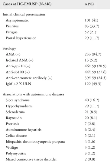
Data from Brazil are scarce. Between 1995 and 2013, 246 patients referred to the Autoimmune and Cholestasis Dis-eases Group at the University of São Paulo were diagnosed with PBC. Table 3 illustrates the relevant clinical presentation and serology data for these patients.
e been -orse clinical
er , -ted with
e
une condition.
esponding topic.
mer s -tients ynaud´s phenomenon is al toid arthritis eas
-tients
TABLE 3 – Clinical and laboratory data on 246 patients diagnosed with PBC at HC-FMUSP (personal communication, data not published)
Cases at HC-FMUSP (N-246) n (%)
Initial clinical presentation Asymptomatic
Pruritus Fatigue
Portal hypertension
101 (41) 83 (33.7) 52 (21) 29 (11.7)
Serology AMA (+) Isolated ANA (+) Anti-gp210 (+) Anti-sp100 (+)
Anti-centromere antibody (+) IgM >2 X ULN
233 (94.7) 13 (5.2) 46/159 (28.9) 44/159 (27.6) 39/159 (24.5) 122 (49.5)
Associations with autoimmune diseases Sicca syndrome
Hypothyroidism Scleroderma Raynaud’s Psoriasis
Autoimmune hepatitis Celiac disease
Idiopathic thrombocytopenic purpura Vitiligo
Polymyositis
Mixed connective tissue disorder
40 (16.2) 29 (11.7) 21 (8.5) 20 (8.1) 7 (2.8) 6 (2.4) 5 (2.1) 4 (1.6) 3 (1.2) 3 (1.2) 2 (0.8)