INTRODUCTION
The classiication of levels of evidence for Brazi lian Society of Hepatology recommendations for the diagnosis, staging, and treatment of hepatocellular carcinoma is based on a modiication of the GRADE approach, as follows: Grade of evidence (according to the GRADE SYSTEM REFERENCE).
A – High quality – Future research is unlikely to change the presented proposition.
B – Moderate quality – Future research may have a signiicant impact on the presented proposition. C – Low or very low quality – Future research is very likely to have a signiicant impact on the presen ted proposition.
BRAZILIAN SOCIETY OF HEPATOLOGY
RECOMMENDATIONS FOR THE DIAGNOSIS
AND TREATMENT OF HEPATOCELLULAR
CARCINOMA
Flair J
CARRILHO
1, Angelo Alves de
MATTOS
2, Alex F
VIANEY
3, Denise Cerqueira P
VEZOZZO
1,
Fábio
MARINHO
4, Francisco J
SOUTO
5, Helma P
COTRIM
6, Henrique Sergio M
COELHO
7,
Ivonete
SILVA
8, José Huygens P
GARCIA
9, Luciana
KIKUCHI
1, Patricia
LOFEGO
10,
Wellington
ANDRAUS
4, Edna
STRAUSS
1, Giovanni
SILVA
11, Isaac
ALTIKES
12,
Jose Eymard
MEDEIROS
13, Paulo L
BITTENCOURT
14and Edison R
PARISE
8ABSTRACT – Hepatocellular carcinoma is a malignancy of global importance and is associated with a high rate of mortality. Recent advances in the diagnosis and treatment of this disease make it imperative to update the recommendations on the management of the disease. In order to draw evidencebased recommendations concerning the diagnosis and management of hepatocellular carcinoma, the Brazilian Society of Hepatology has sponsored a singletopic meeting in in João Pessoa (PB). All the invited pannelists were asked to make a systematic review of the literature and to present topics related to the risk factors for its development, methods of screening, radiological diagnosis, staging systems, curative and palliative treatments and hepatocellular carcinoma in noncirrhotic liver. After the meeting, all panelists gathered together for the discussion of the topics and the elaboration of those recommendations. The text was subsequently submitted for suggestions and approval of all members of the Brazilian Society of Hepatology through its homepage. The present paper is the inal version of the reviewed manuscript containing the recommendations of the Brazilian Society of Hepatology.
HEADINGS – Hepatocelullar carcinoma. Malignant liver tumor. Screening. Diagnosis. Treatment. Carcinoma hepatocellular in non cirrhotic liver.
Declared conflict of interest of all authors: none Disclosure of funding: no funding received
1 Faculdade de Medicina da Universidade de São Paulo, SP, Brasil; 2 Universidade Federal de Ciências da Saúde de Porto Alegre, RS, Brasil; 3 Universidade Federal de
Sergipe, SE, Brasil; 4Hospital Português de Beneficiência, Recife, PE, Brasil; 5Universidade Federal do Mato Grosso, MT, Brasil; 6 Universidade Federal da Bahia, BA,
Brasil; 7Universidade Federal do Rio de Janeiro,RJ, Brasil; 8 Faculdade de Medicina da Universidade Federal de São Paulo, SP, Brasil; 9 Universidade Federal do Ceará,
CE, Brasil; 10 Faculdade de Medicina da Universidade Federal do Espírito Santo, ES, Brasil; 11 Universidade Estadual de São Paulo, SP, Brasil; 12 Hospital Santa Catarina
e Hospital Ipiranga, SP, Brasil; 13 Universidade Federal da Paraíba, PB, Brasil; 14 Hospital Português, Salvador, BA, Brasil.
Correspondence: Flair Carrilho. Rua Prof. Clementino Fraga, 220, 1901 – CEP: 40170-050 – Salvador, Bahia, Brasil. E-mail: [email protected]
Strength of recommendation
1 Factors that inluence the strength of recommen dation include the quality of evidence, patient outcomes, and cost.
2 Varying values and preferences, or greater uncer tainty, and a weak recommendation is probably required. Recommendation is made with a lower degree of conidence, high cost, or high resource utilization.
year(1). HCC is now the most common complication and the leading cause of death in patients with compensated liver cirrhosis(2). The vast majority of HCC cases are associated with cirrhosis. It is estimated that chronic hepatitis B virus (HBV) or hepatitis C virus (HCV) infection is implicated in more than 80% of HCC cases worldwide(3).
From an epidemiological standpoint, HCC is characteri zed by wide geographic variability, with a highly heteroge neous distribution, which is probably related to etiological factors such as prevalence of HBV and HCV infection and exposure to alatoxin B1. The majority of cases (>80%) occur in SubSaharan Africa and East Asia, which are considered highincidence areas(4).
Recent studies conducted in Europe and the United States have demonstrated a rise in HCCrelated mortality, whereas cirrhosisrelated mortality rates have declined or remained stable. In the U.S., HCC is the fastestgrowing cause of can cerrelated death, with an 80% increase in annual incidence over the last two decades(4).
Brazil is considered to have a low incidence of HCC. A study conducted at Hospital das Clínicas da FMUSP repor ted an annual incidence of 3.5% in cirrhotic patients(5). In 2009, a nationwide survey included data from 29 centers for a total of 1,405 patients diagnosed with HCC. The median age was 59 years and 78% were male. Liver cirrhosis was present in 98% of cases and chronic HCV infection was the most common etiology (54%), followed by HBV (16%) and alcoholism (14%)(6).
Risk factors
The main risk factors for HCC are liver cirrhosis, HBV and HCV infection, alatoxin contamination of foods, alco hol abuse, diabetes, obesity, nonalcoholic fatty liver disease (NAFLD), and hereditary hemochromatosis.
Cirrhosis of any etiology is a major risk factor for the development of HCC. Among all cirrhotic patients, those with HCV cirrhosis have the highest risk of developing HCC, followed by those with hemochromatosisrelated cirrhosis. Advanced age, male sex, severity of cirrhosis, and sustained inlammatory activity are predictors of HCC regardless of cirrhosis etiology. In cirrhosis of viral etiology, HBV/HCV or HBV/HDV coinfection increases the risk of HCC, as does comorbid alcohol abuse(7).
In patients with chronic HBV infection, the risk of developing HCC increases with hepatitis progression, C or B genotype (the latter is particularly associated with HCC development in patients under the age of 50 and those without cirrhosis), and elevated viral replication rates (HBV DNA >10,000 copies or 2,000 IU/mL)(812). It bears mention that even inactive carriers of HBV are at risk of developing HCC(13).
In hepatitis C, risk factors include advanced age and ibrosis as well as genotype 1 infection(14, 15), and the syner gistic effects of alcohol and diabetes are likely to be more important(16). Sustained virologic response to therapy sig niicantly reduces the risk of HCC, but does not eliminate it altogether, and the presence of diabetes has been associated
with development of tumors even after cure of infection(17, 18). The B alatoxins are carcinogenic in a wide range of labo ratory animals as well as in humans, and their presence in the diet correlates with the incidence of HCC(19). The molecular events associated with HCC are related to genetic alterations and mutations (such as a p53 gene mutation) triggered by exposure to this toxin. The 249Ser TP53 mutation has been detected in 28% of HCC samples in Brazil, which is a high prevalence rate(20).
Abusive, prolonged alcohol intake is an established risk factor for HCC, both independently (relative risk 1.5–2.0) and in association with HCV and HBV infection(2123). Among alcoholics, the risk of HCC increases linearly with daily intake of >60 g EtOH, and doubles in the presence of comorbid HCV infection(21).
Diabetes, overweight, and obesity are also associated with increased risk of HCC (2426). Predictably, NAFLD – particularly in the steatohepatitis phase – is a risk factor for development of liver cancer(27).
Finally, patients with hereditary hemochromatosis are also at increased risk of developing HCC, particularly those with cirrhosis(28).
Recommendations
Effective antiviral therapy, administered as early as possible, should be recommended to all patients with HBV or HCV infection (category 1A).
Universal immunization against hepatitis B in popula tions at risk of developing HCC (1A).
Address alcohol abuse in patients with chronic liver disease, particularly those with HCV/HBV (1B). Management of diabetes mellitus (2B) and obesity (1A)
in patients with liver disease, and lifestyle modiications in those with NAFLD, may reduce the risk of HCC.
Screening
Some of the characteristics of HCC justify the use of screening strategies: it is a common condition, which carries high rates of morbidity and mortality, in a welldeined at risk population (patients with chronic liver disease). Further more, it is amenable to detection via effective, noninvasive, lowcost diagnostic techniques and curative treatment, which can increase patient survival, is available(29). Several studies have established the eficacy of screening for providing sur vival beneits, as well as the costeffectiveness of screening strategies(3032).
Screening is recommended in:
Noncirrhotic patients: On the basis of incidence, adult patients of Asian or African ethnicity with active HBV infection or a family history of HCC are the groups at greatest risk of developing HCC. European and U.S. guidelines also recommend screening for patients with hepatitis C and bridging ibrosis(29, 34).
In this setting, transient elastography is a promising tool for stratiication of patients at different HCC risk levels(35). There is little information on the incidence of HCC in noncirrhotic patients without chronic viral infection, including those with alcoholic and nonalco holic hepatitis, autoimmune liver disease, hemochro matosis, etc.
Treated patients with chronic viral hepatitis: Patients with chronic HBV hepatitis who remain at risk of developing HCC due to baseline factors and those with HCV infection and advanced cirrhosis or ibrosis, despite sustained virologic response, should be offered screening(13, 17, 18, 36).
The optimal range for HCC surveillance depends on two key characteristics: rate of tumor growth to its limit of de tection and rate of tumor incidence in the target population. On the basis of established knowledge about the mean tumor volume doubling time in HCC(29, 34, 37), a 6month interval be tween screenings is a reasonable choice. Attempts at shorter or longer intervals have not achieved superior effectiveness over the 6month period(38, 39).
Ultrasound plays an important role in the early diagno sis of HCC, and has been adopted as the best – and only – diagnostic modality for screening purposes worldwide. The presence of a liver nodule in patients with chronic liver disease mandates further investigation to rule out or conirm early diagnosis of HCC. Ultrasound is highly recommended for this purpose, as it is a riskfree, nonradioactive, nonin vasive method with good patient acceptability and moder ate relative cost(29, 34). However, it is important to note that ultrasound is highly dependent on the quality of equipment and on operator expertise, which can affect the quality of screening(39, 40). Therefore, in centers that lack expertise for ultrasound examination of the liver, screening of alphafe toprotein (AFP) levels in blood may still be necessary. AFP levels are normal in approximately 40% of tumors, and, under optimal conditions, the addition of AFP testing increases the rate of detection of small lesions by only 6%–8% while substantially increasing the number of falsepositive results, thus increasing the cost of screening(38, 41).
In a Brazilian cohort study, after implementation of a screening program, detection of HCC at a tumor diameter of <3 cm increased from 14% to 32% overall and to 65% in the last period of the study, with 5year survival rates of 50% in patients diagnosed and treated with percutaneous ablation or liver transplantation(42).
Recommendation
Screening should be provided to all patients with cir rhosis of any etiology or those with hepatitis C and advanced ibrosis, even if already receiving antiviral
therapy; and to patients with hepatitis B and a family history of HCC or those of African or Asian descent with active HBV infection (category 1A, 1B).
These patients should undergo screening via abdominal ultrasound, performed by a skilled examiner, every 6 months (1B). Although little additional insight is pro vided by AFP, it is recommended that AFP testing be performed in addition to ultrasound at centers lacking the necessary imaging expertise (2C).
RADIOLOGICAL DIAGNOSIS OF HEPATOCELLULAR CARCINOMA
It is recommended that certain imaging and reporting criteria be followed for radiological diagnosis of HCC.
Computed tomography (CT) should be performed in a multislice scanner, and the following series should be im aged: noncontrast, arterial phase (2030 sec after contrast injection), portal venous phase (6070 sec), and equilibrium phase (180 sec). Contrast should be administered at a high concentration (1.5 mL/Kg body weight) and at a rate of 4 mL/s. Magnetic resonance imaging (MRI) should ideally be performed in a highield scanner (1.5Tesla or greater) and the following phases obtained: T2, T1 gradientecho (GRE) inphase/outofphase; contrastenhanced: arterial, portal venous, and equilibrium (delayed) phases.
Reports should describe the size, location, and vascular ization (hypervascular/fastlow, washout, compare arterial phase to equilibrium phase, which should be thoroughly evaluated) of each nodule. The radiologist should also assess macrovascular invasion of the portal or hepatic veins, distin guish tumor thrombus from bland thrombus, and evaluate extrahepatic tumor extension (metastatic lesions).
Radiological diagnosis
In recent years, a characteristic vascular pattern of HCC was identiied, and noninvasive radiological modalities have progressively replaced liver biopsy for the diagnosis of HCC in a substantial number of cases, particularly in patients with cirrhosis in whom there is a risk of major complications. On dynamic CT or MRI, the typical pattern involves hypervas cularization of the nodule in the arterial phase and contrast washout in the vascular or delayed phase(34). Prospective studies of the discriminant ability of these parameters for noninvasive diagnosis in cirrhotic patients with tumors 0.5–2 cm in diameter found 100% speciicity, but low sensitivity(46). Another study found that sequential use of two dynamic methods(47) not only preserves this high speciicity but also provides signiicant improvements in sensitivity for detection of tumors 1–2 cm in diameter (Figure 1). Therefore, it is recommended that at least two dynamic imaging methods be used to diagnose tumors <2 cm, although recent evidence suggests that, if the tumor exhibits the aforementioned chara cteristic pattern on one imaging modality, conirmation with a second method is unnecessary(34).
liaryspeciic (or liverspeciic) contrast media, which are characterized by hepatocyte uptake and biliary excretion. The agents employed for this purpose are gadoxetic acid and gadobenic acid (meglumine gadobenate). Recent publi cations have shown that liverspeciic contrast agents can be useful in the differential diagnosis of small nodular lesions, to distinguish HCC from benign nodules
Detection and characterization of HCC, particularly in the early stages, is facilitated by use of these contrast media, because the majority of HCC lesions are hypointense on hepatobiliaryphase MRI even if the characteristic vascular pattern is not present (4851). In one study, the adoption of liverspeciic contrast changed the classiication of 11.5% of patients with HCC and classiied by the Barcelona Clinic Liver Cancer (BCLC) criteria as stage 0 or A(52).
Recommendations
Cirrhotic patients with ultrasounddetected nodules <1 cm in diameter and with no diagnosis established by other imaging modalities should be followed every 3–4 (2B).
Nodules >1 cm detected on ultrasound screening in the cirrhotic liver require further investigation by CT or MRI (1B).
If a liver biopsy yields inconclusive results, patients may undergo either a second biopsy or imagingbased followup every 3–6 months (2B).
If imaging indings are typical for HCC, the lesion should be treated as such, regardless of liver biopsy indings (1A).
If indings are not characteristic or the vascular proile is atypical, either a second dynamic imaging study (using another imaging modality) or a biopsy of the lesion should be performed (1B).
Liverspeciic contrast agents can be useful in the differ ential diagnosis of small nodular lesions, to distinguish HCC from benign nodules (2B).
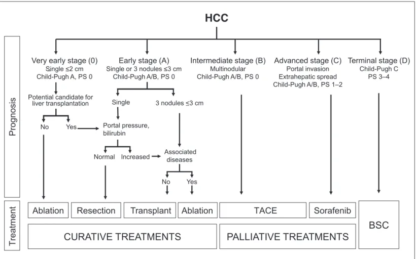
STAGING SYSTEMS FOR HEPATOCELLULAR CARCINOMA
presence of macrovascular invasion, and presence of metas tases; and 3) the general health of the patient. In patients with solid tumors, prognosis is essentially related to tumor stage. In HCC, however, prognostication is far more complex, because the hepatic dysfunction present in the majority of cases also has a signiicant inluence, as does the underlying clinical status and the treatment provided. On the other hand, in addition to tumor stage, the severity of hepatic dysfunc tion also guides the applicability and eficacy of therapy. Serum and tissue biomarkers, which are widely used in the classiication of several tumor types, have yet to be validated as components of HCC staging systems and should not be used for this purpose in routine clinical practice. Studies of molecular and genetic markers are underway and may have future applicability.
Several HCC staging systems are currently available. Those most commonly used are Okuda, TNM, CLIP, CUPI, BCLC, JIS, and GRETCH(5461). Of these, ive have been tested (BCLC, French, CLIP, CUPI, and JIS) and four have been validated (BCLC, CUPI, CLIP, and JIS). Only two (BCLC and CUPI) employ the three main prognostic factors (tumor, hepatic function, and clinical condition), and only the BCLC system determines the optimal treatment modality for each subclass. Comparative studies between these different classiications have concluded in favor of the superiority of
the BCLC system(62, 63). The BCLC classiication has been adopted by the European Association for the Study of the Liver (EASL), the European Organisation for Research and Treatment of Cancer (EORTC)(34), and the American Association for the Study of Liver Diseases (AASLD)(29).
In Brazil, only one study(64) has compared HCC staging systems (Okuda, BCLC, CLIP, CUPI, JIS, French, Tokyo, and TNM). In this study, the BCLC and Okuda systems performed best to predict survival among Brazilian patients with HCC.
The BCLC staging system(55) was proposed in 1999 and, unlike other classiications, is not a scoring system; it employs variables established in a variety of previous studies as inde pendent prognostic factors. The variables taken into account are clinical condition (functional status), tumor characteris tics (size and number of nodules), vascular invasion, portal hypertension, Okuda score, and Child–Pugh classiication, and the system stratiies tumors into a series of risk stages (0, A, B, C, and D). The advantages of this system include the association of subclasses with therapeutic modality, its ability to identify earlystage tumors for treatment curative intent, its introduction of an intermediate classiication (not included in other systems), and its consideration of palliative treatment and identiication of endstage disease in which supportive care alone is indicated (Figure 2).
Recommendation
To adopt the Barcelona Clinic Liver Cancer (BCLC) classiication as the HCC staging system of choice for use in Brazil (1B).
CURATIVE AND PALLIATIVE TREATMENT
Resection
Resection is the main treatment modality for HCC in the noncirrhotic liver, whereas liver transplantation (LTx) is considered the optimal curative treatment for hepatic cirrhosis complicated by early or very earlystage HCC. However, the supply of organs remains insuficient to enable transplantation of all waitlisted patients. This clearly affects overall outcomes for this therapeutic modality, whether due to death on the waiting list or to patient exclusion secondary to disease progression beyond adopted criteria. As a rule, 5year overall survival rates after LTx for HCC are in the region of 70%. However, when analysis is conducted by the intentiontotreat (ITT) principle, estimated survival is far lower. Llovet et al.(65) highlighted this issue and demonstrated a steep decline in 2year overall survival, from 84% to 54%, after considering ITT analysis.
Surgical resection is considered a potentially curative treatment for HCC, and provides good outcomes in select ed patients(66). Technical and technological reinements and improved patient selection now produce 5year survival rates of 50%–70% after resection(67).
The optimal candidates for resection or LTx are patients with very early disease (veHCC), deined as single tumors up to 2 cm in size, and those with earlystage HCC (eHCC), with nodules meeting the Milan criteria (single tumor up to 5 cm in diameter or up to three tumors up to 3 cm in diameter). However, some case series have reported acceptable outcomes after resection in patients with more advanced HCC(68, 69).
Tumor size, number of nodules, hepatic function, and portal hypertension are predictors of prognosis after HCC resection(7072). Surgical resection remains the sole treatment option for patients with veHCC and eHCC when hepatic function and clinical condition permit, largely due to the scarcity of livers for transplantation, because LTx provides superior longterm survival(73).
Selection of the optimal therapeutic modality for HCC in patients with cirrhosis is highly challenging and requires not only adequate assessment of tumor size, number, and location, but also a thorough evaluation of hepatic func tional reserve. Overall, resection is indicated in patients with compensated hepatic function (Child–Pugh A). The presence of portal hypertension, characterized by a venous pressure gradient ≥10 mmHg or the presence of esophageal varices, splenomegaly, and thrombocytopenia, is one of the stron gest predictors of poor survival after resection. Resection is contraindicated in Child–Pugh class C patients. However, it also bears stressing that even Child–Pugh class A patients may decompensate after larger resections(74).
The Model for EndStage Liver Disease (MELD) crite rion, which uses creatinine, INR, and bilirubin as parameters,
has been adopted in the majority of countries (including Brazil) to provide a severitybased priority criterion for LTx in waitlisted patients. However, in 2009, a research group in Bologna, Italy, published a series of 462 cirrhotic patients who had undergone hepatectomy(75). In this case series, patients operated at a MELD score >10 had a 15% rate of postoperative liver failure, regardless of the extent of resection. Other groups later reported similar results using the MELD score as a prognostic indicator of liver failure after resection.
The rate of HCC recurrence after liver resection, which approaches 70% at 5 years, remains the greatest challenge to achieving optimal survival(76, 77). Intrahepatic dissemination of primary HCC appears to account for early recurrences (occurring within 2 years of resection), whereas precancerous liver lesions are probably implicated in late recurrence.
Laparoscopic surgery is a lessinvasive approach that has been increasingly used as an alternative to surgical resection for HCC treatment. It is comparable to open surgery in terms of oncologic success and survival. However, laparoscopic surgery appears to provide advantages including decreased blood loss, lower morbidity, and shorter hospital stay, al though there is no highquality evidence from prospective randomized trials to support this. The availability of lapa roscopic surgery is certainly an additional argument in favor of surgical resection as a treatment modality for HCC.
Recommendations
Resection is the main therapeutic option for HCC in the noncirrhotic liver (1A).
In the cirrhotic liver, HCC resection should be considered in patients with veHCC or eHCC and meeting the Milan criteria, with good liver function (Child–Pugh class A, normal bilirubin, and no portal hypertension) (1B). Use of the MELD score as an indicator of resection
appears promising, but cannot yet be recommended for adoption in routine clinical practice.
Liver transplantation
It is estimated that HCC is the primary indication for LTx in 25% of all cases in Europe and 40% in the United States(78). LTx is still the optimal treatment for HCC, as it provides radical resection (through total hepatectomy) and “cures” both cirrhosis and portal hypertension. Initially, LTx was performed indiscriminately for HCC treatment, with no established criteria, and the recurrence rate was prohibitive, in excess of 50%. In 1996, Mazzafero et al.(79) published a case series in the New England Journal of Medicine of transplant recipients with a single nodule up to 5 cm in size or up to three nodules up to 3 cm in size each, without evidence of vascular invasion or extrahepatic disease. Their 4year survival rate was 74%. These are now known as the Milan criteria.
rates similar to those of patients treated within the Milan criteria. Other expanded criteria have been published in the literature, with similar results. However, a careful assessment of studies that included ITT analysis revealed a substantial reduction in posttransplantation survival with use of ex panded criteria. The recommendations of the international consensus conference for LTx in HCC, led by Professor P.A. Clavien in Zurich(81), were published in 2012. These recommendations cite the Milan criteria as the standard for inclusion of patients on a liver transplant waiting list. Patients with tumors exceeding the Milan criteria may be treated with transarterial chemoembolization (TACE) before LTx. If the tumor is found to meet Milan criteria after a few sessions of TACE (downstaging), transplantation may be indicated and provide good longterm results(82).
Recommendations
The Milan criteria remain the most adequate criteria for selection of HCC patients for liver transplantation. Patients who have been waitlisted for over 6 months
should preferably undergo transarterial chemoemboli zation to help them continue to meet the Milan criteria. Patients who meet the Milan criteria after transarterial chemoembolization (downstaging) may be waitlisted for LTx and achieve good outcomes.
Ablative techniques
Ablative techniques should be considered the irstline treatment of choice for patients with earlystage HCC who are ineligible for surgical resection. Several techniques are effective for ablation of lesions <3 cm in size. Tumor cells can be destroyed by injection of chemical substances (eth anol, acetic acid) or by temperature manipulation (laser, radiofrequency, microwaves). The most widely used ablative techniques include percutaneous ethanol injection (PEI) and radiofrequency ablation (RFA)(34).
PEI consists of an injection of absolute alcohol through a ine needle advanced into the tumor. It is most commonly performed via the percutaneous route, under ultrasound or CT guidance. The presence of intratumoral septation and/ or a capsule limits the curative capacity of this technique, particularly in tumors larger than 2 cm. After PEI, complete necrosis is achieved in >90% of tumors <2 cm, 70% of tumors 2–3 cm in size, and 50% of tumors 3–5 cm in size. The main limitation of this technique is the rate of local recurrence, which may be as high as 43% in lesions >3 cm. Therefore, PEI is indicated in patients with up to three nodules no larger than 3 cm in diameter each (83).
The most common adverse events are pain, fever, and a feeling of alcohol intoxication. A decline in hepatic function is far less likely to occur after PEI than with surgical resec tion or chemoembolization. The mortality rate attributable to the procedure is <1%. PEI is contraindicated in cases of uncontrolled ascites or bleeding diathesis(83, 84).
In recent years, RFA has replaced PEI in some centers. It is also performed percutaneously, under ultrasound or CT guidance, by advancing a needleshaped electrode into the
tumor and applying radiofrequency energy, with the purpose of generating a thermal destruction zone that encompasses the entire tumor and a 1cm margin around it. For tumors 2–5 cm in size, RFA is more effective than PEI in producing complete response, and fewer sessions are required(85).
The primary disadvantage of RFA is its high cost and higher adverse event rate. The mortality rate attributable to the procedure ranges from zero to 0.3%. RFA should be avoided if the tumor is located near the biliary tree or viscera. Furthermore, due to heat dispersion, it may be ineffective in tumors located near major blood vessels(86).
PostPEI or postRFA followup is performed by means of dynamic imaging (CT, MRI, or contrastenhanced ultra sound). It is recommended that followup imaging be per formed 1 month after the last session. Complete response to treatment is deined as absence of contrast enhancement of the lesion, indicating cessation of blood supply and necrosis of the tumor(83).
Randomized controlled trials have shown that RFA is superior to PEI in terms of achieving local disease control. Its impact on survival remains controversial. A Japanese study reported increased survival in patients treated with RFA(87), while a European study found no signiicant improvement(88). Two randomized controlled trials by Lin et al.(89, 90) reported survival advantages in favor of RFA, as compared both to PEI and percutaneous acetic acid injection (PAI), on sub group analysis of patients with tumors >2 cm. The likelihood of local recurrence was signiicantly lower in the RFA arm (vs PEI and PAI) in four studies, whether assessed as primary or as secondary outcomes.
Recommendations
RFA or PEI ablation is the treatment of choice for pa tients with BCLC stage 0/A HCC who are not eligible for surgical resection.
RFA ablation is the preferred method for tumors <5 cm, as it provides superior disease control.
PEI is recommended in cases where RFA is not tech nically feasible, particularly for nodules <3 cm.
Chemoembolization and transcatheter therapies
TACE, drugeluting bead chemoembolization (DAB TACE), and transarterial radioembolization with yttrium90 microspheres (TARE) are regarded as the best options for intraarterial therapy of HCC(91).
TACE is indicated in patients with BCLC stage B (inter mediate) disease, with single tumors >5 cm in diameter or multinodular tumors, as long as there is no vascular invasion or remote involvement. As a rule, it is performed preferential ly in Child–Pugh class A patients, although it should not be ruled out in Child–Pugh class B patients with stilladequate hepatic functional reserve(92, 93).
but the great merit of TACE seems to lie in its ability to slow tumor progression and vascular invasion(92).
There is no evidence from prospective, randomized controlled trials as to how often the procedure should be performed, and no consensus on the optimal embolization agent and/or chemotherapeutic agent(93). However, catheteri zation for TACE should be as selective as possible so as to minimize injury to underlying healthy tissues(96).
The adverse effect proile of this procedure warrants attention. Adverse events are generally similar to those associated with systemic chemotherapy, and the effects of arterial obstruction may be associated with the postembo lization syndrome(97).
A signiicant limitation of TACE is the high rate of tumor recurrence, which limits patient survival. Even in patients with an initial response to treatment, the cumulative likeli hood of tumor recurrence is around 65% (93, 98).
The development of drugeluting polyvinyl chloride beads, used in the DEBTACE technique, has decreased the incidence of side effects, as it reduces passage of the chemotherapeutic agent into the systemic circulation. In addition, as these beads can be calibrated, the degree of arterial obstruction is predictable and the procedure is made more homogeneous, with no loss of eficacy. DEBTACE thus enables administration of high drug concentrations at the level of the tumor, with few systemic effects(99).
The PRECISION V trial, a prospective, randomized, multicenter, phase II study that compared the eficacy of DEBTACE versus conventional TACE, found no difference in response rate between the two procedures, but did ind superior tolerance, less hepatic toxicity, and fewer systemic effects of doxorubicin in the DEBTACE group(100).
A study conducted by Burrel et al.(101) highlighted the good outcomes obtained with this procedure and suggested that its indications could be expanded to create the concept of “treatment stage migration”. In a recent metaanalysis, DEBTACE proved superior to conventional treatment(102).
However, it bears stressing that adequate assessment of treatment outcomes is dificult. Tumor necrosis, as estimated by lack of contrast uptake on dynamic imaging, appears to be the most important parameter. The modiied Response Evaluation Criteria In Solid Tumors (mRECIST) evaluation has been proposed as a tool for outcome assessment. It is important to note that chemoembolization should not be repeated in patients who fail to exhibit signiicant necrosis after two treatment sessions; when followup imaging shows failure to produce signiicant necrosis in sites of tumor pro gression; and when patient assessment indings preclude safe retreatment(103, 104). Authors have proposed that the decision to retreat be based not only on tumor response, but also on the effects of treatment on liver function, as assessed by novel scores(105). Treatment response may also be assessed by measuring reductions in tumor marker levels.
Chemoembolization can be combined with other thera peutic procedures(93, 106).
Recently, transarterial radioembolization with yttrium90 microspheres (TARE) has gained favor in the literature. Pa
tients treated with this technique have shown good responses to treatment, with a mean time to tumor progression of 7.9 months and 17.2 months of survival in Child–Pugh class A patients. Adverse effects were mild, and the 30day mortality rate was 3% (107). A European study(108) reported several ad vantages of this technique, including the fact that it can be performed in advanced HCC (51% of cases were BCLCC); in tumors both with and without vascular invasion (31% had portal thrombosis); in patients ineligible for chemoemboliza tion (47% of cases); and in those with deterioration of liver function. In addition, a multicenter study(109) reported that TARE may translate into increased survival in in patients with advanced disease (BCLCC). It is also interesting to note that TARE is a singlesession procedure and can be performed in an outpatient setting(110).
The role of locoregional therapy as neoadjuvant treat ment to reduce dropout among patients waitlisted for LTx warrants consideration. Therefore, in centers where waitlist time exceeds 6 months, patients should be considered for chemoembolization(111113).
Its potential utility for downstaging of patients whose disease exceeds the Milan criteria should also be conside red(111, 114). Despite debate in the literature concerning the role of downstaging, chemoembolization could be used for this purpose. At least 3 months should be allowed to elapse between successful downstaging and LTx(115).
Recommendations
TACE should be performed as irstline treatment with out curative intent for patients in whom surgery is not indicated, with large or multifocal HCC, no vascular invasion, and no extrahepatic involvement.
DEBTACE is a safer alternative, but its use is limited by its high cost.
Both TACE and DEBTACE could be used to prevent dropout among patients waitlisted for liver transplan tation, as well as for downstaging purposes.
TARE may be an alternative in patients with impaired liver function, portal thrombosis, and contraindications to chemoembolization.
Non-curative treatment: systemic therapy
To date, treatment strategies based on systemic chemo therapy have failed to demonstrate beneit in terms of sur vival and time to tumor progression(116). A recent systematic review of the literature suggests that oxaliplatinbased che motherapy could be beneicial in advanced HCC, although controlled trials are needed(117).
in the AsiaPaciic study(119, 120). Sorafenib remains the only systemic therapy approved for HCC. A subgroup analysis of SHARP trial data suggests that improved survival occurred independently of underlying disease etiology, baseline tumor characteristics, disease stage, and prior therapy(121).
In this context, sorafenib is indicated for patients with ad vanced HCC (BCLCC). In cirrhotic patients with HCC and Child–Pugh class A liver function, treatment is well tolerated and responses have been encouraging(122, 123). However, patients in Child–Pugh class B (score ≥8) exhibited a higher incidence of hepatic dysfunction (ascites, encephalopathy, and jaundice) and decreased survival with this treatment(122, 123).
Combination of other drugs with sorafenib, as well as its joint use with other therapeutic modalities (such as chemo embolization), have thus far failed to produce convincing results(124, 125). A recent study suggests that combined sorafenib therapy and RFA could reduce recurrence rates and prolong mean survival in patients with intermediatesized HCC(126).
The most signiicant and most commonly reported side ef fects of sorafenib therapy are elevated liver enzymes, fatigue, hypertension, the handfoot syndrome, and diarrhea(125). Development of the handfoot syndrome and decrease in AFP level have been associated with favorable response to treatment(123).
Recommendations
To date, treatment strategies based on conventional systemic chemotherapy have failed to show beneit, whether in terms of survival or in reducing time to tumor progression (level of evidence 1B, strength of recommendation A).
Sorafenib is recommended as the sole systemic therapy approved for HCC (level of evidence 1A, strength of recommendation A).
There is no robust evidence to support any beneit from drug combinations or addition of other therapeutic methods to sorafenib therapy (level of evidence 2A, strength of recommendation B).
TREATMENT OF HEPATOCELLULAR CARCINOMA IN THE NONCIRRHOTIC LIVER
It is estimated that, in 7%–20% of cases, depending on the geographic area of assessment, HCC may develop in the noncirrhotic liver (NCHCC). HCC may arise in indi viduals with no evidence whatsoever of liver disease, i.e., in completely normal livers, or in the presence of inlammatory, ibrotic, or degenerative processes that have not yet reached the cirrhotic stage. NCHCC is generally diagnosed at more advanced stages. However, when diagnosis and treatment are provided in a timely manner, patients may have a better prognosis(127, 128).
In patients with NCHCC and no evidence of prior liver, the estimated prevalence is 10%–12%. These cases are often associated with exposure to toxic agents such as alatoxins, chemical agents, or radiation, or with malignant transfor mation of hepatic adenomas(127, 128).
Surgery is the irstline treatment option for HCC in the noncirrhotic liver, with a 5year overall survival rate around 40% for unselected noncirrhotic patients and 64%–85% for selected patients or patients with a nonibrotic liver(129138). The 5year recurrence rate is around 50%(1328). For patients in whom partial liver resection may not be possible, despite the otherwise normal liver parenchyma, or because of recurrence since previous surgery, liver transplantation may be the only alternative providing a chance of cure. A systematic review published in 1999 (138) found poor 5year survival for liver transplantation in these patients (39.4% for the ibrolamellar type and 11.2% for conventional HCC). A recently published retrospective analysis of a large cohort of patients trans planted for NCHCC in Europe found that, in contrast to patients with HCC in a ibrotic or cirrhotic liver, tumor size is not an important risk factor for posttransplant survival in NCHCC, while the presence of macrovascular invasion or hilar lymph node involvement should be considered a contraindication for transplantation. Patient selection is associated with a 5year posttransplant survival rate of 59%.
Carrilho FJ, Mattos AA, Vianey A, Vezozzo DCP, Marinho F, Souto FJ, Cotrim HP , Coelho HSM, Silva I, Garcia JHP, Kikuchi L 1, Lofego P, Andraus W, Strauss E, Silva G, Altikes I, Medeiros JE, Bittencourt PL, Parise ER. Recomendações da Sociedade Brasileira de Hepatologia para diagnóstico e tratamento do carcinoma hepatocelular. Arq Gastroenterol. 2015(Supl 1):214.
RESUMO – O carcinoma hepatocelular é uma neoplasia de importância global e associada a altos índices de mortalidade. Recentes avanços no diagnóstico e tratamento da doença tornaram necessárias que se atualizassem as recomendações sobre o manejo da doença. Para deinir as recomendações sobre o diagnóstico e tratamento do carcinoma hepatocelular, a Sociedade Brasileira de Hepatologia organizou uma reunião monotemática em João Pessoa (PB). Todos expositores foram solicitados a fazer uma revisão sistemática da literatura e apresentar os temas relacionados a fatores de risco para o desenvolvimento de carcinoma hepatocelular, métodos para rastreamento, diagnóstico radiológico e sistemas de estadiamento da doença, tratamen tos curativos e paliativos e carcinoma hepatocelular em fígado não cirrótico. Após o encontro, todos os expositores se reuniram para discussão dos tópicos e elaboração dessas recoemndações. O texto resultante foi ainda submetido a avaliação e aprovação por todos membros da Sociedade através de sua homepage. O documento atual é a versão inal que contêm as recomendaçaoes da Sociedade Brasileira de Hepatologia.
REFERENCES
1. Ferlay J, Shin HR, Bray F, Forman D, Mathers C, Parkin DM. Esti mates of worldwide burden of cancer in 2008: GLOBOCAN 2008. Int J Cancer. 2010;127:2893917.
2. Sangiovanni A, Prati GM, Fasani P, Ronchi G, Romeo R, Manini M e cols. The Natural History of Compensated Cirrhosis Due to Hepatites C Virus: a 17Year Cohort Study of 214 patients. Hepatology. 2006;43: 130310.
3. Okuda H. Hepatocellular carcinoma development in cirrhosis. Best Practice and Research Clinical Gastroenterology. 2007;21:1613. 4. ElSerag HB, Rudolph KL. Hepatocellular carcinoma: Epidemiology
and Molecular Carcinogenesis. Gastroenterology. 2007;132:255776. 5. ParanaguaVezozzo D, et al. Incidence of Hepatocellular Carcinoma in
Cirrhotic Patients in São Paulo, Brasil. Hepatology. 2006;44:504. 6. Carrilho FJ, Kikuchi LOO, Branco F et al. Clinical and epidemiological
aspects of hepatocellular carcinoma in Brazil. Clinics. 2010;65:128590. 7. Fattovich G, Stronffolini T, Zagni I, Donato F. Hepatocellular carcinoma in cirrhosis: Incidence and Risk Factors. Gastroenterology. 2004;127:355. 8. Chen CJ, Yang HI, Su J, Jen CL, You SL, et al. Risk of hepatocellular
carcinoma across a biological gradient of serum hepatitis B virus DNA level. JAMA. 2006;295:6573.
9. Liaw YF, Sung JJ, Chow WC, Farrell G, Lee CZ, et al. Lamivudine for patients with chronic hepatitis B and advanced liver disease. N Engl J Med. 2004;351:152131.
10. Sung JJ, Tsoi KK, Wong VW, Li KC, Chan HL. Metaanalysis: Treatment of hepatitis B infection reduces risk of hepatocellular carcinoma. Aliment Pharmacol Ther. 2008;28:106777.
11. Kao JH, Chen PJ, Lai MY, Chen DS. Hepatitis B genotypes correlate with clinical outcomes in patients with chronic hepatitis B. Gastroenterology. 2000;118:5549.
12. Chan HL, Hui AY, Wong ML, Tse AM, Hung LC, et al. Genotype C hepatitis B virus infection is associated with an increased risk of hepato cellular carcinoma. Gut. 2004;53:14948.
13. Chen JD, Yang HI, Iloeje UH, You SL, Lu SN, et al. Carriers of inactive hepatitis B virus are still at risk for hepatocellular carcinoma and liverre lated death. Gastroenterology. 2010;138:174754.
14. Sun CA, Wu DM, Lin CC, Lu SN, You SL, et al. Incidence and cofactors of hepatitis C virusrelated hepatocellular carcinoma: a prospective study of 12,008 men in Taiwan. Am J Epidemiol. 2003;157:67482.
15. Raimondi S, Bruno S, Mondelli MU, Maisonneuve P. Hepatitis C virus genotype 1b as a risk factor for hepatocellular carcinoma development: a metaanalysis. J Hepatol. 2009;50:114254.
16. Hassan M M, Hwang LY, Hatten CJ, Swaim M, Li D, Abbruzzese JL, Beasley P, Patt YZ. Risk factors for hepatocellular carcinoma: Synergism of alcohol with viral hepatitis and diabetes mellitus. Hepatology. 2002, 36:12061213.
17. Aleman S, Rahbin N, Weiland O, Davidsdottir L, Hedenstierna M, et al. A risk for hepatocellular carcinoma persists longterm after sustained virologic response in patients with hepatitis Cassociated liver cirrhosis. Clin Infect Dis. 2013;57:2306.
18. Hung CH, Lee CM, Wang JH, Hu TH, Chen CH, Lin CY, Lu SN. Impact of diabetes mellitus on incidence of hepatocellular carcinoma in chronic hepatitis C patients treated with interferonbased antiviral therapy. Int J Cancer. 2011;128:234452.
19. Montesano R, Hainaut P, Wild CP. Hepatocellular carcinoma: from gene to public health. J Natl Cancer Inst. 1997;89:184451.
20. Nogueira JA, OnoNita SK, Nita ME et al. 249 TP53 mutation has high prevalence and is correlated with larger and poorly differentiated HCC in Brazilian patients. BMC Cancer. 2009;9:204.
21. Donato F, Tagger A, Gelatti U, Parrinello G, Boffetta P, et al. Alcohol and hepatocellular carcinoma: the effect of lifetime intake and hepatitis virus infections in men and women. Am J Epidemiol. 2002;155(4):32331. 22. Morgan TR, Mandayam S, Jamal MM. Alcohol and hepatocellular
carcinoma. Gastroenterology. 2004;127(5 Suppl 1):S8796.
23. Pereira FE, Gonçalves CS, Zago M da P. The effect of ethanol intake on the development of hepatocellular carcinoma in HBsAg carriers. Arq Gastroenterol. 1994;31:426.
24. Wang P, Kang D, Cao W, Wang Y, Liu Z. Diabetes mellitus and risk of hepatocellular carcinoma: a systematic review and metaanalysis. Diabetes Metab Res Rev. 2012;28:10922.
25. Larsson SC, Wolk A. Overweight, obesity and risk of liver cancer: a metaanalysis of cohort studies.Br J Cancer. 2007;97:10058.
26. Polesel J, Zucchetto A, Montella M, Dal Maso L, Crispo A, et al. The impact of obesity and diabetes mellitus on the risk of hepatocellular carcinoma. Ann Oncol. 2009;20:3537.
27. Michelotti GA, Machado MV, Diehl AM. NAFLD, NASH and liver cancer. Nat Rev Gastroenterol Hepatol. 2013;10:65665.
28. Fracanzani AL, Conte D, Fraquelli M, Taioli E, Mattioli M, et al. In creased cancer risk in a cohort of 230 patients with hereditary hemochro matosis in comparison to matched control patients with nonironrelated chronic liver disease. Hepatology. 2001;33:64751.
29. Bruix J, Sherman M, et al. Management of hepatocellular carcinoma: an update. Hepatology. 2011;53:10202.
30. Zhang BH, Yang BH, Tang ZY. Randomized controlled trial of screening for hepatocellular carcinoma. J Cancer Res Clin Oncol. 2004;130:41722. 31. Trevisani F, et al. Surveillance for early diagnosis of hepatocellular carcino ma: is it effective in intermediate/advanced cirrhosis? Am J Gastroenterol. 2007;102:244857.
32. Thompson Coon J, et al. Surveillance of cirrhosis for hepatocellular carcinoma: systematic review and economic analysis. Health Technol Assess. 2007;11:1206.
33. Yang HI, et al. Hepatitis B e antigen and the risk of hepatocellular car cinoma. N Engl J Med. 2002;347:16874.
34. EaslEortc clinical practice guidelines: management of hepatocellular carcinoma. J Hepatol. 2012;56:90843.
35. Jung KS, et al. Risk assessment of hepatitis B virusrelated hepatocellular carcinoma development using liver stiffness measurement (FibroScan). Hepatology. 2011;53:88594.
36. Sung JJ, et al. Metaanalysis: Treatment of hepatitis B infection reduces risk of hepatocellular carcinoma. Aliment Pharmacol Ther. 2008;28: 106777.
37. Barbara L, et al. Natural history of small untreated hepatocellular carci noma in cirrhosis: a multivariate analysis of prognostic factors of tumor growth rate and patient survival. Hepatology. 1992;16:1327.
38. Trinchet JC, et al. Ultrasonographic surveillance of hepatocellular carcinoma in cirrhosis: a randomized trial comparing 3 and 6month periodicities. Hepatology. 2011;54:198797.
39. Singal A, et al. Metaanalysis: surveillance with ultrasound for earlystage hepatocellular carcinoma in patients with cirrhosis. Aliment Pharmacol Ther. 2009;30:3747.
40. Sato T, et al. Ultrasound surveillance for early detection of hepatocel lular carcinoma among patients with chronic hepatitis C. Hepatol Int. 2009;3:54450.
41. Zhang B, Yang B. Combined alpha fetoprotein testing and ultrasonogra phy as a screening test for primary liver cancer. J Med Screen. 1999;6:108 10.
42. ParanaguáVezozzo DC, et al. Epidemiology of HCC in Brazil: incidence and risk factors in a tenyear cohort. Ann Hepatol. 2014;13:38693. 43. Kojro M, Wanless IR, Alves V, Badve S, Balabaud C, Bedossa P, et
al. The International Consensus Group for Hepatocellular Neoplasia. Pathologic diagnosis of early hepatocellular carcinoma: a report of the international consensus group for hepatocellular neoplasia. Hepatology. 2009;49:65864.
44. Park YN. Update on precursor and early lesions of hepatocellular car cinomas. Arch Pathol Lab Med. 2011 ;135:70415.
46. Forner A, Vilana R, Ayuso C, Bianchi L, Solé M, Ayuso JR, et al. Diagnosis of hepatic nodules 20 mm or smaller in cirrhosis: prospective validation of the noninvasive diagnostic criteria for hepatocellular car cinoma. Hepatology. 2008;47:97–104.
47. Yu NC, Chaudhari V, Raman SS, Lassman C, Tong MJ, Busuttil RW, et al. CT and MRI improve detection of hepatocellular carcinoma, compared with ultrasound alone, in patients with cirrhosis. Clin Gastroenterol Hepatol. 2011;9:161–7.
48. K Sano, T Ichikawa, U Motosugi, H Sou, AM Muhi, M Matsuda, et al. Imaging study of early hepatocellular carcinoma: usefulness of gadoxetic acidenhanced MR imaging Radiology. 2011;261:834–44.
49. Motosugi T Ichikawa, H Sou, K Sano, L Tominaga, A Muhi, et al. Dis tinguishing hypervascular pseudolesions of the liver from hypervascular hepatocellular carcinomas with gadoxetic acidenhanced MR imaging Radiology. 2010;256:151–8.
50. S Kogita, Y Imai, M Okada, T Kim, H Onishi, M Takamura, et al GD EOBDTPAenhanced magnetic resonance images of hepatocellular carcinoma: correlation with histological grading and portal blood low Eur Radiol. 2010;20:2405–13.
51. MéndezSánchez N, Ridruejo E, Mattos AA, ChávezTapia NC, Zapata R, et al. Asociación Latinoamericana para el Estudio del Hígado (ALEH) Guías Clínicas para el Manejo del Carcinoma Hepatocelular. Annals of Hepatology. 2014;13(Supl. 1):s4s404.
52. Jin YJ, Nah SY, Lee JW, Lee JI, Jeong S, Lee DH, Kim YS, Cho SG, Jeon Y. Utility of adding Primovist magnetic resonance imaging to analysis of hepatocellular carcinoma by liver dynamic computed tomography. Clin Gastroenterol Hepatol. 2013;11:18792.
53. Llovet JM, Burroughs A, Bruix J. Hepatocellular carcinoma. Lancet. 2003;362:1907–17.
54. Okuda K, Ohtsuki T, Obata H, Tomimatsu M, Okazaki N, Hasegawa H, Nakajima Y, Ohnishi K. Natural history of hepatocellular carcinoma and prognosis in relation to treatment. Study of 850 patients. Cancer. 1985;56:91828.
55. Llovet JM, Bru C, Bruix J. Prognosis of hepatocellular carcinoma: the BCLC staging classiication. Seminars in Liver Disease. 1999;19:32938. 56. Edge SB, Byrd DR, Compton CC, Fritz AG, Greene FL, Trotti A, editors.
AJCC Cancer Staging Handbook. 7th ed. New York: Springer; 2010. 57. Llovet JM, Bruix J, Fuster J, Castells A, GarcíaValdecasas JC, Grande
L, Franca A et al. Liver transplantation for treatment of small hepa tocellular carcinoma: the TNM classiication does not have prognostic power. Hepatology. 1998;27:1572–7.
58. The Cancer of the Liver Italian Program (CLIP) Investigators. A new prognostic system for hepatocellular carcinoma: a retrospective study of 435 patients. Hepatology. 1998;28:751–5.
59. Leung TW, Tang AM, Zee B, Lau WY, Lai PB, Leung KL, et al. Con struction of the Chinese University Prognostic Index for hepatocellular carcinoma and comparison with the TNM staging system, the Okuda staging system, and the Cancer of the Liver Italian Program staging system: a study based on 926 patients. Cancer. 2002;94:17609. 60. Groupe d’Etude et de Traitement du Carcinome Hépatocellulaire)
(Chevret S, Trinchet JC, Mathieu D, Rached AA, Beaugrand M, Chastang C. A new prognostic classiication for predicting survival in patients with hepatocellular carcinoma.
61. Kitai S, Kudo M, Minami Y, Haji S, Osaki Y, Oka H, et al. Validation of a new prognostic staging system for hepatocellular carcinoma: a comparison of the biomarkercombined Japan Integrated Staging Score, the conventional Japan Integrated Staging Score and the BALAD Score. Oncology. 2008;75:S83–S90.
62. Marrero J, Fontana RJ, Barrat A, Askari F, Conjeevaram HS, Su GL, et al. Prognosis of hepatocellular carcinoma: comparison of 7 staging systems in an American cohort. Hepatology. 2005;41:707–716. 63. Cillo U, Vitale A, Grigoletto F, Farinati F, Brolese A, Zanus G, et al.
Prospective validation of the Barcelona Clinic Liver Cancer staging system. J Hepatol. 2006;44:723–31.
64. Xavier FEB, 2009. Sobrevida de Pacientes com Carcinoma Hepatocelular. Avaliação de Oito Sistemas de Estadiamento, [dissertação]. Ribeirão Preto: Faculdade de Medicina de Ribeirão Preto da Universidade de São Paulo
65. Llovet JM, Fuster J, Bruix J. Intentiontotreat analysis of surgical treat ment for early hepatocellular carcinoma: resection versus transplantation. Hepatology. 1999;30:143440.
66. Taura K, Ikai I, Hatano E, Yasuchika K, Nakajima A, Tada M, et al. Inluence of coexisting cirrhosis on outcomes after partial hepatic resec tion for hepatocellular carcinoma fulilling the Milan criteria: an analysis of 293 patients. Surgery. 2007;142:68594.
67. Rahbari NN, Wente MN, Schemmer P, Diener MK, Hoffmann K, Motschall E, et al. Systematic review and metaanalysis of the effect of portal triad clamping on outcome after hepatic resection. The British journal of surgery. 2008;95:42432.
68. Chen XP, Qiu FZ, Wu ZD, Zhang BX. Hepatectomy for huge hepatocellular carcinoma in 634 cases. World journal of gastroenterology (WJG). 2006;12: 46525.
69. Yang T, Lin C, Zhai J, Shi S, Zhu M, Zhu N, et al. Surgical resection for advanced hepatocellular carcinoma according to Barcelona Clinic Liver Cancer (BCLC) staging. J. Cancer Res.Clinical Oncol. 2012;138:1121. 70. Zhou XD, Tang ZY, Yang BH, Lin ZY, Ma ZC, Ye SL, et al. Experience
of 1000 patients who underwent hepatectomy for small hepatocellular carcinoma. Cancer. 2001;91:147986.
71. Ishizawa T, Hasegawa K, Aoki T, Takahashi M, Inoue Y, Sano K, et al. Neither multiple tumors nor portal hypertension are surgical contraindica tions for hepatocellular carcinoma. Gastroenterology. 2008;134:190816. 72. Ng KK, Vauthey JN, Pawlik TM, Lauwers GY, Regimbeau JM, Belghiti J, et al. Is hepatic resection for large or multinodular hepatocellular car cinoma justiied? Results from a multiinstitutional database. Annals of surgical oncology. 2005;12:36473.
73. Lin S, Hoffmann K, Schemmer P. Treatment of hepatocellular carcinoma: a systematic review. Liver cancer. 2012;1:14458.
74. Belghiti J, Fuks D. Liver resection and transplantation in hepatocellular carcinoma. Liver cancer. 2012;1:7182.
75. Tabrizian P, Roayaie S, Schwartz ME. Current management of he patocellular carcinoma. World journal of gastroenterology (WJG). 2014;20:1022337.
76. Chang WT, Kao WY, Chau GY, Su CW, Lei HJ, Wu JC, et al. Hepatic resection can provide longterm survival of patients with nonearlystage hepatocellular carcinoma: extending the indication for resection? Surgery. 2012;152:80920.
77. Portolani N, Coniglio A, Ghidoni S, Giovanelli M, Benetti A, Tiberio GA, et al. Early and late recurrence after liver resection for hepatocellular carcinoma: prognostic and therapeutic implications. Annals of surgery. 2006;243:22935.
78. Llovet JM, Schwartz M, Mazzaferro V. Resection and liver transplantation for hepatocellular carcinoma. Semin Liver Dis. 2005;25:181200. 79. Mazzaferro V, et al. Liver transplantation for the treatment of small
hepatocellular carcinomas in patients with cirrhosis. N Engl J Med. 1996;334:6939.
80. Yao FY, et al. Liver transplantation for hepatocellular carcinoma: vali dation of the UCSFexpanded criteria based on preoperative imaging. Am J Transplant. 2007;7:258796.
81. Clavien PA, et al. Recommendations for liver transplantation for hepato cellular carcinoma: an international consensus conference report. Lancet Oncology. 2012;13:e1122.
82. Yu CY, et al. Hepatocellular carcinoma downstaging in liver transplan tation. Transplantation Proceedings. 2012;44:4124.
83. Ebara M, Okabe S, Kita K, et al. Percutaneous ethanol injection for small hepatocellular carcinoma: therapeutic eficacy based on 20year observation. J Hepatol. 2005;43:4586.
85. Schwartz M, Roayaie S, Konstadoulakis M. Strategies for the management of hepatocellular carcinoma. Nat Clin Pract Oncol. 2007;4:42432. 86. Livraghi T, Lazzaroni S, Meloni F. Radiofrequency thermal ablation of
hepatocellular carcinoma. Eur J Ultrasound. 2001;13:15966.
87. Shiina S, Teratani T, Obi S, et al. A randomized controlled trial of radiofrequency ablation with ethanol injection for small hepatocellular carcinoma. Gastroenterology. 2005;129:12230.
88. Lencioni RA, Allgaier HP, Cioni D, et al. Small hepatocellular carcinoma in cirrhosis: randomized comparison of radiofrequency thermal ablation versus percutaneous ethanol injection. Radiology. 2003;228:35–240. 89. Lin SM, Lin CJ, Lin CC, Hsu CW, Chen YC. Radiofrequency ablation
improves prognosis compared with ethanol injection for hepatocellular carcinoma < or = 4 cm. Gastroenterology. 2004;127:1714–23.
90. Lin SM, Lin CJ, Lin CC, Hsu CW, Chen YC. Randomized controlled trial comparing percutaneous radiofrequency thermal ablation, percuta neous ethanol injection, and percutaneous acetic acid injection to treat hepatocellular carcinoma of 3 cm or less. Gut. 2005;54:1151–6. 91. Liapi E, Geschwind JF. Intraarterial therapies for hepatocellular carci
noma: where do we stand? Ann Surg Oncol. 2010;17:123446. 92. Llovet JM, Bruix J. Systematic review of randomized trials for unresect
able hepatocellular carcinoma: Chemoembolization improves survival. Hepatology. 2003;37:42942.
93. Lencioni R. Locoregional treatment of hepatocellular carcinoma. Hepatology. 2010;52:76273.
94. Bolondi L, Burroughs A, Dufour JF, Galle PR, Mazzaferro V, et al. Heterogeneity of patients with intermediate (BCLC B) Hepatocellular Carcinoma: proposal for a subclassiication to facilitate treatment deci sions. Semin Liver Dis. 2012;32:34859.
95. Golieri R, Renzulli M, Mosconi C, Forlani L, Giampalma E, et al. Hepatocellular carcinoma responding to superselective transarterial chemoembolization: an issue of nodule dimension? J Vasc Interv Radiol. 2013;24:50917.
96. Golieri R, Cappelli A, Cucchetti A, Piscaglia F, Carpenzano M, et al. Eficacy of selective transarterial chemoembolization in inducing tumor necrosis in small (<5 cm) hepatocellular carcinomas. Hepatology. 2011;53:15809.
97. Guimaraes M, Ulacker R. Locoregional therapy for hepatocellular carcinoma. Clin Liver Dis. 2011;15:395421.
98. Woo HY, Jang JW, Choi JY, Bae SH, You CR, et al. Tumor doubling time after initial response to transarterial chemoembolization in patients with hepatocellular carcinoma. Scand J Gastroenterol. 2010;45:3329. 99. Varela M, Real MI, Burrel M, Forner A, Sala M, et al. Chemoemboliza
tion of hepatocellular carcinoma with drug eluting beads: eficacy and doxorubicin pharmacokinetics. J Hepatol. 2007;46:47481.
100. Lammer J, Malagari K, Vogl T, Pilleul F, Denys A, et al. Prospective randomized study of doxorubicinelutingbead embolization in the treatment of hepatocellular carcinoma: results of the PRECISION V study. Cardiovasc Intervent Radiol. 2010;33:4152.
101. Burrel M, Reig M, Forner A, Barrufet M, de Lope CR, et al. Survival of patients with hepatocellular carcinoma treated by transarterial chemo embolisation (TACE) using Drug Eluting Beads. Implications for clinical practice and trial design. J Hepatol. 2012;56:13305.
102. Huang K, Zhou Q, Wang R, Cheng D, Ma Y. Doxorubicineluting beads versus conventional transarterial chemoembolization for the treatment of hepatocellular carcinoma. J Gastroenterol Hepatol. 2014;29:9205. 103. Bruix J, Reig M, Rimola J, Forner A, Burrel M, et al. Clinical decision
making and research in hepatocellular carcinoma: pivotal role of imaging techniques. Hepatology. 2011;54:223844.
104. Raoul JL, Sangro B, Forner A, Mazzaferro V, Piscaglia F et al. Evolving strategies for the management of intermediatestage hepatocellular carci noma: available evidence and expert opinion on the use of transarterial chemoembolization. Cancer Treat Rev. 2011;37:21220.
105. Sieghart W, Hucke F, Pinter M, Graziadei I, Vogel W, et al. The ART of decision making: retreatment with transarterial chemoembolization in patients with hepatocellular carcinoma. Hepatology. 2013;57:226173 106. Lencioni R, Crocetti L, Petruzzi P, Bozzi E, Della Pina C, et al. Doxo
rubicineluting beadenhanced radiofrequency ablation of hepatocellular carcinoma: a pilot clinical study. J Hepatol. 2008;49:21722.
107. Salem R, Lewandowski RJ, Mulcahy MF, Riaz A, Ryu RK, et al. Radioembolization for hepatocellular carcinoma using Yttrium90 microspheres: a comprehensive report of longterm outcomes. Gastro enterology. 2010;138:5264.
108. Hilgard P, Hamami M, Fouly AE, Scherag A, Müller S, et al. Radioembo lization with yttrium90 glass microspheres in hepatocellular carcinoma: European experience on safety and longterm survival. Hepatology. 2010;52:17419.
109. Sangro B, Carpanese L, Cianni R, Golieri R, Gasparini D, et al. Survival after yttrium90 resin microsphere radioembolization of hepatocellular carcinoma across Barcelona clinic liver cancer stages: a European eval uation. Hepatology. 2011;54:86878.
110. Salem R, Lewandowski RJ.Chemoembolization and radioembolization for hepatocellular carcinoma. Clin Gastroenterol Hepatol. 2013;11:604 11.
111. Forner A, Ayuso C, Isabel RM, Sastre J, Robles R, et al. Diagnosis and treatment of hepatocellular carcinoma. 2009;132:27287.
112. Llovet JM, Bruix J. Novel advancements in the management of hepato cellular carcinoma in 2008. J Hepatol. 2008;48:S20S37.
113. Majno P, Lencioni R, Mornex F, Girard N, Poon RT, et al. Is the treatment of hepatocellular carcinoma on the waiting list necessary? Liver Transpl. 2011;17:S98108.
114. Silva MF, Sherman M. Criteria for liver transplantation for HCC: what should the limits be? J Hepatol. 2011;55:113747
115. Yao FY, Breitenstein S, Broelsch CE, Dufour JF, Sherman M, et al. Does a patient qualify for liver transplantation after the downstaging of hepatocellular carcinoma? Liver Transpl. 2011;17:S109S116. 116. PeckRadosavljevic M. Drug therapy for advancedstage liver cancer.
Liver Cancer. 2014;3:125–31.
117. Petrelli F, Coinu A, Borgonovo K, Cabiddu M, Ghilardi M, Lonati V, Barni S. Oxaliplatinbased chemotherapy: a new option in advanced hepatocellular carcinoma. a systematic review and pooled analysis. Clin Oncol (R Coll Radiol). 2014;26:48896.
118. Chang YS, Adnane J, Trail PA, et al. Sorafenib inhibitis tumor growth and vascularization end induces tumor apoptosis and hypoxia in RCC xenograph models. Cancer Chemotherapy Pharmacol. 2007;59:56174. 119. Josep M. Llovet, Sergio Ricci, Vincenzo Mazzaferro, Philip Hilgard,
Edward Gane, et al. Sorafenib in advanced hepatocellular carcinoma. N Engl J Med. 2008;359:37890.
120. Cheng AL, Kang YK, Chen Z, Tsao CJ, Qin S, et al. Eficacy and safety of sorafenib in patients in the AsiaPaciic region with advanced hepatocellu lar carcinoma: a phase III randomised, doubleblind, placebocontrolled trial. Lancet Oncol. 2009;10:2534
121. Bruix J, Raoul JL, Sherman M, Mazzaferro V, Bolondi L, Craxi A, Galle PR, Santoro A, Beaugrand M, Sangiovanni A, Porta C, Gerken G, Marrero JA, Nadel A, Shan M, Moscovici M, Voliotis D, Llovet JM. Eficacy and safety of sorafenib in patients with advanced hepatocellular carcinoma: subanalyses of a phase III trial. J Hepatol. 2012;57:8219. 122. Shen A, Tang C, Wang Y, Chen Y, Yan X, Zhang C, Liu R, Wei X, Zhu
Y, Zhang H, Wu Z. A systematic review of sorafenib in ChildPugh A patients with unresectable hepatocellular carcinoma. J Clin Gastroenterol. 2013;47:87180.
124. Zhu AX, Rosmorduc O, Evans J, Ross P, Santoro A, Carrilho FJ, et al: Search: a phase III, randomized, double blind, placebocontrolled trial of sorafenib plus erlotinib in patients with hepatocellular carcinoma (HCC). Ann Oncol. 2012;23(Suppl 9):A917.
125. AbdelRahman O, Fouad M. Sorafenibbased combination as a irst line treatment for advanced hepatocellular carcinoma: a systematic review of the literature. Crit Rev Oncol Hematol. 2014;91:18.
126. Kan X, Jing Y, Wan QY, Pan JC, Han M, Yang Y, Zhu M, Wang Q, Liu KH. Sorafenib combined with percutaneous radiofrequency ablation for the treatment of mediumsized hepatocellular carcinoma. Eur Rev Med Pharmacol Sci. 2015;19:24755.
127. Trevisani F, Frigerio M, Santi V, Grignaschi A, Bernardi M. Hepato cellular carcinoma in noncirrhotic liver: a reappraisal. Dig Liver Dis. 2010;42:3417.
128. Alkofer B, Lepennec V, Chiche L. Hepatocellular cancer in the noncir rhotic liver. J Visc Surg. 2011;148:311.
129. Ringe B, Pichlmayr R, Wittekind C, Tusch G. Surgical treatment of hepa tocellular carcinoma: experience with liver resection and transplantation in 198 patients. World J Surg. 1991;15:270–85.
130. Grazi GL, Cescon M, Ravaioli M, et al. Liver resection for hepatocellular carcinoma in cirrhotics and noncirrhotics. Evaluation of clinicopathologic features and comparison of risk factors for longterm survival and tumour recurrence in a single centre. Aliment Pharmacol Ther. 2003;17:119–29.
131. Chang CH, Chau GY, Lui WY, et al. Longterm results of hepatic resec tion for hepatocellular carcinoma originating from the noncirrhotic liver. Arch Surg. 2004;139:320–5.
132. Nagasue N, Ono T, Yamanoi A, et al. Prognostic factor and survival after hepatic resection for hepatocellular carcinoma without cirrhosis. Br J Surg. 2001;88:515–22.
133. DupontBierre E, Compagnon P, Raoul JL, et al. Resection of hepatocel lular carcinoma in noncirrhotic liver: analysis of risk factors for survival. J Am Coll Surg. 2005;201:663–70.
134. Laurent C, Blanc JF, Nobili S, et al. Prognostic factors and longterm survival after hepatic resection for hepatocellular carcinoma originating from noncirrhotic liver. J Am Coll Surg. 2005;201:656–62.
135. Lang H, Sotiropoulos GC, Brokalaki EI, et al. Survival and recurrence rates after resection for hepatocellular carcinoma in noncirrhotic livers. J Am Coll Surg. 2007;205:27–36.
136. Rayya F, Harms J, Bartels M, et al. Results of resection and transplanta tion for hepatocellular carcinoma in cirrhosis and noncirrhosis. Transplant Proc. 2008;40:933–5.
137. Wörns MA, Bosslet T, Victor A, Koch S, HoppeLotichius M, et al. Prog nostic factors and outcomes of patients with hepatocellular carcinoma in noncirrhotic liver.Scand J Gastroenterol. 2012;47:71828.