From the Department of Legal Medicine, Medical Ethics, Social and Occupational Medicine, Hospital das Clínicas, Faculty of Medicine, University of São Paulo.
Received for publication on February 28, 2002.
SEXING THE HUMAN SKULL THROUGH THE
MASTOID PROCESS
Luiz Airton Saavedra de Paiva and Marco Segre
PAIVA LAS et al. – Sexing the human skull through the mastoid process. Rev. Hosp. Clín. Fac. Med. S. Paulo 58(1):15-20, 2003.
OBJECTIVE: The purpose of this study was to evaluate the significance for sex determination of the measurement of the area formed by the xerographic projection of 3 craniometric points related to the mastoid process: the porion, asterion, and mastoidale points.
METHOD: Sixty skulls, 30 male and 30 female, were analyzed. A xerographic copy of each side of the skull was
obtained. On each xerographic copy, the craniometric points were marked to demarcate a triangle. The area (mm2) of the
demarcated triangle for each side of the skull (right (D) and left (E) sides) was determined, and the total value of these measures (T) was calculated.
RESULTS: Concerning the right area of the male and female skulls, 60% of the values overlapped; for the left area, 51.67% overlapped, and for the total area, 36.67% overlapped. The analysis of the differences between the sexes in the areas studied was significant for the 3 areas. Regarding the total area, which is the preferred measurement because of the asymmetry between the sides of the skull, the value of the mean was 1505.32 mm2 for male skulls, which was greater than the maximum
value obtained in the female skulls. The value of the mean for female skulls was 1221.24 mm2, less than the minimum value
obtained for the male skulls.
CONCLUSIONS: This study demonstrates a significant result in the 3 studied areas, (D), (E), and (T). The total area values show less overlapping of values between the sexes, and therefore can be used for sexing human skulls. For the population studied, values of the total area that were greater than or equal to 1447.40 mm2 belonged to male crania (95%
confidence). Values for this area that were less than or equal to 1260.36 mm2 belonged to female crania (95% confidence).
DESCRIPTORS: Forensic medicine. Forensic anthropology. Anthropometry. Mastoid. Sexual characteristics.
INTRODUCTION
Historically, human identification is one of the most challenging subjects that man has confronted. The concept of identity, with few significant varia-tions, is the same as the assertion of Alves1 that identity is a set of
physi-cal characteristics, functional or psy-chic, normal or pathological, that de-fine an individual.
Nowadays, human identification is a universal process based on scientific principles, mainly involving finger-printing, the objective of which is to identify and register individuals for
both civil and criminal identification purposes. According to Arbenz2, the
application of the knowledge of physi-cal anthropology for the purpose of fo-rensic medicine constitutes fofo-rensic anthropology.
The identification of human re-mains, when it is not possible to apply the scientific method of fingerprint identification, demands a forensic
medi-cine investigation. This skillful process, carried out by a coroner using knowl-edge of other professional areas, char-acterizes the medico-legal identifica-tion and is based on the applicaidentifica-tion of knowledge of forensic anthropology.
Reichs3 stated that the application
of some existing methods of study oc-curs through two main approaches: by comment and description of the mor-phology of the bones in question, and by the values obtained using mor-phometry, or in other words, the meas-urement of these bones. A summary of the main differences in bones that present dimorphism between the sexes is presented by Bass4, Ubelaker5,
Stewart6, Rathbum and Buikstra7, and
Krogman and Íscan8.
These authors emphasize the di-morphism of the pelvis and the skull. Krogman and Íscan8 state that
determi-nation of sex, age, and race in a col-lection of 750 skeletons was possible, with levels of reliability of 100% when all the skeleton was present, with 95% reliability when using the pelvis alone, 92% using the skull alone, and 98% using the pelvis and the skull. This clearly demonstrates the impor-tance of these regions—the skeleton, pelvis, and skull—for sex determina-tion in forensic anthropological ex-aminations.
Bass4 says that the skull is
prob-ably the second best region of the skel-eton to determine the sex. Broca (1875), and Hoshi9 have already
sug-gested that when skulls were placed on flat surface, the male skulls rest on the mastoid processes, while the fe-male skulls rest on the occipital condyles or other portions of the skull. A great many researchers have studied the dimorphism of the mastoid proc-ess between the sexes through the use of its measurements, in isolated form or through the product between its val-ues, emphasizing in a general way that the mastoid process is larger in the male.
Many authors, cited by Wahl and Henke10, have highlighted the
impor-tance of the petrous portion of the tem-poral bone and its general preservation in the case of burning. This preserva-tion occurs for two reasons: the
com-pact structure of the petrous portion and its protected position at the base of the skull. Thus, this anatomical re-gion is favorable for sex determination due to its craniometric characteristics. Upon careful examination of the avail-able literature, we can recognize the following:
1 - the importance of the skull for sex determination;
2 - the importance of the temporal bone for anthropological studies due to its robustness and its loca-tion, usually making it possible to examine it in fragmented or burned skulls;
3 - the interest demonstrated by au-thors, since the last century, in the study of the mastoid process for the sex determination, both through its morphological traits (descriptive manner) and its measurement (mor-phometry);
4 - the search for related mathematical values to the mastoid process ob-tained by craniometric techniques that better demonstrate dimorphism between the sexes;
5 - the superior results demonstrated in studies that make use of multi-ple measurements rather than an isolated measurement of the mas-toid process to determine the sex of skeleton;
6 - the significant results demon-strated in studies that make use of the dimorphism between the sexes of the correlation between the sur-face of the mastoid process and the robustness of the mastoid process; 7 - the scarcity of Brazilian national studies utilizing material of our ethnic and biological make up. Thus, the present study, which was carried out using resources generally available to the majority of medical examiner’s offices, is founded on an easily applied methodology and is based on our anthropologic archives.
METHODS
This study involved the use of 60 skulls that were housed in the collec-tion of the Forensic Anthropology Laboratory of the “Setor de Perícias Médico-Legais” of Guarulhos during the period of January to July of 1997. In this study, the skulls (30 males and 30 females) had been obtained through the exhumation of identified cadavers that had anthropological data, including the date of death, sex, age, and color, which is registered in the archives of the Municipal Cem-etery Necropolis of Campo Santo, Guarulhos, São Paulo.
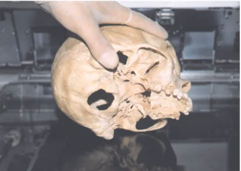
Adult skulls of mature individuals, 18 or more years old, that had no de-struction of the mastoid region or ab-sence of metopic bone in the region of the craniometric points were chosen for the study. A xerographic copy of each side of the skull was obtained through a standardized technique, as demonstrated in figure 1. The skull under study was kept on the copying surface supported by 2 points: a) the lateral surface of the mastoid
process;
b) the zygomatic arc.
The objective of the technique proposed was to obtain a xerographic copy with as little distortion as possi-ble. This was achieved by resting the mastoid process on the surface of the copier.
After the copies were made, each xerographic copy was identified with the identification number of the skull. The device used to obtain the xerographic copies was a XEROX model 5334.
On each xerographic copy, we marked these craniometric points: 1 - Porion – the uppermost lateral
point of the external auditory meatus;
3 - Mastoidale – the lowest point of the mastoid process.
We then drew a triangle linking these three points. The resulting trian-gle was our object of study (Figure 2).
Once demarcated the triangle was transferred to tracing paper and its area calculated.
The values used for the present study, in mm2, were obtained through
the calculation of the area of the de-marcated triangle on each side of the skull, right and left, called right area (D) and left area (E), respectively, and the value of the total of these 2 meas-urements, the total area (T).
The decision to use the value of the total area in the study was based on the evidence obtained by Helmuth11, Schmitt and Saternus12 and
Demoulin13 of the asymmetry of the
mastoid process between the sides of the skull.
RESULTS
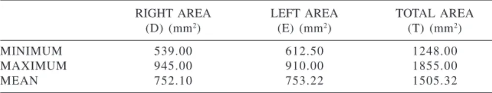
In the group of male skulls, we found the values in Table 1.
In the group of the female skulls, we found the values in Table 2.
The overlapping of the values of the right area (D) between the male and female skulls was 60%. The over-lapping of the values of the left area (E) between the male and female skulls was 51.67%. The overlapping of the values of the total area (T) between the male and female skulls was 36.67%. The analysis of the difference between the male and female skulls, using Stu-dent’s t test, is presented in Table 3.
The values of the mean, the standard error mean and the 95% confidence in-terval for the mean are presented in Figure 3.
DISCUSSION
The objective of this study was to demonstrate that through a practical, easily applied methodology, it is fea-sible for the majority of the medical examiner’s offices to determine the sex of skulls. This can be done using read-ily available resources. Based on a sample of our anthropological ar-chives, we show that the triangle area measurement demarcated through the xerographic projection of cranio-Figure 1 - The standardized technique for obtaining a xerographic copy of the skull.
metrical points related to the mastoid process is useful in the sexing of skulls. Since this study was based on an-thropometric techniques, it surpasses in importance the older studies such as those of Broca14,and Martin15 apud
Hoshi9. It also improves on the
crite-ria reported by Bass4, which were
based only on descriptive anatomical
aspects, as emphasized by Krogman and Íscan8 .
By using a measurement of surface area, or in other words, by using the re-sult of a product between 2 values, our results improve on those of studies by Schultz16 apud Helmuth11, Schäefer17
apud Helmuth11, and Keen18 (1950),
which used only a single measurement.
Thus, this study is in agreement with the conclusions of Helmuth11, Schmitt
and Saternus12, and Demoulin13. The
mastoid region used in this study, be-ing a part of the temporal bone, is rec-ognized as being the most protected and resistant to damage, due to its ana-tomical position at the base of the skull. This has been demonstrated by Kloiber (1953), Wells (1960), Schäefer (1961), Gejval (1963), and Spence (1967), as cited by Wahl and Henke10.
Therefore, compared with the most important historical studies dealing with sex determination of skulls, the present study shows important im-proved results. These results are based on anthroposcopics and anthropomet-ric techniques, and they open paths for further studies based on statistics, which could be of considerable aid to medico-legal investigations.
The required equipment for the ex-ecution of this technique is readily available to the majority of medical examiner’s offices. Any model of pho-tocopy device can be used.
This technique is easy to execute, offers quick results, and dispenses with any type of special training for the medical examiner.
The technique for sexing skulls presented in this study offers a practi-cal alternative to other methods. This technique meets the needs and reali-ties of the forensic investigation in our country today.
ACKNOWLEDGMENT
To Doctor Wilmes Gonçalves Teixeira, Professor of Forensic Medi-cine at the University of Braz Cubas, for suggesting the subject for this study and to Professor Günter Wilhelm Uhlmann, from the Human and Social Sciences Center of the University of Guarulhos, for his assistance in the translation of German language texts, essential for this study.
Table 3 - Analysis of difference of the areas studied using the Student t test.
AREA t Gl SIGNIFICANT at P <.05
RIGTH (D) 6.40 5 8 *
LEFT (E) 7.78 5 8 *
TOTAL (T) 7.92 5 8 *
α = 0.05
Table 2 - Reference values for the study in female skulls.
RIGHT AREA LEFT AREA TOTAL AREA
(D) (mm2) (E) (mm2) (T) (mm2)
MINIMUM 471.50 462.00 942.00
MAXIMUM 742.00 750.00 1475.00
MEAN 608.70 602.54 1211.24
Table 1 - Reference values for the study in male skulls.
RIGHT AREA LEFT AREA TOTAL AREA
(D) (mm2) (E) (mm2) (T) (mm2)
MINIMUM 539.00 612.50 1248.00
MAXIMUM 945.00 910.00 1855.00
MEAN 752.10 753.22 1505.32
RESUMO
PAIVA LAS e col. – Determinação do sexo em crânios humanos através do processo mastóide. Rev. Hosp. Clín. Fac. Med. S. Paulo 58 (1):15-20, 2003.
OBJETIVO: Avaliar a signifi-cância da medida da área formada pela projeção xerográfica de três pontos craniométricos relacionados ao proces-so mastóide, que são, o porion, o asterion e o mastoidale, na determina-ção do sexo em crânios humanos.
MÉTODO: Foram utilizados 60 crânios, sendo 30 masculinos e 30 fe-mininos. De cada crânio foi realizada uma xerocópia de cada lado sendo as-sinalados os pontos craniométricos para demarcação de um triângulo. Fo-ram utilizados os valores em mm3 da
área do triângulo de cada lado (D) e
(E), e o valor correspondente ao somatório dessas duas medidas (T).
RESULTADOS: A sobreposição dos valores da área direita (D) entre os crânios masculinos e femininos foi de 60%, dos valores da área esquerda (E) foi de 51,67% e dos valores da área to-tal (T) foi de 36,67%. A análise da di-ferença dos valores, entre os sexos, mostrou ser significativa nas três áre-as estudadáre-as. No estudo da área total (T), preferida devido à assimetria en-tre os lados da crânio, o valor médio para os crânios masculinos foi 1505,32 mm2, acima do valor máximo
encon-trado em crânios femininos. O valor médio para crânios femininos foi 1211,24 mm2, abaixo do valor mínimo
encontrado em crânios masculinos. CONCLUSÕES: O trabalho mos-tra uma significativa diferença entre os
valores nas três áreas de estudo, (D), (E) e (T). Os valores da área total (T) apresentam menor sobreposição entre os sexos devendo ser preferencialmen-te usados na depreferencialmen-terminação do sexo em crânios. Para a população em estudo, os valores da área total iguais ou su-periores a 1447,40 mm2 significam,
com nível de confiança igual ou supe-rior a 95%, pertencerem os crânios ao sexo masculino. Para os valores da área total iguais ou inferiores a 1260,36 mm2, com nível de confiança igual ou
superior a 95%, pertencem os crânios ao sexo feminino.
DESCRITORES: Medicina legal.
Antropologia forense. Antropo-metria. Mastóide. Características se-xuais.
REFERENCES
1 . ALVES ES – Medicina legal e deontologia. Curitiba, Ed. do Autor, 1965.
2 . ARBENZ GO – Medicina legal e antropologia forense. Rio de Janeiro, Atheneu, 1988.
3 . REICHS KJ – Forensic osteology. Springfield, Thomas, 1986. 4 . BASS WM – Human osteology: a laboratory and field manual
of the human skeleton. Columbia, David R. Evans Editor, 1971.
5 . UBELAKER DH – Human skeletals remains. Chicago, Aldine, 1978.
6 . STEWAR TD – Essentials of forensic anthropology. Springfield, Thomas, 1979.
7 . RATHBUM TA, BUIKSTRA JE – Human identification. Springfield, Thomas, 1984.
8 . KROGMAN WM, ÍSCAN MY – The human skeleton in forensic medicine. 2nd ed. Springfield, Thomas, 1986.
9 . HOSHI H – Sex difference in the shape of the mastoid process in norma occipitalis and its importance to sex determination of the human skull. Okajima’s Folia Anat Jpn 1962; 38: 309-17. 10. WHALL J, HENKE W – Die pars petrosa als diagnostikum für die
multivariat-biometrisch geschlechtsbestimmung von leichenbrandmaterial. Z Morphol Anthropol 1980; 70: 258-68.
11. HELMUTH H – Einige mabe des processus mastoideus beim menschen und seine bedeutung für die geschlechtsbetimmung. Z Morphol Anthropol 1968; 60: 75-84.
13. DEMOULIN F – Importance de certaines mesures crâniennes (en particulier de la longueur sagittale de la mastoide) dans la determination sexuelle des crânes. Bull et Mém de la Soc D’Anthropol 1972; 9: 259 – 64.
14. BROCA P – Instruction craniologique. In: DECHAMBRE D. -Dictionaire encyclopédique des sciences médicales. Paris, Asselin/Masson, 1879; t.22, p.642.
15. MARTIN R – Lerbuch der anthropologie. 2 Aufl Bd 1928; 2: 737-41 apud HOSHI H, 1962. p.309.
16. SCHULTZ AH – Anthropologische untersuchungen na der schädelbasis. Arch Anthrop 1917; 17: 1-103 apud HELMUTH H, 1968. p. 75.
17. SCHAEFER U – Greuzen und möglichkeiten der anthropologischen untersuchung von leichenbränden. In: Bericht über den Internat Kongr f Vor und Frühgeschichte, 5., Hamburg 1958 apud HELMUTH H, 1968. p. 76. 18. KEEN JA – A study of the differences between male and female