Rev Bras Cardiol Invasiva. 2014;22(2):180-2
© 2014 Sociedade Brasileira de Hemodinâmica e Cardiologia Intervencionista. Published by Elsevier Editora Ltda. All rights reserved.
Mid-Ventricular Hypertrophic Obstructive
Cardiomyopathy
Fernando Pivatto Jr.
1, Márcio Mossmann
1, Ana Maria Krepsky
1, Alexandre do Canto Zago
2ABSTRACT
Mid-ventricular hypertrophic obstructive cardiomyopathy is a rare variant form (1%) of hypertrophic obstructive cardiomy-opathy. In this case, we report a patient referred for elective cardiac catheterization due to angina and dyspnea on moder-ate exertion, with no signiicant coronary obstruction, and left ventriculography indicating the presence of mid-ventricular hypertrophic obstructive cardiomyopathy with an intraventricular pressure gradient of 130 mmHg.
DESCRIPTORS: Cardiomyopathy, hypertrophic. Cardiac
cathe-terization.Ventricular outlow obstruction, left.
1 Hospital de Clínicas de Porto Alegre, Porto Alegre, RS, Brazil. 2 Universidade Federal do Rio Grande do Sul, Porto Alegre, RS, Brazil.
Correspondence to: Fernando Pivatto Jr. Serviço de Cardiologia do Hospital de Clínicas de Porto Alegre – Rua Ramiro Barcelos, 2.350 – Rio Branco – CEP: 90035-903 – Porto Alegre, RS, Brazil
E-mail: [email protected]
Received: 02/25/2014 • Accepted: 05/06/2014
RESUMO
Cardiomiopatia Hipertrófica Obstrutiva Médio-Ventricular
A cardiomiopatia hipertróica obstrutiva médio-ventricular é uma variante rara (1%) da miocardiopatia hipertróica obstrutiva. Neste relato de caso, apresentamos uma paciente encaminhada para realização de cateterismo cardíaco eletivo por angina e dispneia aos moderados esforços, sem obstrução coronariana signiicativa e com ventriculograia esquerda, demostrando cardiomiopatia hipertróica obstrutiva médio-ventricular com um gradiente pressórico intraventricular de 130 mmHg.
DESCRITORES: Miocardiopatia hipertróica. Cateterismo
car-díaco. Obstrução do luxo ventricular esquerdo.
Case Report
M
id-ventricular hypertrophic obstructive cardio-myopathy, first described by Falicov et al.1in 1976, is a rare type (1%) of hypertrophic
obstructive cardiomyopathy.2 In this variant, there is a signiicant mid-ventricular hypertrophy associated with a mid-ventricular stenosis, which confers the aspect of a dumbbell to the left ventricle,3 with generation of a pressure gradient between the apical and basal cham-bers, as well as an absence of obstruction of outlow.2
CASE REPORT
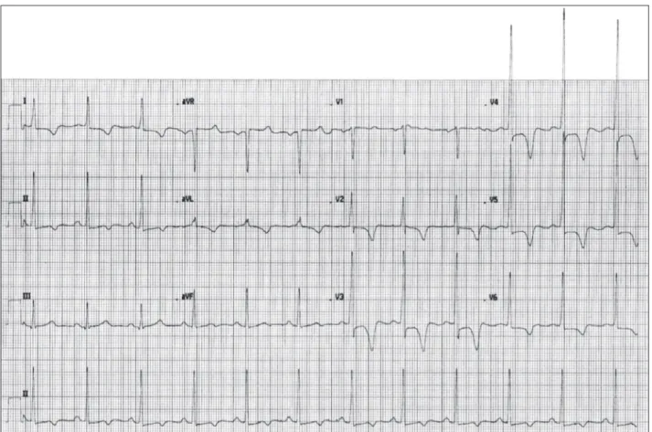
A female patient, 80 years, was referred for elec-tive cardiac catheterization due to angina and dyspnea on moderate exertion. Physical examination revealed mid-systolic murmur without other signiicant indings. An electrocardiogram showed criteria for left ventricular
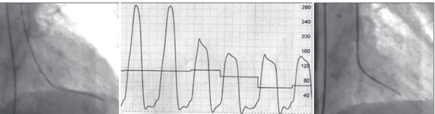
hypertrophy and secondary changes in ventricular repolarization (Figure 1). The coronary angiography showed no signiicant coronary stenoses; however, a left ventriculography in the right anterior oblique projection (30°) revealed mid-ventricular hypertrophic obstructive cardiomyopathy (Figure 2); after analysis of intracavitary pressures, a gradient of 130 mmHg was calculated (Figure 3).
DISCUSSION
Pivatto Jr et al. Mid-Ventricular Hypertrophic Cardiomyopathy
Rev Bras Cardiol Invasiva. 2014;22(2):180-2
181
Apical aneurysms were observed in approximately 25% of patients with mid-ventricular obstructive hypertrophic cardiomyopathy, and were almost unique to this group. Their presence served as a marker of an even worse clinical course.
and without mid-ventricular obstruction, observed that its presence is a strong and independent pre-dictor of sudden death, as well as a determinant of progression to end-stage hypertrophic obstructive cardiomyopathy and to heart failure-related death.
Figure 1 – Electrocardiogram with voltage criteria for left ventricular hypertrophy, secondary changes in ventricular repolarization, and deep inver-sion of T waves.
Figure 2 – Left ventriculography showing severe mid-ventricular hypertrophy, with almost complete mid-ventricular obstruction and apical dilation during diastole (A) and systole (B); these indings are best observed in Figure 2C.
A B
Pivatto Jr et al.
Mid-Ventricular Hypertrophic Cardiomyopathy
Rev Bras Cardiol Invasiva. 2014;22(2):180-2
182
Apical dilation may occur in cases of severe nar-rowing and of progression to a “burned out apex”, with apical aneurysm formation in approximately 10% of patients.3 The pathogenesis of myocardial necrosis remains unknown. It has been suggested that the apical aneurysm may be secondary to after-load and to an increase of apical pressure, as a result of a mid-ventricular obstruction seen in the degenera-tive process of hypertrophic cardiomyopathy. Other possible causes of aneurysm formation are small vessel disease with decreased coronary flow reserve, coronary stenosis due to an increased wall stress in the hypertrophied myocardial segment, decreased coronary perfusion pressure due to the mid-ventricular obstruction, coronary spasm, and decreased capillary/ myocardial fiber ratio.5
If left untreated, the midventricular obstructive hy-pertrophic cardiomyopathy can cause fatal ventricular arrhythmias and sudden death. Beta-blockers are the irst therapeutic choice for hypertrophic obstructive cardiomyopathy, but the treatment of mid-ventricular obstructive hypertrophic cardiomyopathy remains unclear. Dual-chamber pacemaker6 and percutaneous myocardial
ablation7,8 have been proposed as non-surgical treat-ments, but the long-term beneits and safety of these therapeutic options require further study.5 The surgical treatment of midventricular hypertrophic obstructive cardiomyopathy is poorly described in the literature, mostly in the form of case reports.9 Kunkala et al.,9
in a recent study involving 56 patients, described the
results of a transapical approach, noting that this op-tion allows an excellent approach for myectomy, as well as for the relief of the intraventricular gradient and associated symptoms without any complications related to the apical incision were observed with a ive-year survival similar to that expected in the general population (95% vs. 97%).
CONFLICTS OF INTEREST
The authors declare no conlicts of interest.
FUNDING SOURCE
None.
REFERENCES
1. Falicov RE, Renekov L, Bharaki S, Lev M. Mid-ventricular obstruction: a variant of obstructive cardiomyopathy. Am J
Cardiol. 1976;37(3):432-7.
2. Song H, Zhao C, Jinfa J, Yang L, Chen Y. Mid-ventricular hy-pertrophic obstructive cardiomyopathy (MVHOCM) complicated with coronary artery disease: a case report. J Geriatr Cardiol. 2008;5(3):190-2.
3. Noureldin RA, Liu S, Nacif MS, Judge DP, Halushka MK, Abraham TP, et al. The diagnosis of hypertrophic cardiomy-opathy by cardiovascular magnetic resonance. J Cardiovasc Magn Reson. 2012;14:17.
4. Efthimiadis GK, Pagourelias ED, Parcharidou D, Gossios T, Kamperidis V, Theoilogiannakos EK, et al. Clinical charac-teristics and natural history of hypertrophic cardiomyopathy
with midventricular obstruction. Circ J. 2013;77(9):2366-74.
5. Sato Y, Matsumoto N, Matsuo S, Yoda S, Kunimoto S, Saito S. Mid-ventricular hypertrophic obstructive cardiomyopathy presenting with acute myocardial infarction. Tex Heart Inst J. 2007;34(4):475-8.
6. Watanabe H, Kibira S, Saito T, Shimizu H, Abe T, Nakajima
I, et al. Beneicial effect of dual-chamber pacing for a left midventricular obstruction with apical aneurysm. Circ J
2002;66(10):981-4.
7. Tengiz I, Ercan E, Turk UO. Percutaneous myocardial ablation for left mid-ventricular obstructive hypertrophic cardiomyopathy.
Int J Cardiovasc Imaging. 2006;22(1):13-8.
8. Seggewiss H, Faber L. Percutaneous septal ablation for hyper-trophic cardiomyopathy and mid-ventricular obstruction. Eur J Echocardiogr. 2000;1(4):277-80.
9. Kunkala MR, Schaff HV, Nishimura RA, Abel MD, Sorajja P, Dearani JA, et al. Transapical approach to myectomy for midventricular obstruction in hypertrophic cardiomyopathy.
Ann Thorac Surg. 2013;96(2):564-70.