J
ournal of Epilepsy and ClinicalNeurophysiology
J Epilepsy Clin Neurophysiol 2012;18(1):16-20
Awards Works: Expanded Abstract
a Department of Neuroscience and Behavior, Ribeirão Preto School of Medicine, University of São Paulo. b Department of Molecular Biology, São José do Rio Preto Medical School.
c Department of Neurosurgery, Ribeirão Preto School of Medicine, University of São Paulo. d Department of Pathology, RibeirãoPreto School of Medicine, University of São Paulo.
Received Apr. 28, 2012; accepted Apr. 30, 2012.
Trabalho vencedor do
Prêmio Cesare Lombroso
– XXXIV Congresso Brasileiro de Epilepsia – 2012
Different Levels of MT-I/II Between Patients With MTLE
With or Without Seizure Generalization: Does Hippocampal MT-I/II
Affects Seizure Spread, or Does Seizure Spread Promotes
Differential Expression of MT-I/II?
José Eduardo Peixoto-Santosa, Orfa Yineth Galvis-Alonsob, Tonicarlo R. Velascoa,
Ludmyla Kandrataviciusa, João Alberto Assirati Jrc, Carlos Gilberto Carlottic, Renata Caldo Scandiuzzia,
Luciano Neder Serafinid, João Pereira Leitea
Ribeirão Preto School of Medicine, University of São Paulo
ABSTRACT
In the central nervous system, zinc is released along with glutamate during neurotransmission and, in excess, can promote neuronal death. Experimental studies have shown that metallothioneins I/II (MT-I/II), which chelate free zinc, can affect seizures and reduce neuronal death after status epilepticus. Our aim was to evaluate the expression of MT-I/II in the hippocampus of patients with temporal lobe epilepsy (TLE). Hippocampi from patients with pharmacoresistant mesial temporal lobe epilepsy (MTLE) were evaluated for expression of MT-I/II and for neuronal, astroglial, and microglial populations. Compared to control cases, MTLE group displayed widespread increase in MT-I/II expression, astrogliosis and reduced neuronal population. MT-I/II levels did not correlate with any clinical variables, but patients with secondary generalized seizures (SGS) had less MT-I/II than patients without SGS. In conclusion, MT-I/II expression was increased in hippocampi from MTLE patients and our data suggest that it may be associated with different seizure spread patterns.
Keywords: Metallothioneins; zinc homeostasis; gliosis; epilepsy; neuronal density.
RESUMO
Níveis diferentes de MT-I/II entre pacientes com MTLE com ou sem crise generalizada: os níveis hipocampais de MT-I/II afetam o alastramento das crises, ou o alastramento das crises promove expressão diferencial de MT-I/II? No sistema nervoso central, o zinco é liberado juntamente com o glutamato durante a neurotransmissão e, quando liberado em excesso, pode promover morte neuronal. Estudos indicam que as metalotioneínas I/II (MT-I/II), proteínas quelantes de zinco livre, podem afetar parâmetros relacionados às crises e reduzir a morte neuronal subsequente a um status epilepticus. Nosso objetivo foi avaliar a expressão de MT-I/II no hipocampo de pacientes com epilepsia do lobo temporal (ELT). Hipocampos de pacientes com ELT mesial (ELTM) resistente ao tratamento farmacológico foram avaliados para a expressão de MT-I/II e para as populações neuronal e astroglial. Quando comparadas com o grupo controle, pacientes com ELTM apresentaram aumento na expressão de MT-I/II, astrogliose e redução na densidade neuronal. Não foram observadas correlações entre os níveis de MT-I/II e as características clínicas dos pacientes, mas pacientes com crises secundariamente generalizadas apresentaram um aumento menor nos níveis de MT-I/II que os pacientes sem estas crises. Em resumo, um aumento na expressão de MT-I/II é observado em pacientes com ELTM e nossos dados sugerem que o aumento pode estar associado a diferentes padrões de crises epilépticas.
INTRODUCTION
Zinc (Zn2+) is an important modulator of glutamatergic
transmission in the central nervous system (CNS).1-3 Zn2+ is
concentrated in presynaptic vesicles, along with glutamate, and released during normal neurotransmission.4-8
Hippocampal neurons are specially rich in vesicular Zn2+, particularly in the axonal boutons of granule
cells, CA3 and CA1 pyramidal cells and prosubicular neurons.5-7,9,10 In temporal lobe epilepsy (TLE), one of
the most frequent drug-resistant epilepsies in adults, the hippocampus is associated with seizure generation.11,12
The intense neuronal activity during seizures can release high amounts of Zn2+ in the synaptic cleft,13,14 promoting
reactive oxygen species (ROS) production,15 which can
ultimately lead to hippocampal neuronal death.13-17 In fact,
studies in hippocampi from TLE patients who underwent epilepsy surgery have shown neuronal loss,18-20 increased
glial reaction21-24 and reorganization of mossy fibers axon
collaterals into the inner molecular layer of the granule cell dendrites.19,25 This synaptic reorganization of Zn2+
-enriched terminals has been hypothesized to contribute to synchronous firing and epileptiform activity.19
Metallothioneins (MTs) are low molecular weight, cystein-enriched proteins that bound Zn2+ and cadmium.
They can be found in various tissues, in four isoforms.26
Isoforms I, II and III are found in the central nervous system (CNS), where the isoforms I and II are coexpressed in astrocytes and the isoform III is expressed in neurons.27,28
MTs participate in Zn2+ homeostasis, scavenging ROS in the
brain29 and stimulate the expression of several neurotrophic
and antiinflamatory factors.30 Studies on rodent models of
TLE have shown that MT expression is increased in the hippocampal formation shortly after seizures31,32 and that
high levels of MTs I and II are associated with reduced neuronal death after seizure-induced damage.32-34
Since MT-I/II levels may be associated with neuron survival after seizures, we hypothesize that MT-I/II expression is altered in TLE and can be associated with the preservation of neuronal density in the hippocampus of TLE patients. Therefore, in this study we evaluated the immunoexpression of MT-I/II and its correlation with hippocampal neuron density in hippocampi of patients with chronic TLE.
MATERIALS AND METHODS
Patients and clinical data
Patients with drug-resistant epilepsy were evaluated at the University of São Paulo Epilepsy Surgical Centre in Ribeirão Preto (Brazil), according to standard protocols published elsewhere.35 MTLE patients (n=69) were patients
with hippocampal atrophy or with normal hippocampal volume at MRI without other lesions associated with TLE.
For comparison purposes in the neuropathology studies, autopsy controls (Ctrl, n=20) were obtained from autopsy cases without history of neurological diseases, with no sign of CNS pathologies in post mortem pathological evaluation, and with less than 10 hours post mortem.
Medical records of all evaluated patients were assessed for clinical data analysis. The clinical variables investigated were age at death and cause of death for Ctrl patients and age at surgery, epilepsy duration, age at the first recurrent seizure, seizure frequency per month, presence of secondary generalized seizures, and neuropathological evaluation for MTLE patients. This study followed the principles of the Declaration of Helsinki, was registered in Brazilian’s Health Ministry and was approved by our local ethics committee (processes HCRP 9370/2003 and HCRP 2634/2008).
Tissue collection and immunohistochemistry
Hippocampi from surgery or autopsy were cut in coronal sections and placed in 10% (vol/vol) buffered formaldehyde for one week, followed by paraffin embedding. Immunohistochemistry was performed in 8 μm sections at the level of hippocampal body for evaluation of neuronal and astroglial populations and for MT-I/II expression with antibodies against, respectively, NeuN, GFAP and MT-I/II. The sections were submitted to endogenous peroxidase blocking with 4.5% H2O2 in 50 mM phosphate-saline buffer (PSB) pH 7.4, for 15 minutes, followed by microwave antigenic retrieval in 10mM sodium citrate buffer pH 6.0 (for GFAP) or 50 mM Tris-HCl pH 9.6 (for NeuN and MT-I/II). After achieving room temperature, the sections went through blocking free aldehyde groups with Tris-glycine 0.1 M pH 7.4 for 45 minutes, followed by blocking buffer with 5% defatted milk and 15% goat serum (# S-1000, Vector) in Triton buffer (PTB, 20mM phosphate + 0.45M NaCl, pH 7.4, with 0.3% Triton X-100) for four hours. The sections were then incubated with primary antibodies in blocking buffer for 16 hours. We used primary monoclonal antibodies raised in mouse anti-human GFAP (clone 6F2, #M0761, Dako), anti-murine NeuN (clone A60, #MAB377, Chemicon) and anti-equine MT-I/II (clone E9, #M0639, Dako), diluted in blocking buffer at concentrations of 1:500. The primary antibodies were detected using biotinylated rabbit anti-murine IgG (#E0354, Dako), at 1:200 dilution in blocking buffer, for one hour, followed by revelation with avidin-biotin-peroxidase system (Vectastain Elite ABC kit, #PK6100, Vector) and diaminobenzidine as chromogen (DAB, #34001, Pierce Biotechnology). The development times in DAB solution were 10.5 minutes for NeuN and 8 minutes for MT-I/II and GFAP.
Immunohistochemistry analysis
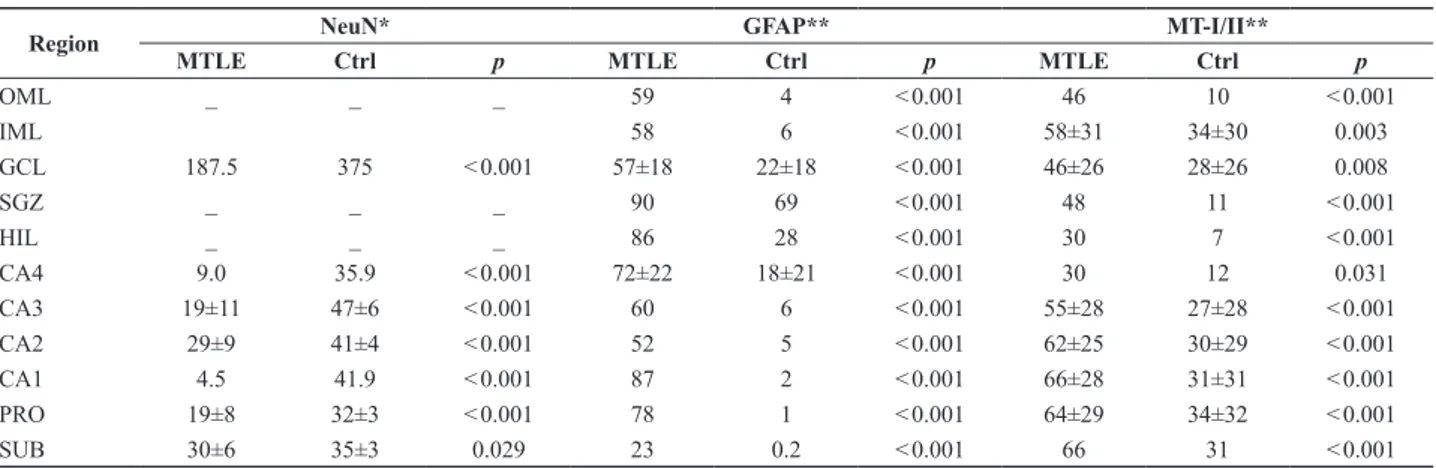
Table 2. Neuronal density and percentage of immunopositive area for GFAP and MT-I/II in patients with MTLE and Ctrl
Region NeuN* GFAP** MT-I/II**
MTLE Ctrl p MTLE Ctrl p MTLE Ctrl p
OML _ _ _ 59 4 < 0.001 46 10 < 0.001
IML 58 6 < 0.001 58±31 34±30 0.003
GCL 187.5 375 < 0.001 57±18 22±18 < 0.001 46±26 28±26 0.008
SGZ _ _ _ 90 69 < 0.001 48 11 < 0.001
HIL _ _ _ 86 28 < 0.001 30 7 < 0.001
CA4 9.0 35.9 < 0.001 72±22 18±21 < 0.001 30 12 0.031
CA3 19±11 47±6 < 0.001 60 6 < 0.001 55±28 27±28 < 0.001 CA2 29±9 41±4 < 0.001 52 5 < 0.001 62±25 30±29 < 0.001 CA1 4.5 41.9 < 0.001 87 2 < 0.001 66±28 31±31 < 0.001
PRO 19±8 32±3 < 0.001 78 1 < 0.001 64±29 34±32 < 0.001 SUB 30±6 35±3 0.029 23 0.2 < 0.001 66 31 < 0.001
* Neuronal density, as thousands of cells per cubic millimeter. ** Percentage of immunopositive area in the amostral area.
Hamamatsu Photonics Model 2400, Japan) attached to an Olympus microscope (Model BX60, Melville, NY), and captured, averaged, and digitized using a frame grabber (Scion Corporation, Frederick, MD) on a Macintosh computer (Model G3, Cupertino, CA). Illuminance was uniformly maintained and regularly checked using optical density standards (Kodak, Rochester, NY). After captured, the image was analyzed using image system software (ImageJ, version 1.37c).
Quantification of the immunohistochemistry was performed with threshold tool, with the investigator blind to the group allocation. After the selection of the region of interest (ROI), the software calculated the immunopositive area by counting all pixels with gray intensity equal or superior to the threshold of staining. A complete protocol for threshold tool can be found at rsbweb.nih.gov/ij/docs/
examples/stained-sections/index.html. The threshold was
defined for each protein evaluated, based on the mean immunopositivity of all control cases. The evaluated regions were outer molecular layer (OML), inner molecular layer (IML), granule cell layer (GCL), subgranular zone (SGZ), the hilus (HIL) and the stratus piramidale of CA4, CA3, CA2, CA1, prosubiculum (PRO) and subiculum (SUB). The characterization of hippocampal regions was based on the Lorente de Nó’s classification.36 Results
were shown as percentage of immunopositive area/total area.
Additionally, neuronal density was evaluated in the NeuN stained sections. Neuronal count was processed in ImageJ 1.37c software with a 520x magnification for granule cell layer and 260x for pyramidal neurons of CA4, CA3, CA2, CA1, prosubiculum and subiculum. Neuronal densities were estimated with the correction of Abercrombie37, which permits to estimate the
neuronal density through mathematical method, and the results were shown as thousands of cells per cubic millimeter.
Statistical analysis
Statistics were carried out in SigmaStat 3.1 software. Tests for normality and homogeneity of variances were performed to define data distribution. For parametric variables, t-test was performed and, for the non-parametric variables, Mann-Whitney test was used. Correlation between MT expression and clinical variables was performed using Pearson’s test. All results were considered significant at p<0.05.
RESULTS
Clinical data
The clinical characteristics of study participants are summarized in Table 1. Patients with MTLE and Ctrl patients have the same age (p=0.175). Epilepsy duration was 25±10 years, and the age at onset was 13±1. MTLE patients had seven seizures by month, being one of those a secondary generalized seizure (SGS).
Table 1. Clinical history of patients with MTLE and Ctrl cases
Group Ctrl MTLE
Age at evaluation* (years) 42±16 38±10 Epilepsy duration (years) _ 25±10 Age at epilepsy onset (years) _ 13±1
Minimal seizure frequency (per month) _ 7
Number of secondary generalizations (per month) _ 1
Frequency of secondary generalization (%) _ 59
* Age of death for Ctrl and age at surgery for TLE.
Immunohistochemistry evaluation
The results are shown as median (for Mann Whitney test) or mean ± standard deviation (for Student’s t-test). OML = outer molecular layer; IML = inner molecular layer; GCL = granule cell layer; SGZ = subgranule zone; HIL = hilus; PRO = prosubiculum; SUB = subiculum.
Tissue alterations and seizures
In MTLE group, patients without SGS had increased MT-I/II immunopositivity, when compared with patients with SGS, in the inner molecular layer (p=0.037), granule cell layer (p=0.018), subgranule zone (p=0.004), CA2 (p=0.039) and CA1 (p<0.043). A trend to increased MT-I/II immunopositivity was observed in the outer molecular layer (p=0.072), CA4 (p=0.076) and subiculum (p=0.068). No differences in neuronal or astroglial populations were observed between MTLE patients with or without SGS. Frequency of seizures did not correlate with NeuN, GFAP or MT-I/II in all hippocampal subfields.
DISCUSSION
In the present study, we found an increased MT-I/II expression in all hippocampal subfields of MTLE patients. In the CNS, MT-I/II are expressed mainly by astrocytes38
and, when the tissue suffers an injury, increased MT-I/II expression is observed in astrocytes and microglias.28,38
We also observed that higher degree of MT-I/II expression was observed in regions with higher astrogliosis. Increased glial population is a common finding in TLE21-24 and is
associated with the degree of neuronal death.22-24,39 In our
study, an increased expression of MT-I/II was observed in astrocytes and in a few neurons of some patients. Thus, our data support the notion that MT-I/II changes are essentially related to astroglial population.
Studies in rodents with kainic acid-induced SE showed an association between MT-I/II expression and neuronal protection. Transgenic mice over-expressing MT-I/II have reduced neuronal death, compared to wild type animals.34
In addition, mice with reduced MT-I/II expression32 or
knockouts for MT-I/II33 had increased neuronal death
following SE, compared to wild type mice. In our study, however, the higher MT-I/II expression was observed in regions with lower neuronal density, indicating that MT-I/II was not associated with neuronal survival. In agreement with our data, an association between the severity of tissue damage and the increase in MT-I/II expression has been reported in mice subjected to soman-induced status epilepticus (SE).31
Data have shown that the increased MT-I/II immunoreactivity observed in animal models of TLE can also be a factor associated with the seizure generation process. Transgenic mice over-expressing MT-I, have increased seizure duration, a tendency to reduced latency, but similar number of seizures after KA administration.34
Since MT-I/II act chelating free Zn2+14,27 and Zn2+
chelation increases tissue excitability and facilitates seizure generation40, excessive MT-I/II levels can reduce free Zn2+
in the synaptic cleft, increasing neuronal excitability and affecting seizure generation. We found no correlation between seizure frequency and MT-I/II expression in TLE.34
In MTLE, we found increased levels of MT-I/II in patients without SGS, when compared with those with SGS. This could indicate that MT-I/II is associated with different seizure spread patterns from the epileptogenic hippocampus to other brain regions. It is important to point out that no difference in neurons or glial cells was observed between MTLE with and without SGS. Studies from different groups also observed no association between changes in the hippocampus and SGS.41-43 All those observations
suggest that the increased MT-I/II expression in patients without SGS is not an effect of gliosis, but it is independently associated with SGS. Further studies with animal models of TLE must evaluate more closely the relationship between MT-I/II expression and seizure susceptibility.
Some limitations of our study must be pointed out. So far, studies about MT-I/II expression in animal models of TLE only evaluated the acute period following SE. Considering that our study was performed in patients with chronic epilepsy, it is difficult to establish comparisons between human and animal data. The lack of correlation between seizure frequency and MT-I/II expression does not exclude an association between seizures and MT-I/II expression. Other seizure characteristics, such as seizure duration and time between the last seizure and the surgery, could better correlate with MT-I/II expression than isolate seizure frequency.
Finally, our study may have translational implications in the future. The role of MTs in antiinflamatory response, neurotrophic factor expression, and protection against ROS and heavy metals make those proteins interesting for clinical applications. Studies have shown that EmtinB, a syntethic peptide that mimics the actions of MTs, attenuates KA-induced seizures and protects neurons from excitotoxic death.30 Further studies with
EmtinB and MTs should be done to evaluate the role of these proteins in neuronal survival and seizure susceptibility.
In summary, our data indicate that increased MT-I/II expression is a plastic alteration of chronic TLE, primarily related to the astrogliosis, a common finding in chronic TLE. Our findings suggest that increase MT-I/II expression may contribute to the control of the brain hyperexcitability.
ACKNOWLEDGMENTS
REFERENCES
1. Peters S, Koh J, Choi DW. Zinc selectively blocks the action of N-methyl-D-aspartate on cortical neurons. Science 1987;236:589-93. 2. Westbrook GL, Mayer ML. Micromolar concentrations of Zn2+
antagonize NMDA and GABA responses of hippocampal neurons. Nature 1987;328:640-643.
3. Rassendren FA, Lory P, Pin JP, Nargeot J. Zinc has opposite effects on NMDA and non-NMDA receptors expressed in Xenopus oocytes. Neuron 1990;4:733-40.
4. Haug FMS. Electron microscopic localization of the zinc in hippocampal mossy fiber synapses by a modified sulphide silver procedure. Histochemie 1967;8:355-68.
5. Frederickson CJ, Hernandez MD, McGinty JF. Translocation of zinc may contribute to seizure-induced death of neurons. Brain Res 1989;480:317-21.
6. Perez-Clausell J. Distribution of terminal fields stained for zinc in the neocortex of the rat. J Chem Neuroanat 1996;11:99-111.
7. Frederickson CJ, Suh SW, Silva D, Frederickson CJ, Thompson RB. Importance of zinc in the central nervous system: the zinc-containing neuron. J Nutr 2000;130:1471S-83S.
8. Brown CE, Dyck RH.Distribution of zincergic neurons in the mouse forebrain. J Comp Neurol 2004;479:156-67.
9. Takeda A, Itoh H, Tamano H, Oku N. Responsiveness to kainate in young rats after 2-week zinc deprivation. Biometals 2006;19:565-72. 10. Amaral D, Lavenex P. Hippocampal Neuroanatomy. The Hippocampus
Book. 1ª ed. Oxford University Press; 2006. p.37-114.
11. Mathern GW, Babb TL, Leite JP, Pretorius K, Yeoman KM, Kuhlman PA. The pathogenic and progressive features of chronic human hippocampal epilepsy. Epilepsy Res 1996;26:151-61.
12. Pitkanen A. Efficacy of current antiepileptics to prevent neuro- degeneration in epilepsy models. Epilepsy Res 2002;50:141-60. 13. Weiss JH, Sensi SL, Koh JY. Zn(2+): a novel ionic mediator of neural
injury in brain disease. Trends Pharmacol Sci 2000;21:395-401. 14. Colvin RA, Fontaine CP, Laskowski M, Thomas D. Zn2+ transporters
and Zn2+ homeostasis in neurons. Eur J Pharmacol 2003;479:171-85. 15. Kim EY, Koh JY, Kim YH, Sohn S, Joe E, Gwag BJ. Zn2+ entry produces
oxidative neuronal necrosis in cortical cell cultures. Eur J Neurosci 1999;11:327-34.
16. Treiber C. Metals on the brain.Sci Aging Knowledge Environ 2005;2005:e27.
17. Frederickson CJ, Koh JY, Bush AI. The neurobiology of zinc in health and disease. Nat Rev Neurosci 2005;6:449-62.
18. Babb TL, Brown WJ, Pretorius J, Davenport C, Lieb JP, Crandall PH. Temporal lobe volumetric cell densities in temporal lobe epilepsy. Epilepsia 1984;25:729-40.
19. Babb TL, Kupfer WR, Pretorius JK, Crandall PH, Levesque MF. Synaptic reorganization by mossy fibers in human epileptic fascia dentata. Neuroscience 1991;42:351-63.
20. Mathern GW, Leite JP, Babb TL, Pretorius JK, Kuhlman PA, Mendoza D et al. Aberrant hippocampal mossy fiber sprouting correlates with greater NMDAR2 receptor staining. Neuroreport 1996;7: 1029-35.
21. Salanova V, Markand O, Worth R, Garg B, Patel H, Asconape J et al. Presurgical evaluation and surgical outcome of temporal lobe epilepsy. Pediatr Neurol 1999;20:179-84.
22. Proper EA, Jansen GH, van Veelen CW, van Rijen PC, Gispen WH, de Graan PN. A grading system for hippocampal sclerosis based on the degree of hippocampal mossy fiber sprouting. ActaNeuropathol (Berl) 2001;101:405-9.
23. Swartz BE, Houser CR, Tomiyasu U, Walsh GO, DeSalles A, Rich JR et al. Hippocampal cell loss in posttraumatic human epilepsy. Epilepsia 2006;47:1373-82.
24. Prayson RA, Yoder BJ. Clinicopathologic findings in mesial temporal sclerosis treated with gamma knife radiotherapy. Ann Diagn Pathol 2007;11:22-26.
25. Sutula T, Cascino G, Cavazos J, Parada I, Ramirez L. Mossy fiber synaptic reorganization in the epileptic human temporal lobe. Ann Neurol 1989;26:321-30.
26. Kille P, Hemmings A, Lunney EA. Memories of metallothioneis. Biochim Biophys Acta 1994;1205:151-61.
27. Aschner M, Cherian MG, Klaassen CD, Palmiter RD, Erickson JC, Bush AI. Metallothioneins in brain – the role in physiology and pathology. Toxicol Appl Pharmacol 1997;142:229-42.
28. Wiese L, Kurtzhals JA, Penkowa M. Neuronal apoptosis, metallothionein expression and proinflammatory responses during cerebral malaria in mice. Exp Neurol 2006;200:216-26.
29. Ebadi M, Brown-Borg H, El RH, Singh BB, Garrett S, Shavali S et al. Metallothionein-mediated neuroprotection in genetically engineered mouse models of Parkinson’s disease. Brain Res Mol Brain Res 2005;134:67-75.
30. Sonn K, Pankratova S, Korshunova I, Zharkovsky A, Bock E, Berezin V et al. A metallothionein mimetic peptide protects neurons against kainic acid-induced excitotoxicity. J Neurosci Res 2010;88: 1074-82.
31. Pazdernik TL, Emerson MR, Cross R, Nelson SR, Samson FE. Soman-induced seizures: limbic activity, oxidative stress and neuroprotective proteins. J ApplToxicol 2001;21Suppl 1:S87-S94.
32. Penkowa M, Molinero A, Carrasco J, Hidalgo J. Interleukin-6 deficiency reduces the brain inflammatory response and increases oxidative stress and neurodegeneration after kainic acid-induced seizures. Neuroscience 2001;102:805-18.
33. Carrasco J, Penkowa M, Hadberg H, Molinero A, Hidalgo J. Enhanced seizures and hippocampal neurodegeneration following kainic acid-induced seizures in metallothionein-I + II-deficient mice. Eur J Neurosci 2000;12:2311-22.
34. Penkowa M, Florit S, Giralt M, Quintana A, Molinero A, Carrasco J et al. Metallothionein reduces central nervous system inflammation, neurodegeneration, and cell death following kainic acid-induced epileptic seizures. J Neurosci Res 2005;79:522-34.
35. Leite JP, Terra-Bustamante VC, Fernandes RM, Santos AC, Chimelli L, Sakamoto AC et al. Calcified neurocysticercotic lesions and postsurgery seizure control in temporal lobe epilepsy. Neurology 2000;55:1485-91.
36. Lorente de Nó R. Studies on the structure of the cerebral cortex. II. Continuation of the study of the ammoniac system. Journal of Psychologie und Neurologie 1934;45:113-77.
37. Abercrombie M. Estimation of nuclear population from microtome sections. Anat Rec 1946;94:239-147.
38. Hidalgo J. Metallothioneins and Brain Injury: What Transgenic Mice Tell Us. Environ Health Prev Med 2004;87-94.
39. Crespel A, Coubes P, Rousset MC, Brana C, Rougier A, Rondouin G et al. Inflammatory reactions in human medial temporal lobe epilepsy with hippocampal sclerosis. Brain Res 2002;952:159-69.
40. Dominguez MI, Blasco-Ibanez JM, Crespo C, Nacher J, Marques-Mari AI, Martinez-Guijarro FJ. Neural overexcitation and implication of NMDA and AMPA receptors in a mouse model of temporal lobe epilepsy implying zinc chelation. Epilepsia 2006;47:887-99. 41. Bernasconi N, Natsume J, Bernasconi A. Progression in temporal lobe
epilepsy: differential atrophy in mesial temporal structures. Neurology 2005;65:223-28.
42. Szabo CA, Lancaster JL, Lee S, Xiong JH, Cook C, Mayes BN et al. MR imaging volumetry of subcortical structures and cerebellar hemispheres in temporal lobe epilepsy. AJNR Am J Neuroradiol 2006;27:2155-60.
43. O’Dwyer R, Silva Cunha JP, Vollmar C, Mauerer C, Feddersen B, Burgess RC et al. Lateralizing significance of quantitative analysis of head movements before secondary generalization of seizures of patients with temporal lobe epilepsy. Epilepsia 2007;48: 524-30.
Corresponding author:
João Pereira Leite
Department of Neuroscience and Behavior
Ribeirão Preto School of Medicine, University of São Paulo Av. Bandeirantes, 3900
ZIP Code 14049-900, Ribeirão Preto, SP, Brazil Phone/Fax: (+55-16)3602-2556