Research Article
Heart Rate Variability in Shift Workers: Responses to
Orthostatism and Relationships with Anthropometry, Body
Composition, and Blood Pressure
Nayara Mussi Monteze,
1Breno Bernardes Souza,
2Henrique José de Paula Alves,
3Fernando Luiz Pereira de Oliveira,
3José Magalhães de Oliveira,
4Silvia Nascimento de Freitas,
1Raimundo Marques do Nascimento Neto,
2Maria Lilian Sales,
5and Gabriela Guerra Leal Souza
61School of Nutrition, Federal University of Ouro Preto, 35400-000 Ouro Preto, MG, Brazil
2School of Medicine, Federal University of Ouro Preto, 35400-000 Ouro Preto, MG, Brazil
3Department of Statistics, Federal University of Ouro Preto, 35400-000 Ouro Preto, MG, Brazil
4Institute of Psychiatry, Federal University of Rio de Janeiro, 22290-140 Rio de Janeiro, RJ, Brazil
5Institute of Cancer of S˜ao Paulo, 01255-000 S˜ao Paulo, SP, Brazil
6Department of Biological Sciences, Federal University of Ouro Preto, 35400-000 Ouro Preto, MG, Brazil
Correspondence should be addressed to Gabriela Guerra Leal Souza; [email protected]
Received 19 February 2015; Revised 3 April 2015; Accepted 8 April 2015
Academic Editor: Giuseppe Biondi-Zoccai
Copyright © 2015 Nayara Mussi Monteze et al. This is an open access article distributed under the Creative Commons Attribution License, which permits unrestricted use, distribution, and reproduction in any medium, provided the original work is properly cited.
In order to investigate the response of heart rate variability (HRV) components to postural change and their association with cardiovascular risk factors in shift workers, a cross-sectional study with 438 Brazilian males rotating shift workers was done. Anthropometric, body composition, and clinical measures were collected. Electrocardiogram was recorded for 3 minutes, in the supine and orthostatic position, and HRV components were extracted. Descriptive analyses showed that mean values of body mass index, waist circumference (WC), waist-to-height ratio, visceral fat area (VFA), and blood pressure (BP) were higher than the reference values. In the regression model, age, WC, VFA, and systolic BP showed negative association with HRV components. These findings suggest the need for determining effective strategies for the evaluation and promotion of health among shift workers focused on the altered variables.
1. Introduction
Heart rate variability (HRV) is a standard noninvasive method that assesses the action of the autonomic nervous system (ANS) on the heart based on variations in the
RR interval between consecutive heartbeats [1]. Analysis of
HRV on electrocardiogram allows for the decomposition of elements that assess the ANS function on the heart period
[2]. A reduction in HRV, and more particularly in vagal
modulation, could be considered an indicator of an imbal-ance in autonomic function and could be associated with an increased risk of morbidity and mortality independent of
the presence of well-established risk factors, such as obesity,
smoking, and sedentary lifestyle [3–5].
The continuous interplay of sympathetic and parasympa-thetic activity is crucial to increase or reduce heart perfor-mance under different circumstances. Under general physio-logical conditions, parasympathetic activity is more intense in the resting state and during repairing functions, and sympathetic activity is more intense in situations requiring
the mobilization of energy [4]. For instance, during postural
change (from supine to orthostatic position), blood pooling occurs in lower parts of the body, which is a normal stimulus that triggers an increase in sympathetic activity in the heart
and the vessels, with a consequent rise in blood pressure
[6]. Such behavior has been found in some studies, which
showed that sympathetic activity was greater in the
ortho-static position (90∘) and parasympathetic activity prevailed
in the supine position (0∘) [7,8]. According to an important
review, HRV at rest (wake-sleep and activity-rest) is reduced
in shift workers compared to nonshift workers [9]; however,
little is known about the causes of these alterations in the shift workers and besides that there is not any study investigating the cardiac autonomic response to postural change in this sample.
Obesity tends to be a strong risk factor for cardiovascular morbidity and mortality in young, middle-aged, and older persons. It has been suggested that ANS dysfunction is an important mediator in the development of obesity associated disease and insulin resistance, although the nature of the link between adiposity and insulin sensitivity is still unclear
[10]. Shift work has been associated with deregulation of
the circadian rhythm, which could change psychological functions, dietary and social habits, and ANS function and
thus contribute to the high risk of cardiovascular diseases [11].
Therefore, it is relevant to investigate the relationship between the HRV components and cardiovascular risk factors (age, obesity, and hypertension) in shift workers.
Therefore, the aims of the present study were to inves-tigate the response of HRV components to postural change and the association of HRV components with age, blood pressure, and obesity indices (anthropometric and body com-position variables) in Brazilian shift workers. The hypotheses underlying the present study were as follows: (1) shift workers exhibit deviations in obesity indices, blood pressure, and HRV components relative to reference values; (2) shift work affects the baroreflex mediated response to postural change (transition of supine position to orthostatic position); and (3) HRV components exhibit negative associations with obesity indices, blood pressure, and age. This study has some relevant points: the sample homogeneity (Brazilian, male, rotating shift, and mine worker) and multiple variables collected on the same sample (HRV components, obesity indices, and blood pressure).
2. Materials and Methods
2.1. Study Design and Sample. This cross-sectional study was conducted with 438 adult Brazilian males who were older than 18 years and worked in shifts. Working in shifts,
according to the International Labour Office (ILO) [12], is
defined as a “method of organization of working time in which workers succeed one another at the workplace so that the establishment can operate longer than the hours of work individual workers” and can be classified into fixed shift (working time can be organized in two or three fixed shifts: the early, late, and night shifts) or rotating shift (workers might be assigned to work shifts that vary regularly over time: from a shift in the morning, to one in the afternoon, to one at
night) [12].
The participants of this study performed rotational shift work as operators of iron ore extraction modern machines
without noise exposure. The work regimen included a six-hour shift followed by 12-six-hour rest. The work shifts and rest periods were rotated, and after each fourth shift the rest period was longer, 36 hours, after which a new cycle began. The full work cycle included four consecutive shifts, in this sequence: 7:00 pm to 1:00 am, 1:00 pm to 7:00 pm, 7:00 am to 1:00 pm, and 1:00 am to 7:00 am.
The study was conducted in the morning of the day with the longest rest period from September 2011 to March 2012. The study complied with the Declaration of Helsinki and was approved by the ethics committee of the institution. All volunteers signed an informed consent form.
2.2. Anthropometric Variables. The volunteers’ weight, height, and circumferences were measured by trained professionals with volunteers in the orthostatic position and wearing light clothing. The waist circumference (WC) was measured at the mid-point between the last costal arch
and the iliac crest [13]. The hip circumference (HC), which
was used to calculate the waist-to-hip ratio (WHR), was
measured at the level of the greater trochanter [13]. The
neck circumference (NC) was measured immediately below
the laryngeal prominence [14]. The WHR and the
waist-to-height ratio (WHtR) were calculated by dividing the WC by the hip circumference and the WC by the height, respectively
[13]. The body mass index (BMI) was calculated by dividing
the body weight by the squared height [13].
2.3. Body Composition Variables. The body fat mass in kilograms (BFkg) and percentage (BF%) and the visceral fat area (VFA) were calculated by means of segmental tetrapolar bioelectrical impedance using a body composition analyzer
InBody model 720 (Biospace Co. Ltd. Factory, Korea) [15].
2.4. Clinical Variables. Blood pressure was assessed using an automated digital sphygmomanometer HEM705CP (Omron, Japan) on the right arm with volunteers in the sitting position and after a five-minute rest period. The measurement was performed three times with one-minute interval between
measurements [16]. The three measurements of the systolic
(SBP) and diastolic (DBP) blood pressure were computed and their means were calculated.
2.5. Heart Rate Variability. One PC-compatible computer controlled data acquisition for electrocardiographic param-eters using the WinCardio (Micromed, version 4.8.2.8) soft-ware program. Electrocardiographic recordings were col-lected at a sampling frequency of 1000 Hz. An off-line peak detection algorithm (derivative plus threshold) was used to estimate fiducial R-wave points, after which the series was screened by hand and corrected for artifacts. Successive RR intervals were estimated in milliseconds and were converted to heart rate (HR) in beats per minute.
of successive RR interval differences (RMSSD) that reflects
parasympathetic activity [2]. The frequency domain method
is a spectral method for analysis of the tachogram that pro-vides basic information about how power (variance)
dis-tributes as a function of frequency [2]. The components
con-sidered in the RR power spectrum in this study were as follows.
(i) High frequency (HF), from 0.15 to 0.4 Hz, reflects
parasympathetic activity [2].
(ii) Low frequency (LF), from 0.04 to 0.15 Hz, could reflect sympathetic activity or a combination of
sympathetic and parasympathetic activity [2]. More
recently studies have proposed that LF reflects only
the parasympathetic activity [17,18].
(iii) LF/HF reflects sympathetic and parasympathetic
bal-ance [2].
Data processing followed the recommendations of the Task Force of the European Society of Cardiology and the
North American Society of Pacing Electrophysiology [2].
MATLAB software (KARDIA) was used to analyze cardiac
parameters [19].
2.6. Procedure. After the employees arrived at the research laboratory and signed the informed consent form, a medical interview was performed to collect information on the use and dosage of medications. Next, body weight, height, WC, HC, NC, BFkg, BF%, and VFA were measured.
Subsequently, after 5 min of resting, blood pressure was measured three times and finally a six-minute electrocar-diogram (ECG) was recorded, with three minutes in the supine position and three minutes in the orthostatic position. Although all 12 leads were recorded, only peripheral (or bipolar limb) lead II was used for ECG processing. The employees had fasted for 12–14 hours.
2.7. Statistical Methods. The description of variables was presented as median, percentile, and interquartile ranges according to normality as tested by the Shapiro-Wilk test.
After logarithmic normalization of HRV variables, the Wilcoxon test was used to establish whether there were sig-nificant differences between the HRV components (logHR, logRMSSD, logHF, logLF, and logLF/HF) in the supine and orthostatic positions.
Spearman’s correlation test was used to investigate the presence of a correlation between each HRV component as assessed in the supine position and obesity indices (anthro-pometric and body composition variables), blood pressure, and age. These correlations were performed only with HRV components in the supine position because these values are more stable and accurate than those in the orthostatic
position [2].
Multiple regression models were fitted to investigate the influence of anthropometric and body composition variables, blood pressure, and age (independent variables) on each HRV component as assessed in the supine position (dependent variables). Analysis of residuals of each model was performed
to assess validity of assumptions of normality, homoscedas-ticity, and independence between observations. The statistics Cook distance and variance inflation factor (VIF) were used to identify outliers and to check for possible multicollinearity. Our analyses considered the wide range of medications
reported as modifiers of HRV [20]. Two different physicians
classified the medications used by each shift worker, and, in the event of disagreement between them, a third physician was consulted. Different groups of medicines are known to affect HRV in different ways and so, in order to identify the possibility of drug bias, all the analyses were redone after exclusion of workers who were taking medications that could influence the evaluated data.
The data were analyzed using Statistica 7.0 (StatSoft Inc.) and R Development Core Team (2013) software. The signifi-cance level was established as 0.05.
3. Results
3.1. Sample Characteristics. The present study comprised 438 individuals working in rotational shifts. Their age, anthro-pometric, body composition, and clinical characteristics are
described inTable 1. The median age of the sample was 34
years. The median values of BMI, WC, WHtR, VFA, SBP, and
DBP were higher than reference values [13–16].
3.2. Response of Heart Rate Variability Components to Postu-ral Change. Comparison of HRV components between the supine and orthostatic positions showed increases in the median HR, LF, and LF/HF and reductions in the median RMSSD and HF in the orthostatic compared to the supine
position (Table 2).
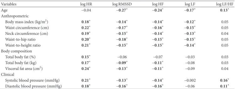
3.3. Relationship between Heart Rate Variability Components and Age, Anthropometric, Body Composition, and Clinical Variables. HR exhibited a positive correlation with all inves-tigated variables except age. RMSSD and HF, which are related to parasympathetic activity, exhibited negative corre-lations with age, BMI, WC, NC, WHR, WHtR, BFkg, VFA, SBP, and DBP. LF, which is related to sympathetic and para-sympathetic activity or to baroreflex function, exhibited neg-ative correlations with age, BMI, WC, WHR, WHtR, and NC. The LF/HF ratio, which is related to autonomic balance, exhibited positive correlations with age, SBP, and DBP. All
correlations are described inTable 3.
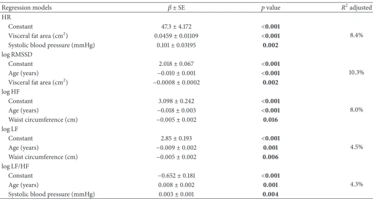
The fitted regression models showed that HRV
compo-nents were influenced by age, WC, VFA, and SBP (Table 4).
More specifically, VFA and SBP contributed to the increase of HR. VFA and age contributed to the reduction of logRMSSD. Age and WC explained the reduction of logHF and logLF. Finally, age and SBP explained the increase of logLF/HF.
Ninety-three of the 438 employees were using some type of medication. Of those 93, 27 were using one of the following groups of medicines, which were not identified as HRV modifiers: proton pump inhibitors, corticosteroids, nonsteroidal anti-inflammatory drugs, oral anticoagulants, and/or statins. The other 66 were using at least one of the following groups, which can potentially influence HRV
Table 1: Sample characteristics.
Variables Median (p25; p75) Quartile range
Age (years) 34.00 (31.00, 39.00) 8.00
Anthropometric
Height (m) 1.74 (1.70, 1.79) 0.09
Body weight (kg) 80.80 (73.60, 89.30) 15.70
Body mass index (kg/m2) 26.70 (24.40, 29.20) 4.80
Waist circumference (cm) 92.00 (85.50, 97.50) 12.00
Hip circumference (cm) 102.50 (98.30, 107.00) 8.75
Neck circumference (cm) 39.25 (37.50, 41.15) 3.65
Waist-to-hip ratio 0.89 (0.86, 0.93) 0.07
Waist-to-height ratio 0.53 (0.49, 0.56) 0.07
Body composition
Total body fat (%) 23.55 (19.20, 28.20) 9.00
Total body fat (kg) 18.95 (14.60, 24.30) 9.70
Visceral fat area (cm2) 121.25 (95.40, 147.70) 52.30
Clinical
Systolic blood pressure (mmHg) 131.67 (122.67, 141.33) 18.70 Diastolic blood pressure (mmHg) 82.33 (76.00, 88.33) 12.30
Table 2: Medians of heart rate variability (HRV) variables in the supine and orthostatic positions.
HRV variables Supine position Orthostatic position 𝑝value
Median (p25, p75) Median (p25, p75)
HR (bpm) 65.9 (59.7, 71.9) 82.3 (74.2, 90.2) <0.0001
RMSSD (ms) 34.3 (23.9, 50.1) 19.9 (13.3, 30.1) <0.0001
HF (ms2) 81.9 (35.7, 168.7) 26.1 (10.5, 62.1) <0.0001
LF (ms2) 103.4 (60.8, 186.4) 120.6 (62.4, 224.1) 0.005
LF/HF 1.3 (0.7, 2.5) 4.7 (2.5, 7.9) <0.0001
HRV: heart rate variability; HR: heart rate; RMSSD: root mean square of successive RR interval differences; HF: high frequency; LF: low frequency; LF/HF: ratio of low frequency and high frequency.Rawdata.The Wilcoxon test was made using log transformation values, but, for ease of interpretation, all medians (plus p25, p75) are given in the original units.
Table 3: Spearman correlation coefficients (Rho) between heart rate variability measures (supine position) and age, anthropometric, body composition, and clinical variables.
Variables log HR log RMSSD log HF log LF log LF/HF
Age −0.04 −0.27∗ −0.24∗ −0.17∗ 0.13∗
Anthropometric
Body mass index (kg/m2) 0.18∗ −0.14∗ −0.14∗ −0.12∗ 0.05
Waist circumference (cm) 0.22∗ −0.17∗ −0.16∗ −0.15∗ 0.05 Neck circumference (cm) 0.19∗ −0.15∗ −0.14∗ −0.13∗ 0.04
Waist-to-hip ratio 0.20∗ −0.18∗ −0.15∗ −0.15∗ 0.05
Waist-to-height ratio 0.21∗ −0.15∗ −0.15∗ −0.14∗ 0.05
Body composition
Total body fat (%) 0.15∗ −0.06 −0.07 −0.03 0.03
Total body fat (kg) 0.17∗ −0.09∗ −0.11∗ −0.08 0.03
Visceral fat area (cm2) 0.24∗ −0.13∗ −0.11∗ −0.09 0.04
Clinical
Systolic blood pressure (mmHg) 0.21∗ −0.13∗ −0.14∗ −0.002 0.16∗
Table 4: Equations using regression analysis for cardiac variables.
Regression models 𝛽 ±SE 𝑝value 𝑅2adjusted
HR
Constant 47.3±4.172 <0.001
8.4% Visceral fat area (cm2) 0.0459±0.01109 <0.001
Systolic blood pressure (mmHg) 0.101±0.03195 0.002
log RMSSD
Constant 2.018±0.067 <0.001
10.3%
Age (years) −0.010±0.001 <0.001
Visceral fat area (cm2) −0.0008±0.0002 0.002
log HF
Constant 3.098±0.242 <0.001
8.0%
Age (years) −0.018±0.003 <0.001
Waist circumference (cm) −0.005±0.002 0.016
log LF
Constant 2.85±0.193 <0.001
4.5%
Age (years) −0.009±0.002 0.001
Waist circumference (cm) −0.005±0.002 0.006
log LF/HF
Constant −0.652±0.181 <0.001
4.3%
Age (years) 0.008±0.002 0.001
Systolic blood pressure (mmHg) 0.003±0.001 0.004
logHF: natural logarithm of power in the high frequency range;logHR, natural logarithm of the heart rate;logLF: natural logarithm of power in the low frequency range;logLF/HF: natural logarithm of the ratio LF (ms2)/HF (ms2);logRMSSD: natural logarithm of the root mean square of successive RR interval differences. Numbers in bold represent𝑝 < 0.05.
channel blocker; angiotensin-converting enzyme inhibitor; angiotensin receptor blocker; and beta-blockers), anticholin-ergics, beta2-agonists, antidepressants, anxiolytics, sedative/ hypnotics, insulin, oral hypoglycemic agents, and/or levothy-roxine. After excluding these 66 employees, we repeated the statistical analyses (data not shown) and noted that the significant results found for the total 438 employees remained. This allowed us to rule out the possibility of drug bias on the findings reported in this study.
4. Discussion
The results of the present study pointed to the presence of cardiovascular risk factors in the sample of Brazilian male rotating shift workers, as shown by anthropometry, body composition, and blood pressure, whose median results were higher than the corresponding normal values. In addition, these alterations were shown to correlate with changes in HRV components in the supine position, indicating increased sympathetic and reduced parasympathetic activity. However, the responses of HRV components to the postural change, a baroreflex mediated response, were adequate.
Obesity and hypertension, in addition to other comor-bidities such as dyslipidemia and diabetes, are disorders that
are frequently found in shift workers [21–23]. In the present
study, median values of BMI, WC, WHtR, and VFA, which are indicators of obesity, and SBP and DBP, which are related
to hypertension, were above the reference values [13–15],
thus corroborating the reports in the literature. The con-stant presence of those disorders in shift workers is mainly
associated with environmental and behavioral changes, as well as deregulation of biological rhythms and the lifestyle to which they are exposed, and these factors may exert a direct influence on their dietary habits, physical activity, sleep quality, mental health, and time available for social
interac-tion [11,22,23].
Our findings showed that HRV components (HF, LF, and LF/HF) were qualitatively lower compared to the mean values
in a review study with healthy adults [24]. In general, the HRV
components vary within a wide range in male shift workers:
HR, 58.2 to 82.8 bpm; LF, 323.7 to 1,212 ms2; RMSSD, 29.9 to
70.2 ms; HF, 181.2 to 558.6 ms2; and LF/HF, 1.6 to 3.4 [21,23,
25,26]. Values for LF and HF in the present study were
sub-stantially lower than the minimum when compared qualita-tively with the values described above. The reason for that dis-crepancy may be associated with the methodological particu-larities of the various studies, including shift regimen (perma-nent or rotating), ECG signal processing (application of Task Force guidelines), and sample characteristics, including age and the presence of cardiovascular risk factors.
Nevertheless, the cardiac autonomic response to pos-tural change was adequate, as the values corresponding to parasympathetic components (RMSSD and HF) were higher in the supine compared to the orthostatic position. Those results corroborate HRV at rest as a marker of
parasympa-thetic control of the heart [4]. An increase in LF and LF/HF in
the orthostatic position relative to the supine position is
con-troversial [17]. Cysarz et al. [6] and Porta et al. [8] found that
and LF/HF increased at a 90∘tilt; the same increase occurred in the present study with voluntary postural change. To our knowledge, this is the first study to investigate the postural changes in a sample of shift workers. Probably, because pos-tural change is a basic process of cardiac autonomic control, mediated by the baroreflex response, the postural control continued to function properly despite the fact that HRV components in the supine position were found to be lower
than expected for healthy adults [24].
Use of RMSSD and HF as indicators of parasympathetic
activity is well established [1,2]. However, the meaning of LF,
and consequently also of LF/HF, is still a subject of debate [6,
17,18,27]. According to classic studies, LF represents mainly
the cardiac sympathetic modulation [5,28]. Nowadays, some
authors have proposed that LF is related to baroreflex
function. According to Goldstein et al. [18], with or without
adjustment for HF power or respiration, LF power seems to provide an index not of cardiac sympathetic tone but of baroreflex function once manipulations and drugs that change LF power or LF/HF do so not by affecting cardiac autonomic outflows directly but by affecting modulation of
those outflows by baroreflex. Piccirillo et al. [27] showed, in a
canine experimental acute myocardial infarction model, that, during congestive heart failure, the reduction of LF power and LF/HF ratio probably reflect diminished sinus node respon-siveness to autonomic modulation or an abnormal baroreflex function. In another study of the same authors, they showed that LF and HF power were significantly lower seven weeks after acute canine myocardial infarction than at baseline
[29]. Other authors have proposed that LF is predominantly
associated with parasympathetic activity based on the find-ings: increases in HF and LF occur after the use of drugs that specifically enhance the cardiac vagal tone; there is similarity of LF values between individuals with cardiac sympathetic denervation and normal baroreflex sensitivity; some vali-dated measures of sympathetic control of the heart, such as plasma epinephrine levels and cardiac norepinephrine
spillover, are not correlated with LF [17, 18]. Therefore,
more studies are necessary to clarify the origins and clinical significance of LF.
Among the variables that exhibited a correlation with HRV, the following remained significant following multiple linear regression analyses: age exhibited negative correlations with logRMSSD, logHF, and logLF and a positive correla-tion with LF/HF; WC exhibited negative correlacorrela-tions with logHF and logLF; VFA exhibited a negative correlation with logRMSSD and a positive correlation with HR; and SBP exhibited positive correlations with HR and LF/HF. These results indicate that, in the present sample, age, WC, VFA, and SBP were the variables with the strongest influence on HRV.
Similar to our study, Kim et al. [26] also observed that
greater age is associated with lower HRV, parasympathetic activity, and baroreflex sensitivity in particular.
The findings about the WC and VFA could indicate that higher values of these anthropometric and body composition parameters are associated with less activity of HRV, specially the components that reflect parasympathetic activity. Similar
results were reported by Ramos and Ara´ujo [30], who found
that cardiac vagal components decrease in parallel with increases in BMI, the sum of skinfold measurements (reflect-ing total body fat), and WC. Additionally, other studies found a relationship between WC, BF, and WHR and HRV vagal
components [5,26,31].
With regard to blood pressure, SBP and DBP exhibited
positive association with HR and LF/HF. Yue et al. [32]
assessed the HRV in hypertensive and nonhypertensive patients and found increased sympathetic and decreased parasympathetic activity in hypertensive patients, compared to normotensive patients. In the same line, Thiyagarajan
et al. [33] found that young adults with prehypertension
had decreased cardiovagal modulation (HF, RMSSD, and SDNN), increased LF/HF ratio, and elevated cardiovascular risk factors comparable to the normotensive group.
Some limitations must be discussed about our findings. First, as the sample is composed only of male shift workers with the same work arrangement of alternating shifts, com-parisons of gender and shift work specificities (permanent or alternating, clockwise or counterclockwise, etc.) could not be performed. Second, although the employees had similar environmental and work conditions (they were all operators of iron ore extraction machinery working in air-conditioned trucks with insulation against sound, water, and dust), data on behavioral factors (e.g., eating habits, physical activity, sleep-wake pattern, and smoking) was not collected. This informa-tion could be useful in determining potential mediators of the associations found. Third, we did not measure breathing dur-ing the electrocardiographic recorddur-ings. Although it is known that breathing (respiratory frequency and volume) potentially
influences the HRV variables, Penttil¨a et al. [34]
demon-strated that RMSSD values are not affected by the respiratory pattern. In the present study, both parasympathetic variables collected (HF and RMSSD) showed the same statistical tendencies; therefore, we can infer that the lack of breathing register most likely did not bias the study results.
5. Conclusion
Our findings showed that the cardiac autonomic response to postural change in shift workers was adequate. Nevertheless, they exhibited alterations in the assessed anthropometric, body composition, clinical, and HRV variables compared to the reference values. Regression analysis showed that age, obesity (WC and VFA), and SBP account for a significant part of the reduction of HRV components in this population. Even when taking into consideration the full complexity and variety of factors that may be involved in those alterations, the need to provide comprehensive healthcare to shift workers is undeniable, including specific occupational strategies to min-imize the impact of these cardiovascular risk factors on their health and ensure good quality of life.
Conflict of Interests
Acknowledgments
The authors are grateful for the excellent technical assistance of Cristina de Oliveira Lisboa Pereira. This work was sup-ported by the Federal University of Ouro Preto (UFOP); the National Council for Scientific and Technological Devel-opment (CNPq); the Coordination for the Improvement of Higher Education Personnel (CAPES); the Foundation for Research Support in Minas Gerais (FAPEMIG); and Gorceix Foundation (Ouro Preto, MG).
References
[1] B. Xhyheri, O. Manfrini, M. Mazzolini, C. Pizzi, and R. Bugia-rdini, “Heart rate variability today,”Progress in Cardiovascular
Diseases, vol. 55, no. 3, pp. 321–331, 2012.
[2] Task Force, “Heart rate variability: standards of measurement, physiological interpretation and clinical use. Task Force of the European Society of Cardiology and the North American Society of Pacing and Electrophysiology,”Circulation, vol. 93, no. 5, pp. 1043–1065, 1996.
[3] J. F. Thayer and R. D. Lane, “The role of vagal function in the risk for cardiovascular disease and mortality,”Biological Psychology, vol. 74, no. 2, pp. 224–242, 2007.
[4] J. F. Thayer, S. S. Yamamoto, and J. F. Brosschot, “The rela-tionship of autonomic imbalance, heart rate variability and cardiovascular disease risk factors,” International Journal of
Cardiology, vol. 141, no. 2, pp. 122–131, 2010.
[5] G.-Y. Chen, T.-J. Hsiao, H.-M. Lo, and C.-D. Kuo, “Abdominal obesity is associated with autonomic nervous derangement in healthy Asian obese subjects,”Clinical Nutrition, vol. 27, no. 2, pp. 212–217, 2008.
[6] D. Cysarz, P. van Leeuwen, F. Edelh¨auser, N. Montano, and A. Porta, “Binary symbolic dynamics classifies heart rate variability patterns linked to autonomic modulations,”Computers in
Biol-ogy and Medicine, vol. 42, no. 3, pp. 313–318, 2012.
[7] K. Efremov, D. Brisinda, A. Venuti et al., “Heart rate variability analysis during head-up tilt test predicts nitroglycerine-induced syncope,”Open Heart, vol. 1, no. 1, Article ID e000063, 2014. [8] A. Porta, E. Tobaldini, S. Guzzetti, R. Furlan, N. Montano,
and T. Gnecchi-Ruscone, “Assessment of cardiac autonomic modulation during graded head-up tilt by symbolic analysis of heart rate variability,”The American Journal of Physiology: Heart
and Circulatory Physiology, vol. 293, no. 1, pp. H702–H708, 2007.
[9] F. Togo and M. Takahashi, “Heart rate variability in occupa-tional health–a systematic review,”Industrial Health, vol. 47, no. 6, pp. 589–602, 2009.
[10] B. G. Windham, S. Fumagalli, A. Ble et al., “The relationship between heart rate variability and adiposity differs for central and overall adiposity,”Journal of Obesity, vol. 2012, Article ID 149516, 8 pages, 2012.
[11] G. Jermendy, J. N´adas, I. Hegyi, I. Vasas, and T. Hidv´egi, “Assessment of cardiometabolic risk among shift workers in Hungary,”Health and Quality of Life Outcomes, vol. 10, article 18, 2012.
[12] International Labour Office (ILO), Conditions of Work and
Employment Programme, International Labour Office (ILO),
Geneva, Switzerland, 2004,http://www.ilo.org/wcmsp5/groups/ public/—ed
protect/—protrav/—travail/documents/publica-tion/wcms 170713.pdf.
[13] WHO, “Waist circumference and waist-hip ratio: report of a WHO expert consultation,” World Health Organization Tech-nical Report Series, 2011.
[14] L. Ben-Noun, E. Sohar, and A. Laor, “Neck circumference as a simple screening measure for identifying overweight and obese patients,”Obesity Research, vol. 9, no. 8, pp. 470–477, 2001. [15] T. G. Lohman, Advances in Body Composition Assessment:
Current Issues in Exercises Science, Human Kinetic Publisher,
Champaign, Ill, USA, 1992.
[16] Sociedade Brasileira de Cardiologia, Sociedade Brasileira de Hipertens˜ao, and Sociedade Brasileira de Nefrologia, “VI Brazilian guidelines on hypertension,”Arquivos Brasileiros de
Cardiologia, vol. 95, no. 1, supplement, pp. 1–51, 2010.
[17] G. A. Reyes del Paso, W. Langewitz, L. J. M. Mulder, A. van Roon, and S. Duschek, “The utility of low frequency heart rate variability as an index of sympathetic cardiac tone: a review with emphasis on a reanalysis of previous studies,”Psychophysiology, vol. 50, no. 5, pp. 477–487, 2013.
[18] D. S. Goldstein, O. Bentho, M.-Y. Park, and Y. Sharabi, “Low-frequency power of heart rate variability is not a measure of cardiac sympathetic tone but may be a measure of modulation of cardiac autonomic outflows by baroreflexes,”Experimental
Physiology, vol. 96, no. 12, pp. 1255–1261, 2011.
[19] P. Perakakis, M. Joffily, M. Taylor, P. Guerra, and J. Vila, “KAR-DIA: a Matlab software for the analysis of cardiac interbeat intervals,”Computer Methods and Programs in Biomedicine, vol. 98, no. 1, pp. 83–89, 2010.
[20] A. L. T. Uusitalo, E. Vanninen, E. Lev¨alahti, M. C. Batti´e, T. Videman, and J. Kaprio, “Role of genetic and environmental influences on heart rate variability in middle-aged men,”The American Journal of Physiology: Heart and Circulatory Physiol-ogy, vol. 293, no. 2, pp. H1013–H1022, 2007.
[21] M. Ishizaki, Y. Morikawa, H. Nakagawa et al., “The influence of work characteristics on body mass index and waist to hip ratio in Japanese employees,”Industrial Health, vol. 42, no. 1, pp. 41– 49, 2004.
[22] Y. Guo, Y. Liu, X. Huang et al., “The effects of shift work on sleeping quality, hypertension and diabetes in retired workers,”
PLoS ONE, vol. 8, no. 8, Article ID e71107, 2013.
[23] M. Kivim¨aki, G. D. Batty, and C. Hublin, “Shift work as a risk factor for future type 2 diabetes: evidence, mechanisms, impli-cations, and future research directions,”PLoS Medicine, vol. 8, no. 12, Article ID e1001138, 2011.
[24] D. Nunan, G. R. H. Sandercock, and D. A. Brodie, “A quan-titative systematic review of normal values for short-term heart rate variability in healthy adults,” Pacing and Clinical
Electrophysiology, vol. 33, no. 11, pp. 1407–1417, 2010.
[25] T.-C. Su, L.-Y. Lin, D. Baker et al., “Elevated blood pressure, decreased heart rate variability and incomplete blood pressure recovery after a 12 hour night shift work,”Journal of
Occupa-tional Health, vol. 50, no. 5, pp. 380–386, 2008.
[26] J. A. Kim, Y.-G. Park, K.-H. Cho et al., “Heart rate variability and obesity indices: emphasis on the response to noise and standing,”Journal of the American Board of Family Practice, vol. 18, no. 2, pp. 97–103, 2005.
[28] F. Lombardi, A. Malliani, M. Pagani, and S. Cerutti, “Heart rate variability and its sympatho-vagal modulation,”Cardiovascular
Research, vol. 32, no. 2, pp. 208–216, 1996.
[29] G. Piccirillo, F. Moscucci, G. D’Alessandro et al., “Myocardial repolarization dispersion and autonomic nerve activity in a canine experimental acute myocardial infarction model,”Heart
Rhythm, vol. 11, no. 1, pp. 110–118, 2014.
[30] P. S. Ramos and C. G. S. Ara´ujo, “Lower cardiac vagal tone in non-obese healthy men with unfavorable anthropometric char-acteristics,”Clinics, vol. 65, no. 1, pp. 45–51, 2010.
[31] M. E. Andrew, L. Shengqiao, J. Wactawski-Wende et al., “Adi-posity, muscle, and physical activity: predictors of perturbations in heart rate variability,”American Journal of Human Biology, vol. 25, no. 3, pp. 370–377, 2013.
[32] W.-W. Yue, J. Yin, B. Chen et al., “Analysis of heart rate variability in masked hypertension,”Cell Biochemistry and Biophysics, vol. 70, no. 1, pp. 201–204, 2014.
[33] R. Thiyagarajan, P. Pal, G. K. Pal et al., “Cardiovagal modulation, oxidative stress, and cardiovascular risk factors in prehyperten-sive subjects: cross-sectional study,”The American Journal of
Hypertension, vol. 26, no. 7, pp. 850–857, 2013.
Submit your manuscripts at
http://www.hindawi.com
Stem Cells
International
Hindawi Publishing Corporationhttp://www.hindawi.com Volume 2014
Hindawi Publishing Corporation
http://www.hindawi.com Volume 2014
INFLAMMATION
Hindawi Publishing Corporation
http://www.hindawi.com Volume 2014
Behavioural
Neurology
Endocrinology
International Journal of Hindawi Publishing Corporationhttp://www.hindawi.com Volume 2014 Hindawi Publishing Corporation
http://www.hindawi.com Volume 2014
Disease Markers
Hindawi Publishing Corporation
http://www.hindawi.com Volume 2014
BioMed
Research International
Oncology
Journal ofHindawi Publishing Corporation
http://www.hindawi.com Volume 2014
Hindawi Publishing Corporation
http://www.hindawi.com Volume 2014
Oxidative Medicine and Cellular Longevity
Hindawi Publishing Corporation
http://www.hindawi.com Volume 2014
PPAR Research
The Scientific
World Journal
Hindawi Publishing Corporationhttp://www.hindawi.com Volume 2014
Immunology Research
Hindawi Publishing Corporation
http://www.hindawi.com Volume 2014
Journal of
Obesity
Journal ofHindawi Publishing Corporation
http://www.hindawi.com Volume 2014
Hindawi Publishing Corporation
http://www.hindawi.com Volume 2014 Computational and Mathematical Methods in Medicine
Ophthalmology
Journal ofHindawi Publishing Corporation
http://www.hindawi.com Volume 2014
Diabetes Research
Journal ofHindawi Publishing Corporation
http://www.hindawi.com Volume 2014
Hindawi Publishing Corporation
http://www.hindawi.com Volume 2014
Research and Treatment
AIDS
Hindawi Publishing Corporation
http://www.hindawi.com Volume 2014
Gastroenterology Research and Practice
Hindawi Publishing Corporation
http://www.hindawi.com Volume 2014
Parkinson’s
Disease
Evidence-Based Complementary and Alternative Medicine
Volume 2014 Hindawi Publishing Corporation