A primary assessment of the endophytic bacterial community in a xerophilous moss
(
Grimmia montana
) using molecular method and cultivated isolates
Xiao Lei Liu, Su Lin Liu, Min Liu, Bi He Kong, Lei Liu, Yan Hong Li
College of Life Science, Capital Normal University, Haidian District, Beijing, China.
Submitted: December 27, 2012; Approved: April 1, 2013.
Abstract
Investigating the endophytic bacterial community in special moss species is fundamental to under-standing the microbial-plant interactions and discovering the bacteria with stresses tolerance. Thus, the community structure of endophytic bacteria in the xerophilous mossGrimmia montanawere esti-mated using a 16S rDNA library and traditional cultivation methods. In total, 212 sequences derived from the 16S rDNA library were used to assess the bacterial diversity. Sequence alignment showed that the endophytes were assigned to 54 genera in 4 phyla (Proteobacteria, Firmicutes, Actinobacteria andCytophaga/Flexibacter/Bacteroids). Of them, the dominant phyla were Proteobacteria (45.9%) and Firmicutes (27.6%), the most abundant genera included Acinetobacter, Aeromonas, Enterobacter,Leclercia,Microvirga,Pseudomonas,Rhizobium, Planococcus, Paenisporosarcina and Planomicrobium. In addition, a total of 14 species belonging to 8 genera in 3 phyla (Proteo-bacteria, Firmicutes, Actinobacteria) were isolated,Curtobacterium, Massilia, Pseudomonas and Sphingomonaswere the dominant genera. Although some of the genera isolated were inconsistent with those detected by molecular method, both of two methods proved that many different endophytic bacteria coexist inG. montana. According to the potential functional analyses of these bacteria, some species are known to have possible beneficial effects on hosts, but whether this is the case inG. montananeeds to be confirmed.
Key words:bacterial diversity, endophytes, moss, molecular method, cultivated isolates.
Introduction
In plant-endophyte interactions, plants provide nutri-ents and residency for the bacteria, while the bacteria in ex-change directly or indirectly improve plant growth and health (Mastrettaet al., 2006). Once inside the plant, endo-phytes either reside in specific plant tissues such as the root cortex or the xylem, or colonize the plant systematically by transport through the vascular system or the apoplast (Quadt-Hallmannet al., 1997). Of the nearly 300 000 plant species on earth, each species is host to one or more species of endophytes (Strobelet al., 2004). The complete descrip-tion of endophytic species has only been enumerated and characterized for a handful of plant species, and the major-ity of these are common higher plants. Few studies have ex-amined the endophytes of bryophytes, which represent the simplest extant land plants and have been classified by
prominent bryologists as “living fossils” (Hornschuhet al., 2002). Consequently, the opportunity to find new and bene-ficial endophytic microorganisms among the diversity of plants in different ecosystems is considerable.
The mosses, one kind of bryophytes, are a diverse group of land plants that usually colonize habitats with ei-ther moist or extremely variable conditions. One of their most important features is their life cycle, which involves alteration between a diploid sporophyte and a dominant free-living haploid gametophyte generation (Opelt and Berg, 2004). Mosses are unique host plants for microorgan-isms in numerous ways. For example, the small size of mosses results in limited availability of the substratum. In addition, most mosses display an extraordinarily high toler-ance to extreme desiccation and can resume normal metab-olism very rapidly after rehydration. Hence, successful microbial colonization requires adaptation to these special
Send correspondence to Y.H. Li. College of Life Science, Capital Normal University, Xisanhuan North Road 105#, Haidian District, 100048 Beijing, China. E-mail: liyh@mail.cnu.edu.cn.
conditions (DoÈbbeler, 1997). Analysis of the epiphytes on the gametophyte ofFunaria hygrometricadetected numer-ous bacterial species on the surface of the phylloid. Among these species, twoMethylobacteriumstrains were found to be able to simulate the well-known effect of cytokinin ap-plication on bud formation inFunariaprotonema and they also promoted the growth of protonemal filaments (Hornschuhet al., 2002). Endophytic methanotrophic bac-teria were also found in the hyaline cells and on the stem leaves ofSphagnummosses; here, they provided carbon for photosynthesis via in situ oxidation of methane to carbon dioxide (Raghoebarsinget al., 2005).
Opelt and Berg (2004) isolated and identified many antagonistic bacteria associated with three moss species (Tortula ruralis, Aulacomnium palustre and Sphagnum rubellum) in the nutrient-poor habitats of the Baltic Sea Coast in Germany. These species belong to nine different genera, among which Burkholderia, Pseudomonas and Serratiawere dominant, but the richness and diversity of antagonistic species were moss species-dependent, and the highest number of species with antagonistic activity was isolated from S. rubellum. Another study examined the function and diversity of bacterial species associated with twoSphagnumspecies (S. fallaxandS. magellanicum) that grow in a temperate mire ecosystem. Species belonging to the genusBurkholderia were predominant in Sphagnum species and this genus was possibly involved in antago-nism/pathogen defense and nitrogen-fixation. The authors concluded thatSphagnumis a reservoir for powerful and extraordinary antagonists and potentially facultative hu-man pathogens (Opelt et al., 2007). Thus, thorough re-search on the bacteria associated with other mosses in different niches would be also useful in discovering bacte-rial resources and helpful in understanding the interactions between mosses and their associated microbes.
Grimmia montanais a xerophilous moss, and has a high tolerance to drought, cold and UV radiation (Yi and Liu, 2007), and can often be found growing in extreme en-vironments. It always lives under extreme desiccation con-ditions and can resume normal metabolism very rapidly after rehydration. In this paper, our aim is to study the di-versity and community structure of its endophytes using 16S rDNA library and culture-dependent approaches, and hope to make a well known on the interactions between endophytes andG. montanaand try to find some bacterial resources with the strong tolerance to the stresses.
Material and Methods
Sampling and surface disinfection
Grimmia montanawere sampled from the surface of one large stone in Beijing Songshan National Nature Re-serve located at an altitude of 890 m, at N: 40°31’00.45” by E:115°49’33.20” on the 19thof April, 2011. About 3 g of
plant material, approximately more than one thousand of
entire plants was collected after absorbing enough water, and then mixed together and immediately transported to the laboratory for surface disinfection as described previously (Liet al., 2010). The plants were first washed many times with tap water to remove attached substratum. Subse-quently, they were immersed in 70% ethanol for 3 min, washed with 15% sodium hypochlorite solution for 10 min, rinsed three times with 70% ethanol for 30 s, and finally washed five times with sterile distilled water. To confirm that the disinfection process was successful, aliquots of the sterile distilled water in the final rinse were used to deter-mine the results of surface disinfection. Bacteria were culti-vated by setting 100mL of the final rinse on R2A and TSA
medium plates, and then examining the plates for bacterial growth after incubation at 28 °C for 3 days. Molecular de-tection of bacterial species was accomplished by 16S rRNA gene PCR detection based on the primers 799f (5’-AACAGGATTAGATACCCTG-3’) and 1492r (5’-GGTTACCTTGTTACGACTT-3’) (Chelius and Triplett, 2001) using the final rinse as template. The 50mL
PCR reaction mixture contained 5mL of the final rinse,
5mL 10x Taq reaction buffer (including 1.5 mM MgCl2),
10 pmol of each primer, 200mM each dNTP, and 1.5 U of
Taq DNA polymerase (Takara Co.). After initial denatur-ation at 94 °C for five minutes, each thermal cycling was as follows: denaturation at 94 °C for one minute, annealing at 53 °C for one minute, and elongation at 72 °C for one min-ute. At the end of 30 cycles, the final extension step was at 72 °C for 15 min. Products of four parallel PCRs were com-bined and electrophoretically separated by 1% agarose. Finally, plant samples were determined to be successfully surface disinfected if no bacterium was identified via culti-vation and PCR. These plants were used for the subsequent analyses.
DNA extraction and amplification of the bacterial 16S rRNA genes
About 2 g of surface-disinfected G. montana was frozen with liquid nitrogen and ground to a fine powder in a sterilized and precooled mortar. Next, the cetyltrimethyl-ammonium bromide (CTAB) procedure was used to extract total DNA as previously described (Xieet al., 1999). The DNA was resuspended in 150mL sterile Milli-Q water. The
primer pair 799f and 1492r was selected to amplify the 16S rDNA of the endophytic bacteria. The PCR reaction mix-ture and programs are the same as described above in the section of surface disinfection. We excised the approxi-mately 730 bp band from a 1% agarose gel, following elec-trophoresis of the DNA, and purified the DNA using the Gel Extraction Kit (Omega Co.), as described by the manu-facturer.
The purified 730 bp PCR products were ligated into the pMD18-T vector (Takara Co.).Escherichia coliTop10 competent cells (Tiangen Co.) were transformed with the ligation products and spread onto Luria-Bertani agar plates with ampicillin (100 mg L-1) for standard blue and white
screening (Sambrooket al., 1989). Randomly selected onies were screened directly for inserts by performing
col-ony PCR with primers RV-M
(5’-GAGCGGATAACAATTTCACACAGG-3’) and M13-47 (5’-CGCCAGGGTTTTCCCAGTCACGAC-3’) for the vector (Takara Co.). Two hundred fifty clones con-taining inserts of the correct size were sequenced using an ABI PRISM 3730 automatic sequencer (Shanghai Sangon Co., Ltd).
Phylogenetic analysis
After being trimmed by cutting the vector sequences using the Editseq program in the DNAStar package (Bur-land, 2000) and removing all the bad sequences as deter-mined by the chimera sequence detection software Mallard 1.02 (www.cardiff.ac.uk/biosi/research/biosoft), all other manually verified nucleotide sequences were submitted to the NCBI GenBank database. Clones of 16S rRNA gene se-quences showing 97% similarity or higher were considered to belong to the same phylotype by sequencher 4.8 (Gene Codes, Ann Arbor, MI) and assigned to an Operational Taxonomic Unit (OTU). Sequences of all phylotypes were compared to the NCBI database using BlastN or aligned by the identify analysis of EzTaxon-e (Kim et al., 2012). Clones with a 16S rDNA sequence similarity larger than 97% were assigned to the same species; those with > 95% identity were assigned to the same genus; those with < 95% were determined to be uncultured bacterial species. Next, those sequences assigned to uncultured bacteria were aligned using Clustal W (Thompsonet al., 1994), and tree constructions were done with the MEGA 5 program pack-age (Tamura et al., 2011) using the neighbor-joining method (Saitou and Nei, 1987) to infer their classification. Bootstrap analysis was performed with 1,000 replicates.
Estimation of the size of the clone library
To estimate the representation of the library, the clone coverage was calculated with the following equation based on the sequencing results:C= (1-n1/N) x 100%, wheren1 represents the number of phylotypes occurring only once andNis the number of clones being examined. Diversity of the clone library was investigated using rarefaction analy-sis. Rarefaction curve was calculated using the Ecosim 7.0 software (Gotelli and Entsminger, 2004).
Isolation of culturable endophytes and determination of CFU
To isolate the endophytes from the plants, 1 mL of sterile 0.85% NaCl was added to 0.5 g (fresh weight) of sur-face disinfectedG. montana and samples were
homoge-nized in a small sterile mortar. The resultant mixture was serially diluted with sterile 0.85% NaCl and plated onto R2A and TSA media (Difco, Detroit, MI). Plates were incu-bated for 3 days at 28 °C, after which Colony-Forming Units (CFU) were counted to calculate the average number of colonies per gram of moss. Isolates obtained by plating were purified and stored at -70 °C in sterile broth contain-ing 40% glycerol.
ARDRA analysis and identification of the isolates by sequencing
1 uL of the bacterial suspension derived from each isolate was used to amplify the 16S rDNA fragments using the primers 27f and 1492r. The PCR reaction mixture and programs are the same as described above in the section on surface disinfection. The approximately 1490 bp band was excised from a 0.8% agarose gel, and purified using the Gel Extraction Kit (Omega Co.) as described by the manufac-turer. Next, the purified products were enzymatically di-gested withHaeIII andHhaI at 37 °C for 4 h, respectively. According to their electrophoresis pattern on a 1.0% aga-rose gel, these isolates were classified into different OTUs. Finally, the PCR products of isolates with different OTUs were sequenced using an ABI PRISM 3730 automatic se-quencer (Shanghai Sangon Co., Ltd). After trimming the low quality nucleotides, the sequence similarities were cal-culated using the EzTaxon-e (Kimet al., 2012).
Results
16S rDNA library analysis of endophytic bacterial community
Bacterial 16S rDNA fragments were amplified from total DNA that was extracted from surface disinfectedG. montana, using the primers 799f and 1492r. The amplified DNA displayed only one distinct and one weak band, of ap-proximately 730 bp and 1000 bp, respectively. The se-quencing result showed that the 730 bp band represented the bacterial 16S rRNA fragment, while the 1000 bp frag-ment was mainly derived from the mitochondria of the mosses. Thus, the purified 730 bp PCR products were used to construct a 16S rDNA clone library for the endophytic bacteria.
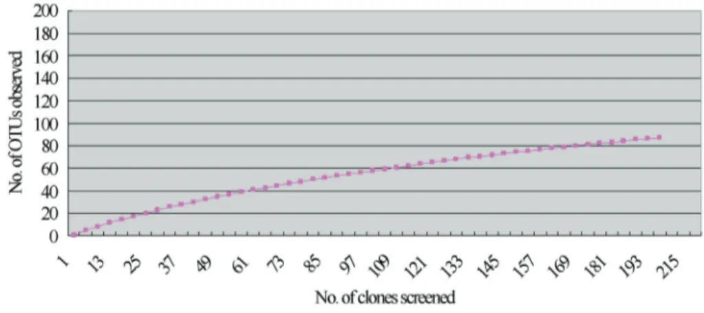
Of 250 clones, two-hundred and twelve individual se-quences were verified. They were determined as 90 phylo-types by sequencher 4.8 and the sequences were deposited in GenBank (Accession No.: JX042330-JX042419). Of them, 48 phylotypes occurring only once, and the calcu-lated coverage of the clone library was 77.4%.The rarefac-tion curve also showed that the clones detected could reflect the main information of endophytes (Figure 1).
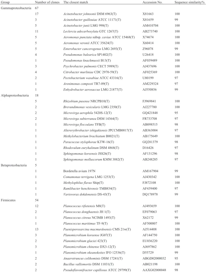
(14.8%) with Actinobacteria, and 23 (11.7%) with Cytophaga/Flavobacterium/Bacteroides(CFB) group. De-tails of all alignments in the clone library are listed in Table 1.
The sequences attributed to Proteobacteria, which in-cludes alpha, beta and gamma classes, made up the largest fraction of the clone library. Of the 90 clones affiliated with Proteobacteria, 67 clones (or 74.4%) exhibited high simi-larity to Gammaproteobacteria. The proportion of clones that grouped with the alpha and beta classes was 20% and 5.6%, respectively. However, there were no sequences with > 95% similarity to genera in the delta or epsilon class. The 67 clones of Gammaproteobacteria were related to four or-ders of bacteria, including Pseudomonadales (34 clones), Enterobacteriales (22 clones), Aeromonadales (10 clones) and Xanthomonadales (1 clone). Of these, the dominant genera include: Acinetobacter, Aeromonas, Citrobacter, Enterobacter,Leclercia,PseudomonasandPsychrobacter; the dominant species were Acinetobacter johnsonii, Acinetobacter junii,Leclercia adecarboxylata,Aeromonas punctataandEnterobacter cancerogenus(Table 1). Alpha-proteobacteria was the second-most abundant subgroup of Proteobacteria in our survey. The 18 clones in this sub-group represented bacteria in four orders (Rhizobiales, Sphingomonadales, Rhodobacterales and Caulobacterales) (Table 1). The dominant genera were Brevundimonas, Microvirga,RhizobiumandSphingomonas. Of the 5 clones affiliated with Betaproteobacteria, four belonged to bacte-rial species in Burkholdebacte-riales and only one was grouped into Methylophilales. All of them were assigned to differ-ent genera, including Bordetella, Comamonas, Methylophilus,RamlibacterandVariovorax(Table 1).
Among the non-Proteobacteria, 54, 29 and 23 clones exhibited high similarity to bacterial species in the phyla Firmicutes, Actinobacteria and CFB respectively (Table 1). In Firmicutes, 43 clones were closely related to bacteria in Bacillales, 9 clones to Clostridiales and only 2 to Lacto-bacillales. The dominant genera included Paenisporosarcina, Planococcus, Planomicrobium, and the most abundant species were Paenisporosarcina macmurdoensis and Planococcus rifietoensis. Of the 29 clones grouped into Actinomycetales of phylum Actino-bacteria, twelve clones were grouped with theArthrobacter genus, while the others grouped with many other genera in-cluding Aeromicrobium and Ornithinicoccus (Table 1). Arthrobacter sulfonivoranswas the most common species. In the 23 clones belonging to the CFB phylum, bacteria oc-curred in four orders, the Sphingobacteriales, Cytopha-gales, Bacteroidales and Flavobacteriales. The dominant genera were Adhaeribacter and Segetibacter, and Segetibacter koreensiswas the most common species.
Finally, the 16S rDNA sequence of 16 clones, showed < 95% similarity to the previously cultivated bacte-ria. The phylogenetic analysis showed that these clones ex-hibited a close relationship with Actinobacteria (4 clones),
Alphaproteobacteria (3 clones), Acidobacteria (3 clones), Bacteroidetes (2 clones), Betaproteobacteria (1 clone) and Firmicutes (3 clones) (Figure 2).
Endophytic bacteria communities detected by cultivation method
The isolation result showed that the number of col-ony-forming units (CFU) as determined for samples grown on R2A medium was higher than the number of CFUs grown on TSA medium. The counts (expressed as g-1fresh
weight) were 2.0*105and 3.3*104on R2A and TSA
me-dium, respectively. Totally 49 isolates were sequenced on the basis of 16S rDNA fragments, the ARDRA analysis re-sulted in the delimitation of 14 OTUs. Based on their 16S rDNA sequences (Genbank no. JX042420 - JX042433), they were assigned to 8 genera in three phyla (Proteo-bacteria, Actinobacteria and Firmicutes). The strains that were successfully cultivated included some genera in the Proteobacteria (Burkholderia, Massilia, Pseudomonas, Spingomonas, Yersinia), and some genera in Firmicutes and Actinobacteria such as Curtobacterium, BrevibacteriumandStreptomyces. The most abundant spe-cies were Curtobacterium flaccumfaciens, Massilia brevitalea,Pseudomonas azotoformansandPseudomonas libanensis(Table 2).
Compared the above bacterial communities with those discovered by 16S rDNA library technique, the culti-vated species only involved in three phyla (Firmicutes, Proteobacteria and Actinobacteria) and no bacteria in group CFB was cultivated. The species and genera discovered by cultivation were much less than those detected by molecu-lar method. In addition, some of genera cultivated also could not be found by molecular method, like Curtobacterium, Massilia, BurkholderiaandYersinia.
Discussion
In this study, we provide a thorough description of the endophytic bacterial community of G. montana, using a combined approach of molecular methods and cultivation-dependent techniques.G. montanaindividuals were sam-pled from stone surfaces poor in nutrient availability and subject to strong stresses, such as a wide range of tempera-tures and extreme drought conditions. As far as we know, ours is the first description to date of the endophytic com-munity of a xerophilous moss species in the Grimmiaceae.
in-formation completely. Both of them proved that many dif-ferent species coexisted in this small host (G. montana).
Compared to published accounts of bacterial commu-nities associated with other moss species growing in peat bog, such asSphagnum, our study revealed the different endophytes inhabiting the tissue ofG. montana. In previous studies,SerratiaandPseudomonasof the Gammaproteo-bacteria,Burkholderiaof the beta subgroup,Methylocella andMethylocapsaof the alpha subgroup (Raghoebarsinget al., 2005) andStaphylococcusof the Firmicutes (Opelt et al., 2007) were reported to be associated withSphagnum species. In this survey, of the Gammaproteobacteria sub-group,Acinetobacter,LeclerciaandAeromonaswere the dominant genera. Rhizobium of the Alphaproteobacteria, Massilia, Burkholderiaand five of other genera of beta-proteobacteria were also detected. In addition, there were also a high proportion of Gram positive bacteria detected in our library. Of them, clones assigned to Firmicutes com-prised 25.5% of the total.Planococcus, Paenisporosarcina, PlanomicrobiumandBacillus were the dominant genera; whileArthrobacterandCurtobacteriumof Actinobacteria were also abundant. The inconsistent endophytic bacterial community inG. montanaandSphagnum species proved that plant species and niches could cooperatively shape the structure of endophytic bacterial communities (Berg and Smalla, 2009).
Analyzing the function of those bacteria dominanted inG. montanawould be helpful to understand the interac-tions between endophytes and hosts. Of gammaproteo-bacteria class, the dominant species Acinetobacter johnsoniihas been reported to produce alkaline and low-temperature lipase (Wang et al., 2011a); Acinetobacter juniiwas considered to be a kind of cellulolytic bacterium that can produce xylanase, cellulose and pectinase (Loet al., 2010; Zhaiet al., 2010) and also could remove (via ac-cumulation) phosphate from synthetic wastewater (Hrenovic et al., 2010); Leclercia adecarboxylata could degrade two and three benzene-ring polycyclic aromatic hydrocarbon compounds (Sarmaet al., 2004; Sarmaet al., 2010); Aeromonas veronii andAeromonas punctatasubsp.
caviae, could produce enzymes such as the amino acid racemase, and xylanase (Caoet al., 2007; Cruzet al., 2008; Silveret al., 2011). As with theSphagnumbacterial com-munities,Pseudomonaswas also the dominant genus in our study. The isolated species Pseudomonas azotoformans (Komeda et al., 2004; Nie et al., 2011) could degrade Cyhalofop-butyl, while Pseudomonas libanensis could produce the biosurfactant viscosin (Dabboussiet al., 1999; Sainiet al., 2008).Rhizobium pusenseof the Alphaproteo-bacteria was first isolated from the rhizosphere of chickpea plants and considered to be a non-symbioticrhizobium. In our survey utilizing a 16S rDNA library, five clones of Rhizobium pusensewere detected, indicating that this spe-cies could be in symbiosis withG. montana.
Of bacteria assigned to Firmicutes, Planococcus rifietensisandPaenisporosarcina macmurdoensiswere the dominant species, which have ever been previously iso-lated from algal or cyanobacterial mats in sulfurous springs (Reddy et al., 2003; Romano et al., 2003). Four Planomicrobiumspecies were also found, which have been previously isolated from coastal sediments (Dai et al., 2005), seafood jeotgal (Yoon et al., 2001) and glaciers (Zhanget al., 2009a); they were considered as the cold tol-erant bacteria (Yanget al., 2011; Zhanget al., 2009a). In addition, Bacillus simplex was isolated by cultivation, which was ever provided to have strong antioxidant activity (Wang et al., 2011b). Among the Actinobacteria, Arthrobacter sulfonivoranscould produce membrane-as-sociated dimethylsulfone- and dimethylsulfoxide-reducta-ses (Borodina et al., 2002); Arthrobacter agilis could release N,N-dimethyl-hexadecanamine (dimethylhexa-decylamine) to directly affect plant morphogenesis (Fong et al., 2001; Velazquez-Becerraet al., 2011) and could con-tribute to membrane stabilization in response to thermal and salt stress by increasing carotenoid accumulation (Fonget al., 2001);Curtobacteriumwas a dominant genus discovered in the cultures, and Curtobacterium flaccumfaciens, as the most dominant species in this group, also was known to reduce symptoms caused by Xylella fastidiosainCatharanthus roseus(Lacavaet al., 2007); the
cultivable Streptomyces griseoplanus could produce anticapsin and Erythromycin-a, and might probably help to resist pathogens in the host (Boecket al., 1971; Thompson et al., 1971).
The dominant species Segetibacter koreensis from CFB phylum was first isolated from ginseng fields in South Korea (Anet al., 2007), whileAdhaeribacter tereus and Adhaeribacter aquaticus were ever isolated from soil (Zhanget al., 2009b) and water biofilms (Rickardet al., 2005), respectively. This is the first time that these species have been found as endophytes, and their possible func-tions remain unclear.
In conclusion, the most important findings of this study were: (1) a high endophytic bacterial diversity and complex community structure were found associated with G. montana,using a combination of molecular and cultiva-tion techniques; (2) community structure differed from that of endophytic communities of Sphagnum mosses, espe-cially in the abundance of Actinobacteria and Firmicutes (higher in G. Montana); and (3) Some bacterial species found endophytically in G. montana are known to have possible beneficial effects on plants, but whether this is the case in G. Montana is not proven. Thus, in order to improve our understanding of the concrete mechanisms through which endophytic bacteria (such as those ofG. montana) adapt to extreme environments and discover new bacterial resources, further work needs to be done in the future.
Acknowledgments
We would like to thank E.B.M. Drummond at the University of British Columbia for her assistance with Eng-lish language and grammatical editing of the manuscript. We also thank professor Guisen Du at Capital Normal Uni-versity for the species identification of moss. This work was funded by the Scientific Research Program of National Natural Science Foundation of China (No. 31100004).
References
An DS, Lee HG, Im WT, Liu OM, Lee ST (2007) Segetibacter koreensis gen. nov., sp nov., a novel member of the phylum Bacteroidetes, isolated from the soil of a ginseng field in South Korea. Int J Syst Evol Microbiol 57:1828-1833. Berg G, Smalla K (2009) Plant species and soil type cooperatively
shape the structure and function of microbial communities in the rhizosphere. FEMS Microbiol Ecol 68:1-13. Boeck LD, Christy KL, Shah R (1971) Production of Anticapsin
by Streptomyces griseoplanus. Appl Microbiol
21:1075-1079.
Borodina E, Kelly DP, Schumann P, Rainey FA, Ward-Rainey NL, Wood AP (2002) Enzymes of dimethylsulfone metabo-lism and the phylogenetic characterization of the facultative methylotrophs Arthrobacter sulfonivorans sp. nov.,
Arthrobacter methylotrophus sp. nov., and
Hyphomicrobium sulfonivorans sp. nov. Arch Microbiol
177:173-183.
Burland TG (2000) DNASTAR’s Lasergene sequence analysis software. Methods Mol Biol 132:71-91.
Cao HP, Yang XL, Wang YH, Li YY (2007) Isolation and growth characteristics of pathogenic Aeromonas punctata caviae from Sturgeons. Chinese J Zool 42:1-6.
Chelius M, Triplett E (2001) The diversity of archaea and bacteria in association with the roots ofZea maysL. Microbial Ecol
41:252-263.
Cruz A, Caetano T, Suzuki S, Mendo S (2008) Aeromonas veronii, a tributyltin (TBT)-degrading bacterium isolated from an estuarine environment, Ria de Aveiro in Portugal (vol 64, pg 639, 2007). Mar Environ Res 66:309-309. Dabboussi F, Hamze M, Elomari M, Verhille S, Baida N, Izard D,
Leclerc H (1999) Pseudomonas libanensis sp. nov., a new species isolated from Lebanese spring waters. Int. J Syst Bacteriol 49:1091-1101.
Dai X, Wang YN, Wang BJ, Liu SJ, Zhou YG (2005)
Planomicrobium chinense sp. nov., isolated from coastal sediment, and transfer of Planococcus psychrophilus and Planococcus alkanoclasticus to Planomicrobium as Planomicrobium psychrophilum comb. nov. and Planomoicrobium alkanoclasticum comb. nov. Int J Syst Evol Microbiol 55:699-702.
DoÈbbeler P (1997) Biodiversity of bryophilous ascomycetes. Biodivers Conserv 6:721-738.
Fong NJC, Burgess ML, Barrow KD, Glenn DR (2001) Caro-tenoid accumulation in the psychrotrophic bacterium
Arthrobacter agilis in response to thermal and salt stress.
Appl Microbiol Biot 56:750-756.
Gotelli NJ and Entsminger GL (2004) EcoSim: Null models soft-ware for ecology. Version 7. Acquired Intelligence Inc. &
Kesey-Bear. Jericho, VT 05465. Available at:
http://garyentsminger.com/ecosim/index.htm.
Hornschuh M, Grotha R, Kutschera U (2002) Epiphytic bacteria associated with the bryophyte Funaria hygrometrica: Ef-fects of methylobacterium strains on protonema develop-ment. Plant Biology 4:682-687.
Hrenovic J, Tibljas D, Ivankovic T, Kovacevic D, Sekovanic L (2010) Sepiolite as carrier of the phosphate-accumulating bacteriaAcinetobacter junii. Appl Clay Sci 50:582-587.
Kim OS, Cho YJ, Lee K, Yoon SH, Kim M, Na H, Park SC, Jeon YS, Lee JH, Yi Het al.(2012) Introducing EzTaxon-e: a prokaryotic 16S rRNA gene sequence database with phylo-types that represent uncultured species. Int J Syst Evol Microbiol 62:716-721.
Komeda H, Harada H, Washika S, Sakamoto T, Ueda M, Asano Y (2004) S-Stereoselective
piperazine-2-tert-butylcarboxa-mide hydrolase from Pseudomonas azotoformans IAM
1603 is a novel L-amino acid amidase. Eur J Biochem 271:1465-1475.
Lacava PT, Li W, Araujo WL, Azevedo JL, Hartung JS (2007) The endophyte Curtobacterium flaccumfaciens reduces
symptoms caused by Xylella fastidiosa in Catharanthus roseus. J Microbiol 45:388-393.
Li YH, Zhu JN, Zhai ZH, Zhang QA (2010) Endophytic bacterial diversity in roots of Phragmites australis in constructed Beijing Cuihu Wetland (China). FEMS Microbiol Lett 309:84-93.
in-digenous cellulolytic bacterium Acinetobacter juniiF6-02
from southern Taiwan soil. Biochem Eng J 53:77-84. Mastretta C, Barac T, Vangronsveld J, Newman L, Taghavi S,
Van der Lelie D (2006) Endophytic bacteria and their poten-tial application to improve the phytoremediation of contami-nated environments. Biotechnol Genet Eng 23:175-207. Nie ZJ, Hang BJ, Cai S, Xie XT, He J, Li SP (2011) Degradation
of cyhalofop-butyl (CyB) by Pseudomonas azotoformans
strain QDZ-1 and cloning of a novel gene encoding CyB-hydrolyzing esterase. J Agr Food Chem 59:6040-6046. Opelt K, Berg C, Berg G (2007) The bryophyte genusSphagnum
is a reservoir for powerful and extraordinary antagonists and potentially facultative human pathogens. FEMS Microbiol Ecol 61:38-53.
Opelt K, Berg G (2004) Diversity and antagonistic potential of bacteria associated with bryophytes from nutrient-poor hab-itats of the Baltic Sea coast. Appl Environ Microb 70:6569-6579.
Zhai QM, Xue WW, Xue YC, Zheng LJ (2010) Optimizations of fermentation for pectinase production with Acinetobacter juniiFM208850. Biotechnology 20:65-69. (in Chinese) Quadt-Hallmann A, Benhamou N, Kloepper JW (1997) Bacterial
endophytes in cotton: Mechanisms of entering the plant. Can J Microbiol 43:577-582.
Raghoebarsing AA, Smolders AJP, Schmid MC, Rijpstra WIC, Wolters-Arts M, Derksen J, Jetten MSM, Schouten S, Damste JSS, Lamers LPM et al. (2005) Methanotrophic
symbionts provide carbon for photosynthesis in peat bogs. Nature 436:1153-1156.
Reddy GSN, Matsumoto GI, Shivaji S (2003) Sporosarcina macmurdoensissp. nov., from a cyanobacterial mat sample
from a pond in the McMurdo Dry Valleys, Antarctica. Int J Syst Evol Microbiol 53:1363-1367.
Rickard AH, Stead AT, O’May GA, Lindsay S, Banner M, Handley PS, Gilbert P (2005)Adhaeribacter aquaticusgen. nov., sp. nov., a Gram-negative isolate from a potable water biofilm. Int J Syst Evol Microbiol 55:821-829.
Romano I, Giordano A, Lama L, Nicolaus B, Gambacorta A (2003) Planococcus rifietensis sp. nov, isolated from algal mat collected from a sulfurous spring in Campania (Italy). Syst Appl Microbiol 26:357-366.
Saini HS, Barragan-Huerta BE, Lebron-Paler A, Pemberton JE, Vazquez RR, Burns AM, Marron MT, Seliga CJ, Gunatilaka AAL, Maier RM (2008) Efficient purification of the bio-surfactant viscosin from Pseudomonas libanensis strain M9-3 and its physicochemical and biological properties. J Nat Prod 71:1011-1015.
Saitou N, Nei M (1987) The neighbor-joining method - a new method for reconstructing phylogenetic trees. Mol Biol Evol 4:406-425.
Sambrook J, Fritsch EF, Maniatis T (1989) Molecular cloning: A laboratory manual.2nd edtion edn: Cold Spring Harbor Lab-oratory Press.
Sarma PM, Bhattacharya D, Krishnan S, Lal BW (2004) Degrada-tion of polycyclic aromatic hydrocarbons by a newly discov-ered enteric bacterium,Leclercia adecarboxylata. Appl En-viron Microb 70:3163-3166.
Sarma PM, Duraja P, Deshpande S, Lal B (2010) Degradation of pyrene by an enteric bacterium, Leclercia adecarboxylata
PS4040. Biodegradation 21:59-69.
Silver AC, Williams D, Faucher J, Horneman AJ, Gogarten JP, Graf J (2011) Complex evolutionary history of the
Table 1- Identity of the clones based on 16S rDNA sequence similarity.
Group Number of clones The closest match Accession No. Sequence similarity%
Gammaproteobacteria 67
17 Acinetobacter johnsoniiDSM 6963(T) X81663 100 3 Acinetobacter guillouiaeATCC 11171(T) X81659 99 5 Acinetobacter juniiLMG 998(T) AM410704 100
11 Leclercia adecarboxylataGTC 1267(T) AB273740 100 5 Aeromonas punctatasubsp.caviaeATCC 15468(T) X74674 100 5 Aeromonas veroniiATCC 35624(T) X60414 100 5 Enterobacter cancerogenusLMG 2693(T) Z96078 99 4 Pseudomonas balearicaSP1402(T) U26418 100
1 Pseudomonas knackmussiiB13(T) AF039489 100 3 Psychrobacter pulmonisCECT 5989(T) AJ437696 100 4 Citrobacter murliniaeCDC 2970-59(T) AF025369 100 2 Pectobacterium wasabiaeATCC 43316(T) U80199 97 1 Arenimonas compostiTR7-09(T) AM229324 97 1 Enhydrobacter aerosaccusLMG 21877(T) AJ550856 99 Alphaproteobacteria 18
5 Rhizobium pusenseNRCPB10(T) FJ969841 100
2 Brevundimonas vesicularisLMG 2350(T) AJ227780 100 2 Microvirga aerophila5420S-12(T) GQ421848 95 2 Microvirga subterraneaDSM 14364(T) FR733708 97 1 Microvirga flocculansTFB(T) AB098515 98 1 Altererythrobacter ishigakiensisJPCCMB0017(T) AB363004 97
1 Methylobacterium brachiatumB0021(T) AB175649 100 1 Paracoccus stylophoraeKTW-16(T) GQ281379 98 1 Rhodovulum euryhalinumDSM 4868(T) D16426 97 1 Sphingomonas koreensisJSS26(T) AF131296 98 1 Sphingomonas molluscorumKMM 3882(T) AB248285 97
Betaproteobacteria 5
1 Bordetella avium 197N AM167904 99
1 Comamonas terrigenaLMG 1253(T) AJ430342 100 1 Methylophilus flavusShip(T) FJ872108 100 1 Ramlibacter henchirensisTMB834(T) AF439400 97
1 Variovorax dokdonensisDS-43(T) DQ178978 99
Firmicutes 54
12 Planococcus rifietensisM8(T) AJ493659 100 2 Planococcus donghaensisJH 1(T) EF079063 97 1 Planococcus citreusNCIMB 1493(T) X62172 99
1 Planococcus maritimusTF-9(T) AF500007 100 13 Paenisporosarcina macmurdoensisCMS 21w(T) AJ514408 100 3 Planomicrobium koreenseJG07(T) AF144750 100 2 Planomicrobium glaciei423(T) EU036220 100 3 Planomicrobium chinenseDX3-12(T) AJ697862 100 1 Planomicrobium okeanokoitesIFO 12536(T) D55729 99 2 Anaerotruncus colihominisDSM 17241(T) ABGD02000032 95 2 Bacillus vallismortisDSM 11031(T) AB021198 100
Group Number of clones The closest match Accession No. Sequence similarity% 2 Robinsoniella peoriensisPPC31(T) AF445285 96
2 Staphylococcus hominissubsp.hominisDSM 20328(T) X66101 100 1 Alkalibacterium kapiiT22-1-2(T) AB294171 98 1 Atopostipes suicloacalisPPC79(T) AF445248 95
1 Finegoldia magnaCCUG 17636(T) AF542227 100 1 Paenibacillus agaridevoransDSM 1355(T) AJ345023 98 2 Roseburia intestinalisL1-82(T) AJ312385 95 Actinobacteria 29
5 Arthrobacter sulfonivoransALL(T) AF235091 99
3 Arthrobacter agilisDSM 20550(T) X80748 100 2 Arthrobacter bergereiCIP 108036(T) AJ609630 100 3 Arthrobacter sulfureusDSM 20167(T) X83409 100 3 Ornithinicoccus hortensisKHI 0125(T) Y17869 98 2 Aeromicrobium erythreumNRRL B-3381(T) AF005021 99
2 Corynebacterium lipophiloflavumDSM 44291(T) ACHJ01000075 100 1 Agrococcus jenensisDSM 9580(T) X92492 100 1 Cellulomonas aerilata5420S-23(T) EU560979 100 1 Geodermatophilus obscurusDSM 43160(T) CP001867 99 1 Microlunatus panaciterraeGsoil 954(T) AB271051 97
1 Nocardioides islandensisMSL 26(T) EF466123 98 1 Sporichthya brevicatenaIFO 16195(T) AB006164 95 1 Streptomyces resistomycificusNBRC 12814(T) AB184166 100 1 Tessaracoccus profundiCB31(T) FJ228690 98 1 Yonghaparkia alkaliphilaKSL-113(T) DQ256087 100
Cytophaga/ Flavobacterium/ Bacteroides
23
6 Segetibacter koreensisGsoil 664(T) AB267478 98 3 Segetibacter aerophilus6424S-61(T) GQ421847 97
2 Adhaeribacter terreusDNG6(T) EU682684 99 1 Adhaeribacter aquaticusMBRG1.5(T) AJ626894 97 1 Adhaeribacter terreusDNG6(T) EU682684 95 2 Bacteroides nordiiWAL 11050(T) AY608697 95 2 Dysgonomonas mossiiDSM 22836(T) ADLW01000023 95
1 Aequorivita sublithincola9-3(T) AF170749 97 1 Cloacibacterium normanenseCCUG 46293(T) AJ575430 99 1 Flavobacterium swingsiiWB 2.3-68(T) AM934651 96 1 Ohtaekwangia koreensis3B-2(T) GU117702 95 1 Parasegetibacter luojiensisRHYL-37(T) EU877263 97
1 Rhodocytophaga aerolata5416T-29(T) EU004198 98 Uncultured bacteria 16
3 Uncultured bacterium EU289421 99
2 Uncultured bacterium JF429066 98
2 Uncultured actinobacterium EF016801 98
1 Uncultured actinobacterium FJ764201 98
1 Uncultured Acidobacteria bacterium EU979093 98
1 Uncultured Acidobacteria bacterium HQ597451 98
1 Uncultured Acidobacteria bacterium JN038624 98
Aeromonas veroniigroup revealed by host interaction and
DNA sequence data. Plos One 6.
Strobel G, Daisy B, Castillo U, Harper J (2004) Natural products from endophytic microorganisms. J Nat Prod 67:257-268. Tamura K, Peterson D, Peterson N, Stecher G, Nei M, Kumar S
(2011) MEGA5: Molecular evolutionary genetics analysis using maximum likelihood, evolutionary distance, and max-imum parsimony methods. Mol Biol Evol 28:2731-2739. Thompson JD, Higgins DG, Gibson TJ (1994) CLUSTAL W:
im-proving the sensitivity of progressive multiple sequence alignment through sequence weighting, position-specific gap penalties and weight matrix choice. Nucleic Acids Res 22:4673-4680.
Thompson RM, Strong FM (1971) Identification of erythro-mycin-a in cultures ofStreptomyces griseoplanus. Biochem Bioph Res Co 43:213-216.
Velazquez-Becerra C, Iveth Macias-Rodriguez L, Lopez-Bucio J, Altamirano-Hernandez J, Flores-Cortez I, Valencia-Cantero E (2011) A volatile organic compound analysis from
Arthrobacter agilisidentifies dimethylhexadecylamine, an amino-containing lipid modulating bacterial growth and
Medicago sativa morphogenesis in vitro. Plant Soil 339:329-340.
Wang HK, Shao J, Wei YJ, Zhang L, Qi W (2011) A novel low-temperature alkaline lipase from Acinetobacter johnsonii LP28 suitable for detergent formulation. Food Technol Biotech 49:96-102.
Wang ZR, Sheng JP, Tian XL, Wu TT, Liu WZ, Shen L (2011) The in vitro antioxidant properties ofBacillus simplexXJ-25 isolated from sand biological soil crusts. Afr J Microbiol Res 5:4780-4786.
Xie Z, Ge S, Hong D (1999) Preparation of DNA from silica gel dried mini-amount of leaves ofOryza rufipogonfor RAPD study and total DNA bank construction. Acta Bot Sin 41:802-807.
Yang X, Chen X, Xu X, Zeng R (2011) Cold-adaptive alkaline protease from the psychrophilicPlanomicrobiumsp. 547:
enzyme characterization and gene cloning. Adv Polar Sci 22:49-54.
Yi YJ, Liu JY (2007) Photochemical analysis of PSII in response to dehydration and
rehydration in mossGrimmia pilifer P. Beauv Acta Ecol Sin 27:5238-5244. (in Chinese)
Yoon JH, Kang SS, Lee KC, Lee ES, Kho YH, Kang KH, Park YH (2001)Planomicrobium koreensegen. nov., sp. nov., a bacterium isolated from the Korean traditional fermented seafood jeotgal, and transfer ofPlanococcus okeanokoites
Table 2- The cultivable endophytic bacteria isolated fromGrimmia montana.
Group No. of isolates The closest match Accession No. Sequence similarity %
Gammaproteobacteria 21
11 Pseudomonas azotoformansIAM1603(T) D84009 99.7 7 Pseudomonas libanensisCIP 105460(T) AF057645 99.5 1 Pseudomonas graminisDSM 11363(T) Y11150 99.9 1 Pseudomonas koreensisPs9-14 (T) AF468452 99.9
1 Yersinia intermediaATCC 29909(T) AF366380 99.4
Alphaproteobacteria 5
1 Sphingomonas aquatilisJSS7(T) AF131295 98.8
2 Sphingomonas azotifigensNBRC 15497(T) AB217471 99.9 2 Sphingomonas melonisDAPP-PG 224(T) AB055863 98.7
Betaproteobacteria 12
1 Burkholderia glathei ATCC 29195(T) Y17052 97.1
11 Massilia brevitaleabyr23-80(T) EF546777 97.9
Actinobacteria 9
6 Curtobacterium flaccumfaciensLMG 3645(T) AJ312209 100 2 Curtobacterium herbarumP 420/07(T) AJ310413 99.3 1 Streptomyces griseoplanusAS 4.1868(T) AY999894 99.9
Firmicutes 2
2 Bacillus simplexNBRC 15720 (T) AB363738 99.9
Group Number of clones The closest match Accession No. Sequence similarity%
1 Uncultured bacterium FJ479325 99
1 Uncultured bacterium FJ534972 94
1 Uncultured bacterium HQ910257 98
(Nakagawa et al., 1996) and Planococcus mcmeekinii
(Jungeet al., 1998) to the genusPlanomicrobium. Int J Syst Evol Microbiol 51:1511-1520.
Zhang DC, Liu HC, Xin YH, Yu Y, Zhou PJ, Zhou YG (2009)
Planomicrobium glacieisp. nov., a psychrotolerant
bacte-rium isolated from a glacier. Int J Syst Evol Microbiol 59:1387-1390.
Zhang JY, Liu XY, Liu SJ (2009) Adhaeribacter terreus sp. nov., isolated from forest soil. Int J Syst Evol Microbiol 59:1595-1598.