Biomineralization processes of calcite induced by bacteria isolated
from marine sediments
Shiping Wei, Hongpeng Cui, Zhenglong Jiang, Hao Liu, Hao He, Nianqiao Fang
School of Marine Sciences, China University of Geosciences, Beijing, China.Submitted: June 24, 2014; Approved: November 16, 2014.
Abstract
Biomineralization is a known natural phenomenon associated with a wide range of bacterial species. Bacterial-induced calcium carbonate precipitation by marine isolates was investigated in this study. Three genera of ureolytic bacteria,Sporosarcina sp.,Bacillus sp.andBrevundimonas sp. were ob-served to precipitate calcium carbonate minerals. Of these species,Sporosarcina sp. dominated the cultured isolates.B. lentusCP28 generated higher urease activity and facilitated more efficient pre-cipitation of calcium carbonate at 3.24±0.25 x 10⁻⁴mg/cell. X-ray diffraction indicated that the dom-inant calcium carbonate phase was calcite. Scanning electron microscopy showed that morphologies of the minerals were dominated by cubic, rhombic and polygonal plate-like crystals. The dynamic process of microbial calcium carbonate precipitation revealed thatB. lentusCP28 precipitated calcite crystals through the enzymatic hydrolysis of urea, and that when ammonium ion concentrations reached 746 mM and the pH reached 9.6, that favored calcite precipitation at a higher level of 96 mg/L. The results of this research provide evidence that a variety of marine bacteria can induce calcium carbonate precipitation, and may influence the marine carbonate cycle in natural environ-ments.
Key words:calcium carbonate precipitation, calcite, marine bacteria, urease.
Introduction
Bacterial calcium carbonate precipitation is a biomi-neralization process, which is a common phenomenon in the bacterial kingdom (Boquet et al., 1973). It can be achieved by two different mechanisms, as either biologi-cally-controlled or biologically-induced mineralization (Mann, 1995). In biologically-controlled mineralization, the organisms, such as magnetotatic bacteria, diatoms and coccolithophores, use specific metabolic and genetic path-ways to control the process (Bazylinski and Moskowitz, 1997). However, calcium carbonate precipitation by bacte-ria is generally regarded as induced mineralization, as the types of minerals produced are dependent on the environ-mental conditions (Brennanet al., 2004). This phenomenon occurs worldwide with numerous bacterial species, in vari-ous environments, such as soils, freshwaters, oceans and saline lakes, found to participate in the precipitation of min-eral carbonates (Douglaset al., 1998; Rivadeneyraet al., 1998; Peckmanet al., 1999; Zamarreñoet al., 2009). These
bacteria play a fundamental role in the calcium biogeochemical cycle, which contributes to the formation of calcium carbonate sediments, deposits and rocks (Chafetzet al., 1991; Paerlet al., 2001).
Biologically-induced mineralization is usually car-ried out in open environments and the process is often linked to microbial cell surface structures and metabolic ac-tivities. Microbial extracellular polymeric substances (EPS) can trap and bind remarkable amounts of calcium to facilitate calcium carbonate precipitation, and most likely also play an essential role in calcium carbonate precipita-tion morphology and mineralogy (Arpet al., 1999; Brais-santet al., 2007; Duprazet al., 2005). The mineralization process associated with microbial metabolic activities usu-ally leads to an increase in environmental alkalinity, thereby facilitating calcium carbonate precipitation (Dou-glas and Beveridge, 1998; Castanieret al., 1999). Among these metabolic activities, the most common is urea hydro-lysis catalyzed by urease enzymes, which commonly oc-curs in large varieties of microorganisms (Mobley and
DOI: http://dx.doi.org/10.1590/S1517-838246220140533
Hausinger, 1989). The microbial urease enzyme hydro-lyzes urea to produce carbonate and ammonia, increasing the pH and carbonate concentration, which then combines with environmental calcium to precipitate as calcium car-bonate (Hammeset al., 2003; Muyncket al., 2010).
Calcite, aragonite and vaterite are three crystal poly-morphs of calcium carbonate in bacterial systems, with cal-cite being the most common and stable bacterial carbonate polymorphs (Rodriguez-Navarro et al., 2012). Bacterial mineralization of aragonite, often representing the meta-stable polymorph, has also been reported (Pedoneet al., 2010). The production of the polymorphs of calcite, ar-agonite and vaterite depend both on their growing environ-ments and bacterial strains. It was reported that different bacteria precipitated different types of calcium carbonate and were mainly either spherical or polyhedral crystalline forms (Cañaveraset al., 2001). Bacterial-induced carbon-ate minerals have often been reported in a large number of bacteria, such as cyanobacteria (Jansson and Northen, 2010), sulphate-reducing bacteria (Warthmann et al., 2000),Bacillus(Goddetteet al., 1992; Betzelet al., 1998; Jørgensenet al., 2000),Myxococcus (Rodriguez-Navarro et al., 2003; Gonzalez-Muñozet al., 2010),Halobacteria (S¢nchez-Rom¢net al., 2011) and Pseudomonas (Jha et al., 2009). Grothet al.(2001) tested the crystal-producing ability among cave bacteria and found that all produced cal-cite except forBacillus sp., which precipitated vaterites. Rodriguez-Navarroet al.(2003) reported thatM. xanthus was able to induce precipitation of calcite and vaterite. Emerging evidence suggests that bacteria do not directly influence calcium carbonate morphology or polymorph se-lection (Chekrounet al., 2004; Bosaket al., 2005; Rodri-guez-Navarro et al., 2012). The morphological features instead may be influenced by the composition of the culture medium, the specific bacterial outer structures and their chemical nature, which might be crucial for the bacterial crystallization process (Gonzalez-Muñozet al., 2010).The aim of this study was to identify calcium carbonate produc-ing bacteria in marine sediment and to characterize the CaCO3crystals produced.
Materials and Methods
Bacteria isolation and culture conditions
Calcium carbonate precipitating strains were isolated from Beidaihe marine sediment (119°31’18.89” N and 39°50’11.90” E). The sample was suspended in a filter ster-ilized saline solution (0.85% NaCl), diluted appropriately and plated on calcium carbonate precipitation media (CCP) containing (per liter) 20 g of urea, 2.12 g NaHCO3, 10 g NH4Cl, 3 g of Nutrient broth, 30 mM CaCl2, 20 g agar, pH 8.5. The plates were then incubated at 28 °C for 7 days, and the appearing colonies were assessed under a stereo-microscope. The positive individual colonies were finally selected based on their visual crystal formation and purified
by repeated streaking on the calcium carbonate precipita-tion media with CaCl2removed.
DNA extract, PCR amplification and sequencing
Bacterial genomic DNA was extracted from pure cul-ture with the fast spin kit (Invitrogen) following the manu-facturer’s instructions. Amplification of 16S rRNA gene was performed in 50 mL of reaction mixture containing 0.25 mM each primer of 27f (5’-GTTTGATCCTG GCTCAG-3’) and 1492r (5’-TACCTTGTTACGACTT-3’), 0.2 mM dNTP, 1.5 mM MgCl2, 5mL of Taq buffer, and 5 U Taq DNA polymerase (Invitrogen, USA), 10-20 ng template DNA. PCR was then performed on a thermalcycler under the following conditions: 95 °C for 5 min, 35 cycles of 50 s at 95 °C, 50 s at 45 °C and 1.5 min at 72 °C, followed by a final extension for 10 min at 72 °C. The PCR products were visualized on an agarose gel, and the bands with the corrected size were excised and purified using the Wizard SV gel purification protocol (Promega, USA). The partial 16S rRNA fragment was sequenced on an ABI 3730 automated DNA sequencer (Applied Bio-systems).
Phylogenetic analysis
Phylogenetic affiliation of each 16S rRNA sequence was initially queried by BLAST search to suggest the clos-est relatives against the GenBank database. The sequences were then aligned with their relatives using Clustal W, and phylogenetic trees were constructed from a matrix of pair-wise genetic distances by the maximum-parsimony algo-rithm of the MEG 4 software. Three partial sequences of 16S rRNA genes from the strains, CP16, CP23 and CP28, isolated from Beidaihe marine sediment, have been depos-ited in the GenBank database under accession numbers: KF378645, KF378646, KF378647, respectively.
Urease activity assay
All the isolates were tested for their urease activity on the urea agar media containing 1.0 g of pancreatic digest gelatin, 1.0 g of dextrose, 5.0 g of sodium chloride, 2.0 g of monosodium phosphate, 20.0 g of urea, 12.0 mg of phenol red, 15.0 g of agar, and the final pH was adjusted to 6.8 (Hammeset al., 2003; Chahalet al., 2011). 0.5mL cell sus-pension of each candidate strain (106cells/mL) was inocu-lated on the urea agar media, and the plates were incubated at 28 °C for 1-2 days. The urease activity was resolved on the media to the extent of the indication of the pink-red color, which specifically represents the generation of alka-line conditions that are attributed to the production of am-monia via urease activity on urea. An Escherichia coli strain was chosen as the negative control.
Test for calcium carbonate solubilization
capabil-ity of calcium carbonate on the media (CCS) containing (per liter) 0.5 g of yeast extract, 10 g of dextrose, 5 g of CaCl2, 0.5 g of (NH4)2SO4, 5 g of Ca3(PO4)2, 0.2 g of KCl, 0.1 g of MgSO4, 0.0001 g of MnSO4and 0.0001 g of FeSO4, 20 g agar, pH 7.0, and grown at 28 °C for 5 days. The solubilization capability of calcium carbonate was quanti-fied by measuring the diameter of the clear halo around a colony.
Calcium carbonate precipitation and collection
For calcium carbonate precipitation and collection, bacteria were grown aerobically in 100 mL of liquid cal-cium carbonate precipitation media in 500 mL Erlenmeyer flasks and incubated at 28 °C for 60 h. The control con-sisted of uninoculated liquid calcium carbonate precipita-tion medium. At each time point and after the incubaprecipita-tion, the whole culture was centrifuged at 10,000 g for 1 min. The pellet, which included calcium carbonate precipitate and the bacteria cells, was resuspended in 50 mL TE buffer (10 mM Tris, 1 mM EDTA pH 8.5). Lysozyme was added at a final concentration of 1 mg/mL and the cell suspension was incubated at 37 °C for 1 h to digest the bacteria cell wall. The cell debris was removed by centrifugation and the pellet was washed with sterile distilled water (pH 8.5), then air dried at 37 °C for 24 h. The pellet was weighed to esti-mate the amounts of carbonate crystals precipitated by the different strains and subjected to the following analyses.
X-ray diffraction analysis (XRD)
X-ray diffraction (XRD) was used to determine the mineralogy of calcium carbonate precipitation induced by different bacteria. The collected dry precipitation of cal-cium carbonate was crushed using a mortar and pestle, then homogenized with ethanol. The powdered sample was back-packed into an aluminum sample holder and analyzed using XRD on a Panalytical X’Pert PRO MPD (Cu-K|Á) at the Nuclear Industry Geological Analysis and Testing Re-search Center (Beijing, China). Instrument parameters were set to 40-kV accelerating voltage and 35-mA current. Scans were run from 20° to 60° 2qat a scanning speed of 0.01 °/s. The peak in the d (112) was used to determine the calcite minerals.
Scanning electron microscopy (SEM)
Morphology of calcium carbonate precipitation was observed by scanning electron microscopy (SEM Hitachi S-450). The collected carbonate crystals were mounted di-rectly into the SEM stubs and sputter-coated with a gold/palladium mixture (Hitachi HUS-5GB coating unit). Scanning was performed under the condition of accelerat-ing voltage at 25 kV.
Cell number, pH and chemical analytic methods
In order to determine the correlation of calcium car-bonate formation to the parametric changes during the
growth phase ofB. lentusCP28, parameters such as cell number, pH and ammonia were monitored at constant time intervals. At each time point of post incubation, a 0.5 mL aliquot of the culture was taken from the flask, appropri-ately diluted, then spread on the nutrition broth agar (per li-ter, 5 g of enzymatic digest gelatin, 3 g of beef extract, 15 g of agar) and incubated at 28 °C for 24 h to determine the cell numbers. Calcium carbonate precipitation was deter-mined as described above and the supernatant was used to determine the pH and the concentration of ammonia. pH was measured using a pH indicator (PB-10, Sartorius AG). Ammonia released in the medium as a result of urea hydro-lysis was determined by the spectrophotometric method (Natarajanet al., 1995).
Results
Isolation of bacteria involved in inducing calcium carbonate precipitation
Twenty strains were isolated from calcium carbonate precipitation agar plates, all of which could induce the pre-cipitation of calcium carbonate under those conditions. Mi-croscopy revealed that precipitation started with a scattered white spot circling the bacteria colony, then developed into a hard gray-white crystal covering the colony with an encir-cling scattered white spot appearing after 7 days. Based on the morphological differences of crystal formed on the agar plate, the strains were divided into two types, those demon-strating either strong induction or weak induction of cal-cium carbonate. Five and fifteen colonies, belonging to the two types respectively, were isolated and selected for the further studies.
Phylogenetic analysis of the isolated candidate strains
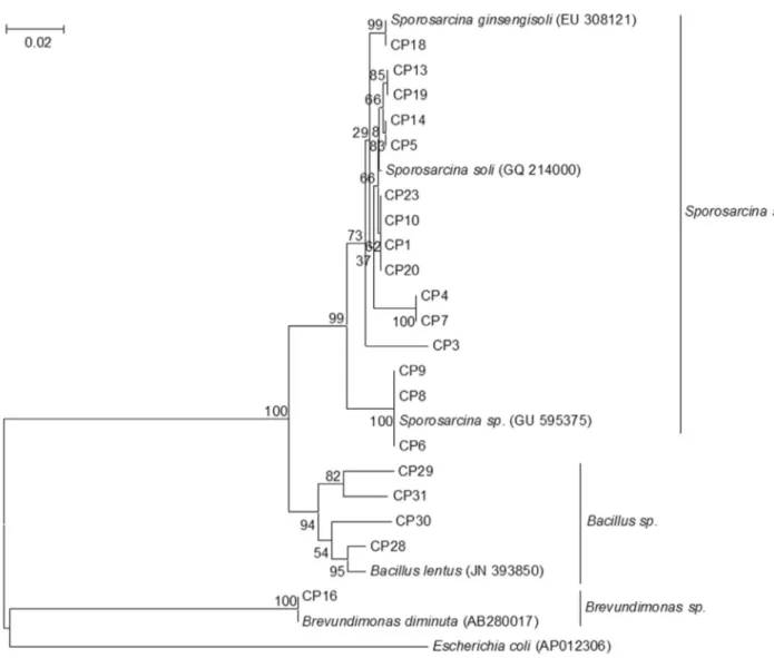
A total of twenty isolates were identified and charac-terized by sequencing of 16S rDNA. These sequences were BLAST searched against the GenBank database using the BLASTN program. Twenty isolates belonging to three gen-era were identified, and had the closest relatives belonging toSporosarcina sp.,Bacillus sp. andBrevundimonas sp. (Figure 1). Sequences related toSporosarcina sp. (occu-pied 75% of total sequences based on 98% of sequence sim-ilarity) dominated the cultured isolates, which included 15 isolates (CP1, CP3 to CP10, CP13, CP14, CP18 to CP20, CP23), followed byBacillus sp.(occupied 20% of total se-quences based on 98% of sequence similarity), which in-cluded 4 isolates (CP28 to CP31) andBrevundimonas sp. (occupied 5% of total sequences based on 99% of sequence similarity) which was comprised of a single isolate (CP16).
Characterization of three phylogenetic distinct strains
their growth rate, capability of inducing calcium carbonate precipitation, urease activity, and calcium carbonate solu-bilization ability.B. lentusCP28 grew faster and faciliated more calcium carbonate precipitation than the strains ofB. diminutaCP16 andS. soliCP23 (Figure 2). After 60 h of in-cubation, the cell number of CP16, CP23 and CP 28 were 2.78±0.38 x 10⁶, 2.38±0.28 x 10⁶and 2.87±0.42 x 10⁶ cells/mL, respectively. The masses of the precipitates of the three strains were 842±80, 456±70 and 931±98 mg/L, re-spectively. CP28 was the most efficient strain at inducing calcium carbonate precipitation when calculations were based on the mass of precipitation per cell, with CP28 capa-ble of inducing calcium carbonate precipitation at 3.24± 0.25 x 10⁻⁴mg/cell.
Microbial-induced calcium carbonate precipitation by urea hydrolysis was investigated extensively. The bacte-rium converts urea into ammonia by producing the enzyme
urease, thus increasing the environmental pH and subse-quently inducing calcium carbonate precipitation. All 20 of the isolated strains possessed the urease activity when tes-ted in the urea agar media. Among of testes-ted strains, CP23 and CP28 generated higher urease activity than CP16, whereas theE. colistrain did not show any purple color sur-rounding the inoculated site, which indicates a lack of urease activity. This urease activity assay result, together with the result in Figure 2, implied that the mass of calcium carbonate precipitation was directly linked to the urease ac-tivity, with higher urease activity causing more calcium carbonate precipitation. Therefore, strains CP16, CP23 and CP28 were chosen for further analyses.
of CP23. However, no calcium carbonate formed when plates were inoculated withE. coli control. To determine whether the isolates play roles in the deterioration of
lime-stone by solubilizing calcium carbonate, carbonate-solu-bilization capability was tested on the calcium carbonate solubilization media. Both CP16 and CP23 dissolved cal-cium carbonate and formed a clear, circular halo around the inoculation site. However, CP28 did not form similar halos. These data suggested that the mechanism of calcium car-bonate precipitation induced by bacteria may change with variations in environmental conditions.
SEM and XRD analyses of microbically-induced calcium carbonate precipitation
Precipitations collected on the 7th day were analyzed by XRD. The results showed that all three strains, B. diminutaCP16,S. soliCP23,B. lentusCP28, induced the formation of calcite (Figure 3), which was the only crystal assayed in the XRD spectra.
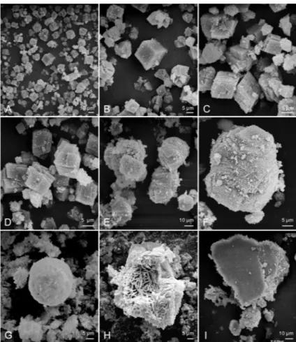
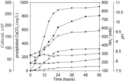
Morphologies of crystals induced by CP16, CP23 and CP28 were observed under SEM microscopy, and the re-sults showed the morphologies of crystals induced by CP16, CP23 and CP28 were similar. Basically, five differ-ent morphologies of crystals, the cubic crystal, the rhombic crystal, the polygonal plate-like crystal, the spherical crys-tal, and the irregular shaped cryscrys-tal, were observed (Figu-re 4). The cubic, the rhombic and the polygonal plate-like crystal were the three main shapes of crystals induced by the isolates, while the spherical and the irregular shaped crystals were less common (Figure 4A). Different morpho-logies of crystal showed the different properties of aggre-gated minerals described as follows. The surfaces of cubic shaped crystals were smooth (Figure 4B). The rhombic-shaped (Figure 4C and 4D) and the polygonal plate-like (Figure 4E and 4F) crystals generally presented well-defined faces and edges with accumulation of plate-like structures. The spherical crystals were formed by accumu-lation of granular composition with a rough surface (Figu-Figure 2- Comparison of the growth rate and capability of inducing
cal-cium carbonate precipitation among the strains ofB. diminutaCP16,S. soliCP23 andB. lentusCP28.
Table 1- Characterization of the isolates on precipitation and dissolution of CaCO3.
Strains CaCO3precipitation on CCP media
CaCO3dissolution on CCS media
E. coliCK -ª -ª
B. diminutaCP16 + -ª
S. soliCP23 +++ +
B. lentusCP28 ++++ -ª
Notes: + and -ª indicate the relative degree of precipitation or dissolution of CaCO3amongE. coliand the tested ureolytic bacteria.
Figure 3- XRD spectra of the calcium carbonate crystals induced by bacteria. C, calcite. From top to bottom:B. diminutaCP16,S. soliCP23 andB. lentus
re 4G). The irregular porous shaped crystals appeared to be amorphous with many tiny holes inside (Figure 4H). An-other kind of irregular crystals with smoothed surfaces were also occasionally observed (Figure 4I).
Chemical process of calcium carbonate precipitation induced byB. lentusCP28
Based on our observation and analyses, all the strains induced calcite precipitation in the liquid media. To deter-mine the correlation of calcium carbonate formation with the metabolic parameter changes on the growth phase ofB. lentusCP28, several parameters, including pH, cell num-ber, ammonium ion concentration and mass of calcium car-bonate, were monitored. The amount of calcium carbonate precipitation appeared to maintain a positive correlation with the growth of B. lentus CP28 (Figure 5). The pH quickly increased from the initial pH of 8.3 to 9.4 in the first 12 h of inoculation. While in the stage of log phase growth, B. lentusCP28 maintained robust growth, and the concen-tration of ammonium ions, which is believed to have con-tributed to the rise of pH, increased to 608 mM. Relatively more calcium carbonate precipitation was precipitated dur-ing this period. When the growth ofB. lentusCP28 was in the stationary phase after 12 h of incubation, the pH
gradu-ally increased to 9.6 from 9.4 and the ammonium ion concentration slightly increased to 746 mM. During this pe-riod, the rate of calcium carbonate precipitation was lower than that of the first phase. The calcium carbonate precipi-tation tended to reach plateaus, with a production of 96 mg/L in this phase. However, in the control experiment without bacteria, the pH of the media increased only slightly from 8.3 to 8.4, and the concentration of ammo-nium ions remained relatively stable. With such slight alka-linity of the media, only trace calcium carbonate precipita-tion was collected in the control experiments (Figure 5).
Discussion
Previous studies demonstrated that a great diversity of microbial genera took part in calcium carbonate precipi-tation in various natural environments (Wright and Oren, 2005), such as soils, freshwater, oceans and saline lakes (Douglaset al., 1998; Rivadeneyraet al., 1998; Peckmanet al., 1999; Zamarreñoet al., 2009). Our research showed that a variety of bacteria inhabiting the marine sediments could also induce calcium carbonate precipitation. Three genera of bacteria, Sporosarcina, Bacillus and Brevundimonas, were identified as precipitating calcium carbonate.S. soli, rather thanB. lentusandB. diminuta, was
Figure 4- SEM micrographs revealing the different morphologies of calcite crystals induced byB. lentusCP28. A) morphologies of crystals; B) the cubic
found to be the dominant cultured species. In previous stud-ies, Sporosarcina pasteurii (formerly known as Bacillus pasteuriifrom older taxonomies) andBacillus subtiliswere frequently reported to be isolated from various environ-ments and studied for calcium carbonate biomineralization or for being a limestone consolidant (Stocks-Fischeret al., 1999; Fujitaet al., 2000; Bachmeieret al., 2002; Achalet al., 2009; Zamarreñoet al., 2009; Okwadha and Li, 2010). B. diminuta was found to be the most effective carbo-natogenic bacterium isolated from decayed building stones (Jroundi et al., 2010; Rodriguez-Navarro et al., 2012), whileB. lentusfrom soil, marine waters and sediments was often used as a producer of alkaline protease in industry (Goddetteet al., 1992; Betzelet al., 1988; Jørgensenet al., 2000). Biomineralization of calcium carbonate facilited by B. lentuswas also reported. Our data showed that the bacte-rium could strongly induce calcium carbonate precipitation in comparison to the other strains in our experiments. With considerable researches, broad ranges of bacteria were found to be involved in the process of calcium carbonate biomineralization. It is thought that calcium carbonate bio-mineralization is not necessarily linked to any particular group of organisms but rather a general phenomenon in the bacterial world (Boquetet al., 1973; Ehrlich, 1998).
Considerable research on carbonate precipitation by bacteria has been performed using ureolytic bacteria (Ham-meset al., 2003; Ai-Thawadi, 2011), which by means of urea hydrolysis produce ammonia and carbonate ion, lead-ing to an increase in pH, and thus favorlead-ing calcium carbon-ate precipitation. Our data showed that urease activity was present in all the isolates when tested on the urease activity assay media. The strain ofB. lentusCP28 exhibited higher urease activity and more rapid growth and crystallization of
calcium carbonate aggregation than strains B. diminuta CP16 andS. soliCP23. This observation coincides with the observations by Hammeset al.(2003), who reported a di-versity of urease genes in the genomes of ureolytic bacteria and proposed that their high affinities and specific rates were the basase of rapid crystal formation. Urea is an or-ganic nitrogenous compound present in coastal environ-ments and introduced by the excretion of certain terrestrial and aquatic animals. Biotic urease activity is widespread in the environment and includes the actions of bacteria, yeasts and filamentous fungi (Mobley and Hausinger, 1989). Urease hydrolyses the substrate urea, which creates an al-kaline environment to facilitate calcium carbonate precipi-tation in the natural settings, and thus partially contributes to the marine lithifications. With removal of urea from the experimental media, strainsB. diminutaCP16 andS. soli CP23 were switched to metabolizing glucose and probably produced organic acids to dissolve calcium carbonate (Ta-ble 1). This phenomenon was observed in cave isolates, demonstrating their abilities to precipitate and dissolve cal-cium carbonate (Bankset al., 2010). Therefore, we inferred that the processes of both precipitation and dissolution of calcium carbonate are dynamic processes in the natural ma-rine sediment system, with both processes depending on the availability of urea and other organic substances.
(Rivadeneyraet al., 1996). Zamarreñoet al.(2009) demon-strated that Pseudomonas D2 and F2 have a remarkable ability to induce the precipitation of primarily calcite and vaterite, similar to the results obtained by Rodriguez-Navarroet al.(2003) and Muyncket al.(2008), who used Myxococcus xanthusandBacillus sphaericusin their work, respectively. Acinetobacter B14 induced more precipita-tion of vaterite than calcite (Zamarreño et al., 2009), Deleya halophliainduced precipitation of aragonite (Riva-deneyra et al., 1996), whereas Lysinibacillus sphaericus INQCS 414 precipitated only vaterite (Shirakawaet al., 2011). In contrast, our research isolates of B. diminuta CP16,S. soliCP23 andB. lentusCP28 predominantly in-duced calcite precipitation. Nevertheless, this finding is consistent with the results from Liet al.(2011) and Achalet al.(2009), who reported usingBacillus sp.andS. pasteurii MTCC 1761, respectively, to induce calcium carbonate precipitation. Despite extensive studies on bacterial carbo-natogenesis, little is known about what causes bacteria to precipitate different carbonate polymorphs. Besides the ob-servation that the particular bacterial species used has an important influence on the type of carbonate precipitation, the composition of the culture medium is also believed to be one of the determinants (Rivadeneyraet al., 1996, 1998; Gonzalez-Munozet al., 2010;). It has also been reported that the specific amino acid sequence in the urease enzyme of bacteria may be responsible for the carbonate polymorph selection. Higher concentration of Asp and Glu in the urease of B. pasteurii favored the formation of vaterite, while calcite was the predominant precipitate when using urease ofCanavalia ensiformiswith a lower concentration of Asp and Glu (Sondi and Salopek-Sondi, 2005). Kawa-guchi and Decho (2002) reported that specific proteins in extracellular polymeric substances (EPS) ofSchizothrix sp. influenced aragonite and calcite polymorph selection. These previous studies suggest that the polymorph selec-tion is a complex process involving a variety of abiotic and biotic factors.
Our results showed that B. diminuta CP16, S. soli CP23 andB. lentusCP28 induced similar morphologies of crystals. The cubic, rhombic and polygonal plate-like crys-tals were the dominant cryscrys-tals compared with the less common spherical and irregularly shaped crystals. This re-sult is consistent with that Tourney and Ngwenya (2009) observed from a strain ofBacillus licheniformisS-86. Ac-cording to Liet al.(2010), the crystal morphologies of pre-cipitates produced byBacillus sp. mainly showed cubic and polyhedral shapes. Zamarreñoet al.(2009) reported that microbial calcite crystals presented a variety of morpho-logies depending on the type of isolate. Pseudomonas putidaF2 induced nailhead and spheroidal crystals, Pseu-domonas aeruginosa D2 induced pseudo-ellipsoidal and pseudo-cubic crystals, whereasAcinetobacter juniiB14 in-duced semi-spheroidal, pseudo-hexagonal prism and nail-head spar crystals in the same growth medium.
The process of calcium carbonate precipitation is usu-ally governed by four key factors: (1) calcium concentra-tion, (2) concentration of dissolved inorganic carbon, (3) pH, (4) the availability of nucleation sites (Muyncket al., 2010). The first three factors can be influenced by bacteria, most notably, the creation of an alkaline environment. Moreover, the bacteria can also provide the crystal nucle-ation sites for calcium carbonate precipitnucle-ation (Hammes and Verstraete, 2002). In the process of calcium carbonate precipitation, bacterial precipitation caused faster precipi-tation rates than chemical precipiprecipi-tation (Stocks-Fischeret al., 1999). As shown in Figure 5, calcium carbonate precip-itation was clearly correlated with the growth ofB. lentus CP28, which utilized ureases to hydrolyze urea and gener-ate carbongener-ate and ammonia, and result in an increase in pH. With the rise of pH, more ammonium ion was released and a considerable quantity of calcium carbonate was precipi-tated. On the other hand, it is also possible that with the bac-terial cell number increase more crystal nucleation sites were available, favoring the calcium carbonate precipita-tion (Stocks-Fisheret al., 1999). In the bacterium-free con-trol, both pH and ammonium ion concentration kept increasing slightly while little calcium carbonate precipita-tion was collected. The kinetics of microbial calcium car-bonate precipitation are similar to those of reported by Stocks-Fisheret al.(1999). The data obtained from the bac-terial process of calcium carbonate precipitation induced by B. lentusCP28 provides straightforward evidence to under-stand microbial calcium carbonate precipitation.
In summary, in this paper we have clearly shown that three species of bacteria isolated from marine sediment par-ticipate in microbial calcium carbonate precipitation through hydrolysis of urea. Mineralogical analysis of the induced calcium carbonate precipitation shows that calcite is the dominant carbonate polymorph, and morphologies of crystals are mainly cubic and rhombic. These results sug-gest that production of carbonate polymorph is not specifi-cally related to any bacterial species, but is rather influ-enced by complicated environmental factors such as the pH, the composition of the media, etc.
Acknowledgments
The authors acknowledge James Hurley of Depart-ment of Plant Pathology and Microbiology, Texas A & M University, for making a critical reading and revision of this paper. This research were supported by the Fundamental Research Funds for the Central Universities (2652012138) and National Natural Science Foundation of China (41030853).
References
Ai-Thawadi SM (2011) Ureolytic bacteria and calcium carbonate formation as a mechanism of strength enhancement of sand. J Adv Sci Eng Res 1:98-114.
Arp G, Thiel V, Reimer Aet al.(1999) Biofilm exopolymers con-trol microbialite formation at thermal spring discharging into the alkaline Pyramid Lake, Nevada, USA. Sediment Geol 126:159-176.
Bachmeier KL, Williams AE, Warmington JRet al.(2002) Urea-se activity in microbiologically-induced calcite precipita-tion. J Biochechnol 93:171-181.
Bang SS, Galimat JK, Ramakrishan V (2001) Calcite precipita-tion induced by polyurethane-immobilized Bacillus pasteurii. Enzyme Microb Technol 28:404-409.
Banks ED, Taylor NM, Gulley Jet al.(2010) Bacterial calcium carbonate precipitation in cave environments: A function of calcium homeostasis. Geomicrobiology J 27:444-454. Bazylinski DA, Moskowitz BM (1997) Microbial
biominera-lization of magnetic iron minerals: microbiology, magne-tism and environment significance. Rev Mineral Geochem 35:181-223.
Betzel C, Dauter Z, Dauter Met al.(1988) Crystallization and pre-liminary X-ray diffraction studies of an alkaline protease fromBacillus lentus. J Mol Biol 204:803-804.
Boquet E, Boronat A, Ramos-Cormenzana A (1973) Production of calcite (calcium carbonate) crystals by soil bacteria is a general phenomenon. Nature 246:527-529.
Bosak T, Newman DK (2005) Microbial kinetic controls on cal-cite morphology in supersaturated solutions. J Sediment Res 75:190-199.
Braissant O, Decho AW, Dupraz Cet al.(2007) Exopolymeric substance of sulfate-reducing bacteria: interactions with cal-cium at alkaline pH and implication for formation of carbon-ate minerals. Geobiology 5:401-411.
Brennan ST, Lowenstein TK, Horita J (2004) Seawater chemistry and the advent of biocalcification. Geology 32:473-476. Cañaveras JC, Sacchez-Moral S, Soler Vet al.(2001)
Microor-ganisms and microbially induced fabrics cave walls. Geo-microbiology J 18:223-240.
Castanier S, Le M|tayer-Levrel G, Perthuisot JP (1999) Carbonate precipitation and limestone genesis-the microbiologist point of view. Sedimentary Geol 126:9-23.
Castanier S, Le M|tayer-Levrel G, Perthuisot JP (2000) Bacterial roles in the precipitation of carbonate minerals. In: Micro-bial sediments. Springer, Heidelberg, Germany. pp 32-39. Chafetz HS, Rush P, Utech NM (1991) Microenvironmental
con-trols on mineralogy and habitat of CaCO3precipitates: an
example from an active travertine system. Sedimentology 38:107-126.
Chahal N, Rajor A, Siddique R (2011) Calcium carbonate precipi-tation by different bacterial strains. Afr J Biotechnol 10:8359-8372.
Chekroun KB, Rodríguez-Navarro C, Gonzalez-Muñoz MTet al. (2004) Precipitation and growth morphology of calcium car-bonate induced byMyxococcus xanthus: implication for rec-ognition of bacterial carbonates. J Sediment Res 74:868-876.
Douglas S, Beveridge TJ (1998) Mineral formation by bacteria in natural microbial communities. FEMS Microbiol Ecol 26:79-88.
Dupraz C, Visscher PT (2005) Microbial lithification in marine stromatolites and hypersaline mats. Trends Microbiol 13:429-438.
Fujita Y, Ferris EG, Lawson RDet al.(2000) Calcium carbonate precipitation by ureolytic subsurface bacteria. Geomicrobiol J 17:305-318.
Giralt S, Julia R, Klerkx JJ (2001) Microbial biscuits of vaterite in Lake Issyk-Kul (Republic of Kyrgyzstan). J Sediment Res 71:430-435.
Goddette DW, Paech C, Yang SSet al.(1992) The crystal struc-ture of theBacillus lentusalkaline protease, subtilisin BL, at 1.4 Å resolution. J Mol Biol 228:580-595.
Gonzalez-Muñoz MT, Rodriguez-Navarro C, Martinez-Ruiz Fet al. (2010) Bacterial biomineralization: new insights from Myxococcus-induced mineral precipitation. Geological So-ciety, London Special Publications 336:31-50.
Groth I, Schumann P, Laiz Iet al.(2001) Geomicrobiological study of the Grotta dei Cervi, Porto Badisco, Italy. Geomi-crobiol J 18:241-258.
Hammes F, Verstraete W (2002) Key roles of pH and calcium me-tabolism in microbial carbonate precipitation. Rev Environ Sci Biotechnol 1:3-7.
Hammes F, Boon N, Villiers JDet al. (2003) Strain-specific ureolytic microbial calcium carbonate precipitation. Appl Environ Microbiol 69:4901-4909.
Jansson C, Northen T (2010) Calcifying cyanobacteria-the poten-tial of biomineralization for carbon capture and storage. Curr Opin Biotechnol 21:1-7.
Jha BK, Pragash MG, Cletus Jet al.(2009) Simultaneous phos-phate solubilization potential and antifungal activity of new fluorescent pseudomonad strains, Pseudomonas aeruginosa, P. plecoglossicidaand P. mosselii. World J Microbiol Biotechnol 25:573-581.
Jroundi F, Fern¢ndez-Vivas A, Rodriguez-Navarro Cet al.(2010) Bioconservation of deteriorated monumental calcarenite stone and identification of bacteria with carbonatogenic ac-tivity. Environ Microbiol 60:39-54.
Jørgensen PL, Tangney M, Pedersen PEet al.(2000) Cloning and sequencing of an alkaline protease gene fromBacillus lentus and amplication of the gene on theB. lentuschromosome by improved technique. Appl Environ Microbiol 66:825-827. Kawaguchi T, Decho AW (2002) A laboratory investigation of
cyanobacterial extracellular polymeric secretions (EPS) in influencing CaCO3 polymorphism. J Cryst Growth
240:230-235.
Li W, Liu L, Chen Wet al.(2010) Calcium carbonate precipita-tion and crystal morphology induced by microbial carbonic anhydrase and other biological factors. Process Biochem 45:1017-1021.
Li W, Liu LP, Zhou PPet al.(2011) Calcite precipitation induced by bacteria and bacterially produced carbonic anhydrase. Curr Sci 100:502-508.
Mann S (1995) Biomineralization and biomimetic materials chemistry. J Mater Chem 5:935-946.
Mobley HL, Hausinger RP (1989) Microbial ureases: signifi-cance, regulation, and molecular characterization. Micro-biol Mol Biol Rev 53:85-108.
Muynck WD, Belie ND, Verstraete W (2010) Microbial carbon-ate precipitation in construction mcarbon-aterials: A review. Ecol Eng 36:118-136.
Natarajan KR (1995) Kinetic study of the enzyme urease from Dolichos biflorus. J Chem Educ 72:556-557.
Okwahha GDO, Li J (2010) Optimum conditions for microbial carbonate precipitation. Chemosphere 81:1143-1148. Paerl HW, Steppe TF, Reid RP (2001) Bacterially mediated
pre-cipitation in marine stromatolites. Environ Microbiol 3:123-130.
Peckman J, Paul J, Thiel V (1999) Bacterially mediated formation of diagenetic aragonite and native sulphur in Zechstein car-bonate (Upper Permian, central Germany). Sediment Geol 126:205-222.
Pedone VA, Folk RL (2010) Formation of aragonite cement by nannobacteria in the Great Salt Lake, Utah. Geology 24:763-765.
Rivadeneyra MA, Ramoss-Cormenzana A, Delgado G et al. (1996) Process of carbonate precipitation by Deleya halophila. Curr Microbiol 36:308-313.
Rivadeneyra MA, Delgado G, Ramos-Cormenzana Aet al.(1998) Biomineralization of carbonates byHalomonas eurihalina in solid and liquid media with different salinities: crystal for-mation sequence. Res Microbiol 149:277-287.
Rodriguez-Navarro C, Jroundi F, Schiro Met al.(2012) Influence of substrate mineralogy on bacterial mineralization of cal-cium carbonate: implication for stone conservation. Appl Environ Microbiol 78:4017-4029.
Rodriguez-Navarro C, Rodriguez-Gallego M, Chekroun KBet al. (2003) Conservation of ornamental stone by Myxococcus
xanthus-induced carbonate biomineralization. Appl Environ Microbiol 69:2182-2193.
S¢nchez-Rom¢n M, Romanek CS, Fern¢adez-Remolar DCet al. (2011) Aerobic biomineralization of Mg-rich carbonates: Implications for natural environments. Chem Geol 281:143-150.
Shirakawa MA, Cincotto MA, Atencio Det al.(2011) Effect of culture medium on biocalcification byPseudomonas putida, Lysibacillus sphaericusandBacillus subtilis. Braz J Micro-biol 42:499-507.
Sondi I, Salopek-Sondi B (2005) Influence of the primary struc-ture of enzymes on the formation of CaCO3polymorphs: a
comparison of plant (Canavalia ensiformis) and Bacterial (Bacillus pasteurii) urease. Langmuir 21:8876-8882. Stocks-Fischer S, Galinat JK, Bang SS (1999) Microbiological
precipitation of CaCO3. Soil Biol Biochem 31:1563-1571.
Tourney T, Ngwenya BT (2009) Bacteria extracellular polymeric substance (EPS) mediate CaCO3morphology and
polymor-phism. Chem Geol 262:138-146.
Warthmann R, Van Lith Y, Vasconcelos Cet al.(2000) Bacte-rially induced dolomite precipitation in anoxic culture ex-periments. Geology 28:1091-1094.
Wright DT, Oren A (2005) Nonphotosynthetic bacteria and the formation of carbonates and evaporates through time. Geomicrobiol J 22:27-53.
Zamarreño DV, Inkpen R, May E (2009) Carbonate crystals pre-cipitated by freshwater bacteria and their use as a limestone consolidant. App Environ Microbiol 75:5981-5990.
Associate Editor: Valeria Maia de Oliveira