Cells from Patients with a History of Urothelial Cell
Carcinoma
Joa˜o Paulo de Castro Marcondes1*, Maria Luiza Cotrim Sartor de Oliveira1, Alisson M. Gontijo2, Joa˜o
Lauro Viana de Camargo1, Daisy Maria Fa´vero Salvadori1
1UNESP – Univ. Estadual Paulista, Faculdade de Medicina Botucatu SP, Brazil,2Centro de Estudos de Doenc¸as Croˆnicas, Faculdade de Cieˆncias Me´dicas (FCM), Universidade Nova de Lisboa, Lisboa, Portugal
Abstract
Bladder cancer is one of the most common genitourinary neoplasms in industrialized countries. Multifocality and high recurrence rates are prominent clinical features of this disease and contribute to its high morbidity. Therefore, more sensitive and less invasive techniques could help identify individuals with asymptomatic disease. In this context, we used the micronucleus assay to evaluate whether cytogenetic alterations could be used as biomarkers for monitoring patients with a history of urothelial cell carcinoma (UCC). We determined the frequency of micronucleated urothelial cells (MNC) in exfoliated bladder cells from 105 patients with (n = 52) or without (n = 53) a history of UCC, all of whom tested negative for neoplasia by cytopathological and histopathological analyses. MNC frequencies were increased in patients with a history of UCC (non-smoker and smoker/ex-smoker patientsvsnon-smoker and smoker/ex-smoker controls;p,0.001), in non-smoker UCC patients (vs non-smoker controls; p,0.01), and in smoker/ex-smoker controls (vs non-smoker controls; p,0.001). Patients with a history of recurrent disease also demonstrated a higher MNC frequency compared to patients with non-recurrent neoplasia. However, logistic regression using smoking habits, age and gender as confounding factors did not confirm MNC frequency as a marker for UCC recurrence. Fluorescentin situhybridization analysis (using a pan-centromeric probe) showed that micronuclei (MN) arose mainly from clastogenic events regardless of UCC and/or smoking histories. In conclusion, our results confirm previous indications that subjects with a history of UCC harbor genetically unstable cells in the bladder urothelium. Furthermore, these results support using the micronucleus assay as an important tool for monitoring patients with a history of UCC and tumor recurrence.
Citation:Marcondes JPdC, de Oliveira MLCS, Gontijo AM, de Camargo JLV, Salvadori DMF (2014) Genetic Instability Persists in Non-Neoplastic Urothelial Cells from Patients with a History of Urothelial Cell Carcinoma. PLoS ONE 9(1): e86162. doi:10.1371/journal.pone.0086162
Editor:Harriet Wikman, University Medical Center Hamburg-Eppendorf, Germany ReceivedJuly 24, 2013;AcceptedDecember 6, 2013;PublishedJanuary 22, 2014
Copyright:ß2014 Marcondes et al. This is an open-access article distributed under the terms of the Creative Commons Attribution License, which permits unrestricted use, distribution, and reproduction in any medium, provided the original author and source are credited.
Funding:Fundac¸a˜o de Amparo a` Pesquisa do Estado de Sa˜o Paulo (FAPESP,#05/55594-6, to M. L. C. S. O.), Conselho Nacional de Desenvolvimento Cientı´fico e Tecnolo´gico (CNPq,#301079/2009-9, to D. M. F. S.) and Coordenac¸a˜o de Aperfeic¸oamento de Pessoal de Nı´vel superior (CAPES, fellowship to J. P. C. M.). The funders had no role in study design, data collection and analysis, decision to publish, or preparation of the manuscript.
Competing Interests:The authors have declared that no competing interests exist. * E-mail: jpcastromarcondes@uol.com.br
Introduction
Bladder cancer is one of the most common genitourinary neoplasms in industrialized countries, and cigarette smoking is the main risk factor for this disease, as smokers have an approximate 3-fold increased risk of disease [1]. Urothelial cell carcinomas (UCC) account for 90% of all urinary bladder tumors, and multifocality and high recurrence rates are important clinical features of this disease [2,3]. Another critical feature of UCC is the high morbidity associated with the periodic surgical procedures required to investigate tumor recurrence and eventual resection [4]. Tumor grade, stage, size, and multifocality may predict progression and recurrence, although in some patients, these variables fail to accurately predict progression and recurrence because tumors of similar grade and stage can still differ significantly in their biology [5]. The development of cytogenetic markers has increased the sensitivities of UCC diagnosis and prognosis, but the precise characterization of atypical urinary cells remains a challenge for pathologists, especially in cases of low-grade tumors [6].
washings of patients with a history of UCC and/or smoking, but this increase was not statistically significant when compared to urothelial cells from a control population, which was likely due to the relatively small sample size [13]. This finding, together with recent data from Aroraet al.[10], prompted us to investigate the utility of a MN assay to detect genetic instability in urothelial cells with normal cytology in a new and larger cohort of patients with recurrent UCC. Finally, to improve our understanding of the genetic mechanisms involved in UCC, we also performed fluorescent in situ hybridization (FISH) in urothelial cells using a human pan-centromeric probe to gain insight into the mechanism by which MN originate in these cells.
Materials and Methods
1. Subjects
This study was approved by the Ethics Committee for Human Research of Botucatu Medical School, UNESP (Document No 276/2005-CEP), and signed informed consent was obtained from all subjects recruited for the present study.
The screening for micronucleated urothelial cells (hereafter referred to as MNCs) was conducted on 52 patients (43 males and 9 females) with a history of UCC and a negative cytopathological diagnosis for neoplasia. The reference group (controls) consisted of 53 patients (32 males and 21 females) with no history of UCC who were scheduled for cystoscopy to investigate other urinary tract complaints (e.g., hematuria, dysuria, pollakiuria, nocturia, vesical and kidney stones, cystitis, neurogenic bladder). UCC history was confirmed from previous positive biopsies, cytological analyses, and medical records. Tumors were classified as low or high grade according to the WHO classification [14]. The UCC patient and control groups were aged 38–89 years and 35–81 years, respectively. The studied population included 3 self-identified ethnic groups, as defined by The Brazilian Institute of Geography and Statistics [15]: white (96.4%), yellow (Asians; 1.8%), and brown (mixed race; 1.8%). Both the patient and control groups were randomly recruited at the Clinical Hospital in Botucatu, Sa˜o Paulo State (22u53909?S, 48u26942?W). The city of Botucatu is a Schistosoma haematobiumnon-endemic region. Personal data for all individuals were recorded in a detailed questionnaire that was administered immediately before or after cystoscopy. The collected data included ethnicity, age, gender, smoking and alcohol consump-tion habits, medical data, and histories of relevant exposures to
organic solvents, dyes, diesel exhaust, pesticides, and X-ray radiation. Subjects were considered smokers if they smoked at least 100 cigarettes in their life and currently smoke every day or some days; ex-smokers were subjects who had stopped smoking at least 1 year prior to sample collection [16]. Alcohol consumption was classified in 3 subjective categories [13]: non-drinker, defined as no alcohol consumption or social drinking; mild drinker, defined as the consumption up to 1 cup (,3.4 fl.
oz) of an alcoholic beverage per day or more than 1 cup on weekends; or heavy drinker, defined as the consumption of more than 1 L of a light alcoholic beverage (beer, wine, or cider) or 2 cups of hard liquor (Brazilian cachac¸a, vodka, or whiskey) per day for at least 6 years. Subjects were considered ‘‘exposed to toxic substances’’ (pesticides, solvents, and diesel exhaust) if they were occupationally exposed or had been exposed for at least 2 years [13].
2. Bladder washes
Bladder washes (bladder barbotage) were obtained by intraves-ical administration of a 0.9% saline solution. Two aliquots of 15 mL were collected per patient; one of the aliquots was subjected to Giemsa staining for MN examination, and the other was used for FISH. For Giemsa staining, bladder washing samples were centrifuged at 1,500 rpm for 10 min, and 200mL of the
obtained cell suspensions were cytocentrifuged at 1,400 rpm for 10 min. For FISH, 200mL of the cell suspensions were fixed 3 times in a methanol: acetic acid (3:1) solution. Slides were stored at room temperature for 2 weeks and then at220uC until staining and analysis.
3. Micronucleus test
The frequency of MNCs in 5% Giemsa-stained slides was determined by light microscopy at a magnification of 1,0006
following the criteria established by Lehucher-Michelet al.[12]. Those subjects with frequencies of Giemsa-stained MNCs above 1%were selected for FISH MN scoring. The probes used for all human centromeres were directly labeled with FITC (PAHC0001-G, Qbiogene, USA), and FISH slides were pro-cessed according to the probe manufacturer’s protocol with slight modifications. After the hybridization step, DAPI in antifade solution was used to counterstain DNA (Vector Laboratories, USA), which enabled the simultaneous observation of the total DNA and hybridization signals. MN were examined for fluorescence using a fluorescence microscope at 1,0006 magni-fication with the Case Data Manager software (Applied Spectral Imaging, CA, USA). Cells with non-FITC-labeled MN (centro-mere-negative MN; MNC2) were assumed to contain acentric chromosome fragments (Figure 1a), whereas FITC-labeled MN (centromere-positive MN; MNC+) contained whole chromo-somes (Figure 1b). The frequencies of MNC, MNC+, and MNC2 cells per 1,000 cells were calculated for each subject. Due to the large number of degenerate and occasionally scarce urothelial cells, the number of cells analyzed by Giemsa staining ranged from 500 to 1,000 (n = 17, 500–700 cells; n = 21, 701– 999; n = 67, 1,000 cells) per subject. For FISH analysis, the number of analyzed cells also ranged from 500 to 1,000 (n = 4, 500–700 cells; n = 2, 701–999; n = 31, 1000 cells).
4. Statistical analysis
The number of urothelial cells and MNC were adjusted according to a generalized linear model that considered the MN frequency (MNC, MNC+, and MNC2) as the dependent variable. A binomial distribution was used to adjust the model to consider UCC history, smoking history, and the interaction between UCC Figure 1. FISH-stained urothelial cells with probes for all
chromosome centromeres (green spots). a) Urothelial cells counterstained with DAPI, and the red arrow indicates a micronucleus (MN) with no centromeric signal (MNC2). b) Urothelial cells counter-stained with DAPI, and the yellow arrow indicates a MN with centromeric signal (MNC+). Photomicrographs were acquired at 1,0006magnification.
and smoking history. The same model was adjusted for gender, age, number of cigarettes per day, duration of smoking, alcohol consumption, and toxic substance exposure. Logistic regression analysis adjusted for demographic variables was used to provide an estimated risk for tumor recurrence in relation to MN frequency. The associations between tumor invasiveness, tumor grade, and tumor recurrence in patients with and without a history of smoking were analyzed using the chi square test. Comparisons of the simple frequencies of gender, tumor invasiveness, tumor grade, tumor recurrence, alcohol consumption, and toxic substance exposure were performed using the test of proportions. All analyses were performed using SAS for Windows (v. 9.2), and a pvalue,0.05 was considered significant.
Results
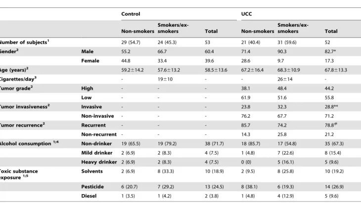
The demographic and medical data are presented in Table 1. The number of males (n = 43; 82.7%) with a history of UCC was significantly higher (p,0.05) than the corresponding number of females (n = 9; 17.3%). For male and female patients, the average age was 67.8613.3 years (35–89 years); 31 (59.6%) patients were smokers or ex-smokers, and 21 (40.4%) were non-smokers. No statistically significant differences were observed in the number of non-smoker and smoker/ex-smoker patients. Similarly, there was no statistically significant difference between the number of patients with a history of high-grade (n = 23, 44.2%) or low-grade tumors (n = 29, 55.8%), even when considering smoking habits Table 1.General characteristics of the study population.
Control UCC
Non-smokers
Smokers/ex-smokers Total Non-smokers
Smokers/ex-smokers Total
Number of subjects1 29 (54.7) 24 (45.3) 53 21 (40.4) 31 (59.6) 52
Gender2 Male 55.2 66.7 60.4 71.4 90.3 82.7*
Female 44.8 33.4 39.6 28.6 9.7 17.3
Age (years)2 59.2
614.2 57.6613.2 58.5613.6 67.2616.4 68.3610.9 67.8613.3
Cigarettes/day3 - 19
610 - - 26614
-Tumor grade2 High - - - 38.1 48.4 44.2
Low - - - 61.9 51.6 55.8
Tumor invasiveness2 Invasive - - - 23.8 32.3 28.8**
Non-invasive - - - 76.2 67.7 71.2
Tumor recurrence2 Recurrent - - - 85.7 74.2 78.8#
Non-recurrent - - - 14.3 25.8 21.2
Alcohol consumption1;4 Non-drinker 19 (65.5) 19 (79.2) 38 (71.7) 18 (85.7) 17 (54.8) 35 (67.3) Mild drinker 2 (6.9) 2 (8.3) 4 (7.5) 1 (4.8) 7 (22.6) 8 (15.4)
Heavy drinker 2 (6.9) 2 (8.3) 4 (7.5) 0 (0) 5 (16.1) 5 (9.6)
Toxic substance exposure1;5
Solvents 2 (6.9) 8 (33.3) 10 (18.9) 2 (9.5) 8 (25.8) 10 (19.2)
Pesticide 6 (20.7) 7 (29.2) 13 (24.5) 8 (38.1) 6 (19.3) 14 (26.9)
Diesel 1 (3.5) 1 (4.2) 2 (3.8) 1 (4.8) 4 (12.9) 5 (9.6)
UCC, patients with history of urothelial cell carcinoma;1No. (%);2data are presented as percentages (%);3mean
6standard deviation; *p,0.05, compared to female patients; **p,0.01, compared to non-invasive tumors;#p
,0.001, compared to non-recurrent tumors;4data not available for 7 controls (6 non-smokers and 1 smoker/ ex-smoker) and 4 UCC patients (2 non-smokers and 2 smokers/ex-smokers);5data not available for 1 smoker/ex-smoker control and 6 UCC patients (2 non-smokers and 4 smokers/ex-smokers).
doi:10.1371/journal.pone.0086162.t001
Table 2.Urothelial MNC frequency in subjects with and without a history of bladder UCC.
Groups Number of subjects Number of cells analyzed Number of MNC %MNC1 pvalue
Control 53 54,804 107 1.9
-Non-smokers 29 30,639 40 1.3
-Smokers/ex-smokers 24 24,165 67 2.8a ,0.001a
UCC 52 44,949 135 3.0b ,0.001b
Non-smokers 21 19,052 70 3.7c,d
,0.05c, ,0.001d
Smokers/ex-smokers 31 25,897 65 2.5e,f ,0.01e,.0.05f
1MNC per 1,000 cells; a, smoker/ex-smoker controlsvs.non-smoker controls; b, UCC patients (non-smokers
+smokers/ex-smokers)vs.controls (non-smokers+smokers/ ex-smokers); c, non-smoker UCC patientsvs.smoker/ex-smoker UCC patients; d, non-smoker UCC patientsvs.non-smoker controls; e, smoker/ex-smoker UCC patients vs.non-smoker controls; f, smoker/ex-smoker UCC patientsvs.smoker/ex-smoker controls.
(p.0.05). Regardless of smoking history, the number of subjects diagnosed with invasive tumors (n = 15, 28.8%) was significantly lower compared to the number of subjects with non-invasive tumors (n = 37, 71.2%) (p,0.01). Additionally and regardless of smoking history, the number of patients with recurrent tumors was significantly higher (n = 41, 78.8%) than the number of patients with non-recurrent tumors (n = 11, 21.2%) (p,0.001). Regarding alcohol consumption and exposure to toxic substanc-es, no significant difference was detected between UCC patients and controls (p.0.05). In the control group, 32 (60.4%) patients were male, and 21 (39.6%) female; 29 (57.4%) patients were non-smokers, and 24 (45.3%) were smokers or ex-smokers. The average age was 58.5613.6 years (35–82 years).
The MNC frequency (3.0%) in patients with a history of UCC was significantly higher (p,0.001) than the frequency in the control group (1.9%) (Table 2). However, when considering smoking history, a statistically significant (p,0.001) difference was detected only between non-smoker UCC patients (3.7%) and
non-smoker controls (1.3%). In both the UCC patient and control groups, a statistically significant difference was observed between non-smokers and smokers/ex-smokers (Table 2). Indeed, it is important to notice that there was no statistically significant difference in the MNC frequency of smokers/ex-smokers from the UCC group, when compared with the control group (p.0.05). No interference was observed in MNC frequencies when alcohol consumption and toxic substance exposure were included in the binomial analysis.
FISH data were obtained from 37 subjects (21 UCC patients and 16 controls) with greater than 1% Giemsa-stained MNC. Although the binomial analyses demonstrated significant differ-ences, data were interpreted qualitatively because of the low number of MNC+ and MNC2 samples. Overall, 75% (range, 66.6–81.5%) of the scored MNC samples stained negative for centromeres (MNC2). In fact, the largest increase in MNC2
status compared to centromere-positive MNCs (MNC+) was observed for smoker/ex-smoker UCC patients (81.5% MNC2
vs.18.5% MNC+) (Table 3).
When demographic and clinical features were considered (Table 4), significant (p,0.05) interference was detected in MNC frequency for age and tumor recurrence status in the UCC patient group. Patients older than 60 years showed higher MNC frequencies compared to patients less than 60 years of age (3.5% vs. 2.1%, respectively; p,0.05); patients with recurrent tumors also had higher MNC frequencies compared to patients with non-recurrent tumors (3.4%vs.1.7%, respectively;p,0.05). Importantly, 70.3% of patients with recurrent tumors (n = 29) were older than 60 years of age. In addition, logistic regression analysis did not reveal a relationship between high MNC frequencies and increased tumor recurrence risk in smokers/ex-smokers UCC patients (p.0.05). Age, gender and smoking habit were considered as potential confounding factors in the regression model.
Discussion
In this study, we used the micronucleus test to evaluate chromosomal damage in patients with a history of UCC, in an attempt to introduce this test as an auxiliary biomarker for bladder cancer monitoring. We observed a significant increase in MNC Table 3.Frequencies of centromere-positive (MNC+) and
centromere-negative (MNC2) urothelial MNCs from subjects with and without a history of bladder UCC.
Groups
Number of
subjects Total number of
MNC MNC1 MNC+(%) MNC2(%)
Control 16 14,783 20 5 (25) 15 (75)
Non-smokers 8 7,229 4 1 (25) 3 (75)
Smokers/ex-smokers
8 7,554 16* 4 (25) 12* (75)
UCC 21 20,150 39 9 (23.1) 30** (76.9)
Non-smokers 8 8,000 12 4 (33.3) 8 (66.6)
Smokers/ex-smokers
13 12,150 27 5 (18.5) 22** (81.5)
1MNC, micronucleated urothelial cells; *p
,0.05, compared to non-smoker controls; **p,0.01, compared to % MNC+within the UCC group. doi:10.1371/journal.pone.0086162.t003
Table 4.Urothelial MNC frequency (Giemsa staining) in subjects with and without a history of UCC stratified according to demographic, histological and clinical variables.
Variables Number of subjects %MNC1
Control UCC Control UCC
Gender male 32 43 1.8 2.8
female 21 9 2.2 3.8
Age #60 years old (35–60 years) 29 16 1.9 2.1
.60 years old (61–89 years) 24 36** 1.9 3.5*
Tumor grade low grade - 29 - 2.9
high grade - 23 - 3.2
Tumor recurrence2 non-recurrent - 11 - 1.7
recurrent - 41 - 3.4
Tumor invasiveness non-invasive - 37 - 3.1
invasive - 15 - 2.7
1MNC – micronucleated cells per 1000 cells; *p,0.05, compared to UCC patients less than 60 years of age;2Logistic regression analysis (considering smoking habit, age and gender as confounding factors) did not show significant difference, p.0.05.
frequency in patients with a history of UCC, but with cytological and histopathological diagnoses negative for neoplasia. Our data both corroborate and bring statistical support to the trend of increased MN in UCC, which we previously detected in an independent study with a smaller group of patients with a history of UCC [13]. Furthermore, the present data are highly consistent with increased levels of primary DNA damage and aneuploidy, which we previously detected using the alkaline single-cell gel (comet) assay [13]. Similarly, a recent study reported p53 protein expression in more than 25% of exfoliated urothelial cells from histologically normal urothelium of patients with a history of UCC, which also indicates that this ‘‘normal’’ urothelium is genomically unstable [17]. Additionally, urothelial cells were also analyzed for chromosomal aberrations using high-resolution comparative genomic hybridization and, they did not find numerical alterations of the chromosomes 7, 17 and 9p21 regions, but did find high frequencies of gains in 11p12 and losses in 16p12 [17]. It is important to emphasize that an increased MNC frequency was not predictive of UCC recurrence when a logistic regression was carried out including age, gender and smoking habits as confounding factors. Therefore, taken together our data strongly support the idea that genetic instability can be detected prior to a diagnosis of cellular atypia and highlight MN examination as an important tool for monitoring patients with a history of UCC and recurrent tumors.
To gain insight into the mechanism of MN formation in cytologically normal urothelial cells from patients with a history of UCC, we used FISH with a pan-centromeric probe to evaluate aneugenic and clastogenic events. In the control and UCC patient groups, the frequencies of centromere negative MN (MNC2) were consistently higher than the frequencies of centromere positive MN (MNC+). This result suggested that MN mainly arose from chromosomal, and that this tendency was either stabilized or mildly exacerbated in patients with a history of UCC. In fact, Del Rey et al. [18] suggested that MN resulting from acentric fragments (chromosome breakage or cyclin D1 gene amplification) or from whole chromosomes might contribute to genomic instability in UCC tumor samples. Similarly, a high frequency of acentromeric MN was observed in the peripheral lymphocytes of patients with different types of cancer prior to treatment [11].
Our data from control subjects also confirm previous reports in which smoking history was associated with significant increases in
MN frequencies [12,13,19] and primary DNA lesions [20]. Interestingly, we did not observe the same effect in smoker/ex-smoker UCC patients. Thus, as previously proposed [21], we suggest that the pathological status may mask the effects of cigarette smoking on MNC frequency. Cigarette smoking has also been associated with other genetic alterations found in bladder cancer patients.TP53mutations, TP16promoter hypermethyla-tion, and the overexpression of various oncogenes and growth factors have been detected in tumor and normal tissues from patients with a history of smoking [22–25]. Furthermore, increases in p53 protein levels were observed in the normal urothelium of current and previous smokers [26]. Therefore, in urothelial cells, mutations or other alterations in the p53 pathway caused by cigarette smoking could be involved in the high MNC frequencies observed in our study as well as the increased risk of bladder cancer development and recurrence [12,13,19,20].
Few studies have addressed the aneugenic or clastogenic effects of cigarette smoking in target and surrogate cells. The available data have shown a trend towards no differences between these 2 mechanisms of MN formation [27]. Despite the relatively small sample size, especially after stratification into subgroups, our data demonstrate that subjects with a history of UCC harbor and accumulate genetically unstable cells in the bladder urothelium that may represent early precursors of new UCC or subclones from previous UCC. Furthermore, we showed that the majority of MN detected in urothelial cells arose from chromosomal breakage. Therefore, micronucleus test may provide an informative auxiliary biomarker to monitor bladder cancer.
Acknowledgments
The authors are grateful to Dr. Samuel M. Cohen (University of Nebraska Medical Center) for critically reviewing this manuscript and to Prof. Dr. Jose´ Eduardo Corrente (UNESP) for help with statistical analyses.
Author Contributions
Conceived and designed the experiments: JPCM DMFS AMG MLCSO JLVC. Performed the experiments: JPCM. Analyzed the data: JPCM DMFS AMG. Contributed reagents/materials/analysis tools: DMFS MLCSO JLVC. Wrote the paper: JPCM DMFS AMG.
References
1. Zeegers MP, Kellen E, Buntinx F, van den Brandt PA (2004) The association between smoking, beverage consumption, diet and bladder cancer: a systematic literature review. World J Urol 21: 392–401.
2. Denzinger S, Mohren K, Knuechel R, Wild PJ, Burger M, et al (2006) Improved clonality analysis of multifocal bladder tumors by combination of histopathologic organ mapping, loss of heterozygosity, fluorescence in situ hybridization, and p53 analyses. Human Pathol 37: 143–151.
3. Frau DV, Usai P, Dettori T, Caria P, De Lisa A, et al. (2006) Fluorescence in situ hybridization patterns in newly diagnosed superficial bladder lesions and corresponding bladder washings. Cancer Genet Cytogenet 169: 21–26. 4. Reznikoff CA, Sarkar S, Ju¨licher KP, Burger MS, Puthenveettil JA, et al. (2000)
Genetic alterations and biological pathways in human bladder cancer pathogenesis. Urol Oncol 5: 191–203.
5. Youssef RF, Lotan Y (2011) Predictors of outcome of non-muscle-invasive and muscle-invasive bladder cancer. Scientific World Journal 11: 369–381. 6. Pajor G, Somogyi L, Melegh B, Alpar D, Kajtar B, et al. (2011) Urovysion:
Considerations on modifying current evaluation scheme, including immuno-phenotypic targeting and locally set, statistically derived diagnostic criteria. Cytometry A 79: 375–382.
7. Jefford CE, Irminger-Finger I (2006) Mechanisms of chromosome instability in cancers. Cr Rev Oncol Hematol 59: 1–14.
8. Fenech M (2000) The in vitro micronucleus technique. Mutat Res 455: 81–95. 9. Bonassi S, El-Zein R, Bolognesi C, Fenech M (2011) Micronuclei frequency in peripheral blood lymphocytes and cancer risk: evidence from human studies. Mutagenesis 26: 93–100.
10. Arora SK, Dey P, Saikia UN (2010) Micronucleus in atypical urothelial cells. Diagn Cytopathol 38: 811–813.
11. Baciuchka-Palmaro M, Orsie`re T, Duffaud F, Sari-Minodier I, Pompili J, et al. (2002) Acentromeric micronuclei are increased in peripheral blood lymphocytes of untreated cancer patients. Mutat Res 520: 189–198.
12. Lehucher-Michel MP, Di Giorgio C, Amara YA, Laget M, Botta A (1995) The micronucleus assay in human exfoliated urothelial cells: effect of smoking. Mutagenesis 10: 329–332.
13. Gontijo AM, Marcondes JP, Elias FN, de Oliveira ML, de Lima RO, et al. (2002) DNA damage in cytologically normal urothelial cells of patients with a history of urothelial cell carcinoma. Environ Mol Mutagen 40: 190–199. 14. Eble JN, Sauter G, Epstein JI, Sesterhenn IA (2004) World Health Organization:
Tumors of the urinary system and male genital organs. Oxford, UK: IARC Press.
15. Instituto Brasileiro de Geografia e Estatı´stica (2008) Caracterı´sticas E´ tnico-raciais da Populac¸a˜o – um estudo das categorias de classificac¸a˜o de cor ou rac¸a. Brasil: IBGE. 95 p.
16. Centers for Disease Control and Prevention (2013)Tobacco Control State Highlights 2012.Atlanta: U.S. Department of Health and Human Services, Centers for Disease Control and Prevention, National Center for Chronic Disease Prevention and Health Promotion, Office on Smoking and Health.
18. Del Rey J, Prat E, Ponsa I, Lloreta J, Gelabert A, et al. (2010) Centrosome clustering and cyclin D1 gene amplification in double minutes are common events in chromosomal unstable bladder tumors. BMC Cancer 10: 280. 19. Burgaz S, I˙s¸can A, Bu¨yu¨kbingo¨l ZK, Bozkurt A, Karakaya AE (1995) Evaluation
of micronuclei in exfoliated urothelial cells and urinary thioether excretion of smokers. Mutat Res 335: 163–169.
20. Gontijo AM, Elias FN, Salvadori DM, Oliveira ML, Correa LA, et al. (2001) Single-cell gel (comet) assay detects primary DNA damage in nonneoplastic urothelial cells of smokers and ex-smokers. Cancer Epidemiol Biomark Prev 10: 987–993.
21. Duffaud F, Orsie`re T, Digue L, Villani P, Volot F, et al. (1999) Micronucleated lymphocyte rates from head-and-neck cancer patients. Mutat Res 439: 259–266. 22. LaRue H, Allard P, Simoneau M, Normand C, Pfister C, et al. (2000) P53 point mutations in initial superficial bladder cancer occur only in tumors from current or recent cigarette smokers. Carcinogenesis 21: 101–106.
23. Marsit CJ, Karagas MR, Danaee H, Liu M, Andrew A, et al. (2006) Carcinogen exposure and gene promoter hypermethylation in bladder cancer. Carcinogen-esis 27: 112–116.
24. Wallerand H, Bakkar AA, de Medina SG, Pairon JC, Yang YC, et al. (2005) Mutations in TP53, but not FGFR3, in urothelial cell carcinoma of the bladder are influenced by smoking: contribution of exogenous versus endogenous carcinogens. Carcinogenesis 26: 177–184.
25. Gabriel U, Li L, Bolenz C, Steidler A, Kra¨nzlin B, et al. (2012) New insights into the influence of cigarette smoking on urothelial carcinogenesis: smoking-induced gene expression in tumor-free urothelium might discriminate muscle-invasive from nonmuscle-invasive urothelial bladder cancer. Mol Carcinog 51: 907–915. 26. Mothersill C, O’Malley K, Colucci S, Murphy D, Lynch T, et al. (1997) p53 protein expression and increased SSCP mobility shifts in the p53 gene in normal urothelium cultured from smokers. Carcinogenesis 18: 1241–1245.