Predominant overexpression of CD25/FOXP3, IFN-
γ
,
and suppressive cytokines in high-grade lesion samples
infected with human papillomavirus
Predominância de superexpressão de CD25/FOXP3, IFN-
γ
e citocinas supressoras em
amostras de lesão de alto grau infectadas pelo papilomavírus humano
Camila M. Bonin; Cacilda T. J. Padovani; Alda Maria T. Ferreira; Leandro S. Ávila; Ana Paula Machado; Thiago T. M. Prata; Carlos Eurico S. Fernandes; Inês Aparecida Tozetti
Universidade Federal de Mato Grosso do Sul (UFMS), Mato Grosso do Sul, Brazil.
First submission on 20/06/16; last submission on 11/11/16; accepted for publication on 28/11/16; published on 20/02/17
ABSTRACT
Introduction: Human papillomavirus (HPV) persistent infection is the leading cause of cervical cancer and its precursor lesions, and the inappropriate immune response is among the factors that contribute to viral persistence. This may be inluenced by regulatory T (Treg) cells and the production of immunosuppressive cytokines, such as transforming growth factor beta (TGF-β) and interleukin-10 (IL-10).
Objective: We established the proile of the predominant response, Th1 or immunosuppressive response, in the tissue microenvironment, by detecting interferon-gamma (IFN-γ), TGF-β, and IL-10, as well as the co-expression of IL-2 receptor alpha (CD25) and forkhead box P3 (FOXP3). Methods: Seventy-four samples from uterine cervix biopsies that underwent HPV deoxyribonucleic acid (DNA) detection and histopathology analysis were immunostained to detect CD25/FOXP3, IFN-γ and suppressive cytokines in lymphocytes. Results: The microenvironment of high-grade squamous intraepithelial lesion (HSIL) samples with high numbers of viral particles (≥ 10,000 copies/ml) contained high numbers of CD25/FOXP3+, TGF-β+, IL-10+, and IFN-γ+ cells. Conclusion: The co-expression of CD25/FOXP3 and the expression of TGF-β, and IL-10 in HSIL samples suggest the existence of Treg cells in these locations, although IFN-γ expression was observed in several cells in these samples. Our data suggest that this cytokine could be related to immunosuppressed microenvironment maintenance, favoring the persistent HPV infection and the progression to carcinoma.
Key words: immunohistochemistry; immunomodulation; papillomavirus infections.
INTRODUCTION
Persistent infection with certain human papillomavirus (HPV)(1), high-risk oncogenic types (HR-HPV) 16 and 18, is the
leading cause of cervical cancer and its precursor lesions(2). The viral
persistence is inluenced by several factors(3), including the
deiciency of cell-mediated immune response and the cytokine proile of the Th1 helper T cell response (Th1 response), which is crucial to eliminate HPV(1, 4, 5).
Interferon-gamma (IFN-γ), the signature cytokine of the Th1 response proile(5), plays an essential role in defending against
viruses and intracellular pathogens, and inducing inlammatory responses(6). The production of high levels of IFN-γ is typically
associated with effective host defense against viral infection(7),
because the production of IFN-γ is indicative of local activation of cell-mediated immunity, commonly seen in viral infections or cancer(8). IFN-γ is a dominant cytokine that polarizes Th cells for
the Th1 phenotype and inhibits the development of Th2 cells(4).
This polarization plays a crucial role in the host defense against viral infection and tumor development(9).
The predominance of Th1 cells with the production of IFN-γ has been associated with elimination of HPV infection and regression of squamous intraepithelial lesions (SIL)(9). Scott et al.
(2009)(1) showed that high levels of IFN-γ messenger RNA (mRNA)
in cervical samples were signiicantly associated with the decreased likelihood of developing high-grade squamous intraepithelial lesions (HSIL)(1). Likewise, lower levels of IFN-γ mRNA were
found in HSIL than in low-grade squamous intraepithelial lesion (LSIL)(10). This decrease may be related to the presence of cells
with immunoregulatory properties, capable of interfering with antigen presentation(11) and with the activation and proliferation
of antigen-speciic T cells(12).
Among the various cell subsets with immunoregulatory properties, regulatory T (Treg) cells are strongly associated with neoplastic progression. They are considered as a predictive factor for poor prognosis when present in high density in tumor microenvironments(13). Such cells can be phenotypically characterized
by the expression of the alpha chain of the interleukin 2 receptor, IL-2Rα (CD25) and the transcription factor forkhead box protein P3 (FOXP3)(14), which are essential components in the generation,
maintenance, development, and function of Treg cells(15).
One way by which Treg cells perform their function is through the production of immunosuppressive cytokines such as interleukin-10 (IL-10) and transforming growth factor beta (TGF-β). They are then able to trigger the anergy of T cells and allow cancer progression through the local production of these cytokines(16, 17). Thus, the presence of Treg cells can also be
associated with the detection of IL-10 and TGF-β in the tissue microenvironment.
Taking into account that HPV infection is restricted to the epithelium(18), the understanding of the active immune
mechanisms in the infected tissue microenvironment is fundamental. Thus, in the present study, we aimed to detect the co-expression of the markers CD25/FOXP3 and the expression of IFN-γ, IL-10, and TGF-β in lymphocytes, by performing immunohistochemistry, to help elucidate the prevailing T helper response proile (Th1 or immunosuppressive response) in the HPV-infected microenvironment, and to provide a better understanding of the pathological processes associated with persistence or regression of the infection, or progression to the malignant form.
METHODS
Samples
This is a cross-sectional study in which seventy-four biopsy samples of uterine cervix were collected from June 2010 to June 2011 at the Cancer Prevention Center of Campo Grande (MS), Brazil. These samples were selected in a non-probability sampling by convenience. A irst fragment of these samples was previously subjected to HPV viral load quantiication (VL) and viral typing. Quantiication was done by real-time polymerase chain reaction (qPCR) with SYBR Green using LightCycler®. Copy number
quantiication was determined in deoxyribonucleic acid (DNA) extraction volume. This, in turn, was classiied as follows: VL0 (negative for HPV), VL1 (1,050 to < 10,000 viral copies/ml) and VL2 (≥ 10,000 viral copies/ml) (Table 1). For viral typing, the PCR end point was used followed by restriction fragment length polymorphism (RFLP) analysis(19). The primers used were those
described by Payan et al. (2007)(20).
A second fragment of the same samples was embedded in parafin and sent to histopathology and immunohistochemistry. Based on the histological analysis, the samples were classiied as LSIL (CIN I), HSIL (CIN II, III), carcinoma, and negative for intraepithelial lesion and malignancy (NILM) (Table 1).
Immunohistochemistry (IHC)
The IHC methods used to stain the samples for IL-10, TGF-β, and IFN-γ, and co-stain for CD25 and FOXP3 markers involved the use of antigen retrieval in damp heat and buffer (10 mM Tris and 1 mM EDTA, pH 9) for IL-10 and TGF-β cytokines as well as CD25
and FOXP3 markers. The IFN-γ antigen retrieval was performed
using citrate buffer (10 mM, pH 6). The primary antibodies used in the simple labeling reaction were anti-IL-10 (Invitrogen, clone
945A2A5/cod. AHC9102, Carlsbad, CA, USA), anti-TGF-β (Spring
Bioscience, ref: E11264, Pleasanton, CA, USA), and anti-IFN-γ (eBioscience, clone: MD- 1, San Diego, CA, USA). For the double staining, the antibodies used were human anti-IL-2R/CD25 (eBioscience, clone: B10-B) and anti-FOXP3 (eBioscience, clone: 236A/E7).
The Universal LSAB Kit/HRP (Dako, Carpinteria, CA, USA) was used as the detection system for simple marking, and diaminobenzidine (DAB) (Dako, Carpinteria, CA, USA) was used as the chromogen. The Vectastain ABC system was employed for marking multiple antigens, enzyme substrate NovaRED (Vector Laboratories, Burlingame, CA, USA), and plated DAB (Ni-DAB) (Vector Laboratories, Burlingame, CA, USA) were used for the detection of the double staining. Counterstaining was performed TABLE 1 − Distribution of histopathological indings according to the viral load
Histopathology
NILM LSIL HSIL CA Total
n % n % n % n % n %
VL
VL 0a 6 8.1 0 0 0 0 0 0 6 8.1
VL 1b 0 0 4 5.4 1 1.3 0 0 5 6.8
VL 2c 0 0 5 6.75 46 62.2 12 16.2 63 85.1
Total 6 8.1 9 12.2 47 63.5 12 16.2 74 100
NILM: negative for intraepithelial lesion and malignancy; LSIL: low-grade squamous intraepithelial lesion; HSIL: high-grade squamous intraepithelial lesion; CA: carcinoma; VL: viral load; a: negative for HPV; b: 1,050 to < 10,000 viral copies/ml; c: ≥ 10,000 viral
using hematoxylin. Human tonsil tissue stained using the primary antibody was used as a positive control. A negative control was obtained by replacing the primary antibody by phosphate buffer (pH 7.4) containing 1% albumin.
Quantitative analysis
The quantiication of the immunostained cells to establish cytokines expression (IL-10, TGF-β and IFN-γ) and Treg cells was performed by evaluating 10 random ields including only stromal area of cervical tissue. According to the presence of immunomarked cells, the histological sections were classiied in low (when there was up to three stained cells per ield) and high (when there were more than three stained cells per ield) scores(21).
The biopsy sections were analyzed by two independent observers, previously calibrated (κ = 0.98), and the inal result of discordant cases was obtained by common analysis to achieve a consensus.
Statistical analysis
Statistical analysis was performed using SPSS software version 17.0 and BioEstat version 5.0. The frequency analysis of histology and viral load in accordance with the intensity of expression of the markers was compared by Pearson’s chi-squared test for contingency tables. When signiicant, Fisher’s exact test was applied.
Ethical considerations
This study was approved by the Ethics Research Committee of Universidade Federal do Mato Grosso do Sul (UFMS), Brazil, protocol number 87527, August 30, 2012.
RESULTS
We observed that all samples with lesions were positive for HPV (Table 1). Among these, the most prevalent HPV types were HPV 16 (48.5%, 33/68), HPV 18 (41.2%, 28/68) and HPV 45 (14.7%, 10/68). In all HSIL and carcinoma samples mainly 16 and 18 HR-HPV were detected. HR-HPV16 was present in 44.7% (21/47) of the HSIL samples and 50% (6/12) of carcinoma samples, while HPV 18 was found in 48.6% (22/47) of the HSIL samples and 50% of the carcinoma samples too (Table 2).
The distribution of histopathological indings according to the expression of IFN-γ, TGF-β, IL-10, and co-expression of CD25/ FOXP3 is presented in Table 3.
TABLE 2 − Distribution of HPV genotypes according to the histopathological
indings in samples of cervical biopsies (n = 68)
Genotypes LSIL HSIL CA
n % n % n %
HPV 16 5 55.6 14 29.8 6 50
HPV 18 - - 18 38.3 6 50
HPV 33 - - 2 4.3 -
-HPV 45 3 33.3 3 6.4 -
-HPV 11 and 16 1 11.1 1 2.1 - -HPV 11 and 33 - - 1 2.1 - -HPV 11 and 45 - - 1 2.1 - -HPV 16 and 18 - - 3 6.4 - -HPV 16 and 45 - - 3 6.4 - -HPV 6 and 18 - - 1 2.1 -
-Total 9 100 47 100 12 100
HPV: human papillomavirus; LSIL: low-grade squamous intraepithelial lesion; HSIL: high-grade squamous intraepithelial lesion; CA: carcinoma.
TABLE 3 − Distribution of histopathological indings according to the
co-expression of CD25/FOXP3 and the expression of TGF-β, IL-10 and IFN-γ
Histopathology Total χ2 p
NILM
n (%) LSIL n (%)
HSIL n (%)
CA
n (%) n (%)
CD25/ FOXP3
Lowa 4 (6) 4 (6) 15 (22.4) 1 (1.5) 24 (35.8)
9.59 0.022
Highb 1 (1.5) 3 (4.5) 28 (41.8)* 11 (16.4) 43 (64.2)
Total 5 (7.5) 7 (10.4) 43 (64.2) 12 (17.9) 67 (100)
TGF-β
Lowa 3 (4.2) 5 (6.9) 5 (6.9) 0 (0) 13 (18.1)
16.722 0.001
Highb 3 (4.2) 4 (5.6) 41 (56.9)* 11 (15.3) 59 (81.9)
Total 6 (8.3) 9 (12.5) 46 (63.9) 11 (15.3) 72 (100)
IL-10
Lowa 0 (0) 4 (5.6) 7 (9.7) 0 (0) 11 (15.3)
8.981 0.03
Highb 5 (6.9) 5 (6.9) 39 (54.2)* 12 (16.7) 61 (84.7)
Total 5 (6.9) 9 (12.5) 46 (63.9) 12 (16.7) 72 (100)
IFN-γ
Lowa 5 (7.2) 3 (4.3) 14 (20.3) 3 (4.3) 25 (36.2)
6.333 0.096
Highb 1 (1.4) 6 (8.7) 30 (43.5) 7 (10.1) 44 (63.8)
Total 6 (8.7) 9 (13) 44 (63.8) 10 (14.5) 69 (100)
NILM: negative for intraepithelial lesion and malignancy; LSIL: low-grade squamous intraepithelial lesion; HSIL: high-grade squamous intraepithelial lesion; CA: carcinoma; CD25: interleucina-2 receptor alpha; FOXP3: forkhead box P3; TGF-β: transforming growth factor beta; IL-10: interleukin-10; IFN-γ: interferon-gamma; a: small quantities of
immunomarked cells; b: large quantities of immunomarked cells; *p < 0.05.
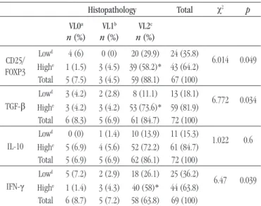
The expression of IFN-γ, TGF-β, IL-10, and co-expression of CD25/FOXP3 was then analyzed in relation to the distribution of the viral load (Table 4). We observed that a large number (high) of immunostained cells for all markers prevailed in VL2 samples (≥ 10,000 viral copies/ml), of which 58% (40/69), 73.6% (53/72), 72.2% (52/72) and 58.2% of samples analyzed showed a large number (high) of cells expressing IFN-γ (p = 0.039), TGF-β (p = 0.034), IL-10 (p = 0.6) and CD25/FOXP3 (p = 0.049), respectively. We emphasize that the difference in the number of samples used among the IHC markers in this study was due to the scarcity
TABLE 4 −Distribution of viral load according to the intensity of expression of
CD25/FOXP3, TGF-β, IL-10 and IFN-γ
Histopathology Total χ2 p
VL0
a
n (%)
VL1b
n (%)
VL2c
n (%)
CD25/ FOXP3
Lowd 4 (6) 0 (0) 20 (29.9) 24 (35.8)
6.014 0.049
Highe 1 (1.5) 3 (4.5) 39 (58.2)* 43 (64.2)
Total 5 (7.5) 3 (4.5) 59 (88.1) 67 (100)
TGF-β
Lowd 3 (4.2) 2 (2.8) 8 (11.1) 13 (18.1)
6.772 0.034
Highe 3 (4.2) 3 (4.2) 53 (73.6)* 59 (81.9)
Total 6 (8.3) 5 (6.9) 61 (84.7) 72 (100)
IL-10
Lowd 0 (0) 1 (1.4) 10 (13.9) 11 (15.3)
1.022 0.6
Highe 5 (6.9) 4 (5.6) 52 (72.2) 61 (84.7)
Total 5 (6.9) 5 (6.9) 62 (86.1) 72 (100)
IFN-γ
Lowd 5 (7.2) 2 (2.9) 18 (26.1) 25 (36.2)
6.47 0.039
Highe 1 (1.4) 3 (4.3) 40 (58)* 44 (63.8)
Total 6 (8.7) 5 (7.2) 58 (63.8) 69 (100)
VL: viral load; CD25: interleukin-2 receptor alpha; FOXP3: forkhead box P3; TGF-β: transforming growth factor beta; IL-10: interleukin-10; IFN-γ: interferon-gamma; a:
negative for human papillomavirus; b:1,050 to < 10,000 viral copies/ml; c: ≥ 10,000
viral copies/ml; d: small quantities of immunomarked cells; e: large quantities of
immunomarked cells; *p < 0.05.
of material. Some samples did not have enough material for all markers. We also emphasize that the patients with no cytological abnormalities were not subjected to biopsy, what justiies the small quantity of NILM samples (n = 6).
DISCUSSION
In the present study, high expression of CD25/FOXP3, TGF-β, IL-10 and IFN-γ was observed in the microenvironment of tissue samples with high-grade lesions, as well as in samples with high levels of HPV viral copies. Cytokine expression and Treg cells were not evaluated in the epithelial area, since the IL-10 expression and Treg cells had been evaluated in this location by our group in another study(22).
Among the analyzed samples, there was a predominance of HPV 16 and 18 in high-grade lesions and carcinoma samples. Our results support the notion that high-risk oncogenic HPV infection, mainly HPV 16 and 18, is the major cause of cervical cancer and its precursor lesions(2).
In most cases, the production of high levels of IFN-γ by activated Th1 cells is associated with the control on the progression of HPV-associated lesions(9), because IFN-γ plays an essential role
in defending against viruses and intracellular pathogens, and inducing inlammatory responses(6). Moreover, this cytokine also
16.4% (11/67), 15.3% (11/72), 16.7% (12/72) and 10.1% (7/69) were cells expressing CD25/FOXP3, TGF-β, IL-10 and IFN-γ, respectively.
FIGURE − Immunohistochemical double staining of CD25/FOXP3 and simple staining of
TGF-β, IL-10 and IFN-γ
A) positive control human tonsil stained with anti-human CD25 and anti-human FOXP3 antibodies; B) negative control human tonsil with omission of the primary antibodies, showed no staining; C) HSIL, with CD25+ FOXP3+ cells in large quantities; D) positive
control human tonsil stained with anti-human TGF-β antibodies; E) negative control human tonsil with omission of the primary antibodies, showed no staining; F) HSIL, with TGF-β+ cells in large quantities; G) positive control human tonsil stained with
anti-human IL-10 antibodies; H) negative control anti-human tonsil with omission of the primary antibodies, showed no staining; I) HSIL, with IL-10+ cells in large quantities; J) positive
control human tonsil stained with anti-human IFN-γ antibodies; K) negative control human tonsil with omission of the primary antibodies, showed no staining; L) HSIL, with IFN-γ + cells in large quantities. All figures are presented in the same magnification (400×)
CD25: interleukin-2 receptor alpha; FOXP3: forkhead box P3; TGF-β: transforming growth factor beta; IL-10: interleukin-10; IFN-γ: interferon-gamma; HSIL: high-grade squamous intraepithelial lesion.
A
D
G
J
B
E
H
K
C
F
I
increases expression of major histocompatibility complex (MHC) class I on tumor cells, thereby increasing the immunogenicity of these cells(23). However, the development of high-grade lesions
induced by HPV was shown to be associated with a decrease in the level of IFN-γ(1). A study assessing mRNA levels of sub-epithelial
IFN-γ on precancerous samples infected by HPV16 showed a signiicant decrease in IFN-γ mRNA levels in precancerous lesions when compared to normal tissue. This study also showed that the
level of IFN-γ mRNA was signiicantly lower in CIN II and CIN
III (HSIL) than in CIN I (LSIL), indicating that IFN-γ expression decreased according to the degree of the lesion(10).
In contrast with these indings, in our study, the analysis of IFN-γ expression and the histopathology indings (p = 0.096) indicate a large number (high) of cells expressed IFN-γ in 43.5% of the HSIL samples. Although this result was not signiicant, we suggest that high expression of IFN-γ in the HSIL samples can be linked with immunosuppressive mechanisms that promote progression to carcinoma. Some authors noted the invariant NKT cells (iNKT) are capable of suppressing the local immune environment in persistent HR-HPV infected cervical tissues by producing IFN-γ to induce high-grade CIN(24).
IFN-γ is also involved in the induction(25) and migration of Treg
cells, in particular Treg CXCR3+ cells to the site of inlammation(26).
In fact, the CXCR3 ligand production (CXCL9, CXCL10, and CXCL11) involved in the recruitment of CXCR3+ cells is induced by IFN-γ(27, 28).
Thus, the high expression of IFN-γ in the samples with high-grade lesions, carcinoma and with high viral load observed in our study, may have induced the recruitment of Treg cells to these sites, hindering the elimination of HPV and the tumor cells. However, evidence to support this suggestion cannot be provided for an experimental model, considering the characteristics of this virus in vitro culture.
The presence of Treg cells has been associated with viral persistence, the development of high-grade lesions, and their progression to cervical cancer(1, 8, 29). This association may be
justiied by the fact that Tregs CD4+CD25+FOXP3+ negatively
regulate the immune response at the site of HPV infection, hampering the elimination of the viruses and neoplastic cells(1, 29).
The predominance of large numbers (high) of cells co-expressing CD25/FOXP3 in high-grade lesion samples (41.8%, p = 0.022) suggests that the increase of this cell population promotes local immunosuppression, facilitating lesion progression to cervical cancer. These indings are consistent with previous studies, which reported that these cells predominate in high-grade lesions and invasive cancer(8, 30).
The high expression of FOXP3 is associated with the persistent high-risk oncogenic HPV(1). Kojima et al. (2013)(31) have shown
that the high prevalence of Treg FOXP3 in precursor lesions of cervical cancer inversely correlates with the spontaneous regression of the lesions(31), demonstrating that Treg cells may
be responsible for the viral persistence and progression of the lesion. In the present study, we observed a predominance of large numbers (high) of CD25/FOXP3 positive cells in samples with high viral load (VL2) (p = 0.049). This reinforces the negative
role played by these cells in viral clearance, i.e., their inluence on the maintenance and persistence of high levels of viral copies and subsequent progression to neoplasia.
Other immunosuppressive components, associated with the persistent HPV infection and the development of lesions to cervical cancer, are TGF-β and IL-10(8, 32). These cytokines are part of one
of the main immunosuppressive mechanisms of Treg cells(33),
capable of suppressing the Th1 response(34).
Besides being one of the cytokines involved in the immunosuppression performed by Tregs(35), TGF-β is also involved
in the maintenance of peripheral Treg cells (adaptive/induced). Once TGF-β is secreted, it induces the expression of FOXP3 in CD4+
CD25- FOXP3- naïve peripheral T cells, converting them into FOXP3+
Treg cells(36, 37). In this context, the large number of cells expressing
TGF-β in HSIL (56.9%; p = 0.001) observed in our study, added to
the large number of cells expressing CD25/FOXP3 in the same lesion, suggesting that the transformation of CD4+CD25-FOXP3- T cells naïve
in FOXP3+ occurred, allowing for the maintenance of induced Treg
cells in these locations. Furthermore, the association between the expression of TGF-β and FOXP3 and the histopathological indings (p < 0.05) may suggest that, in HSIL and carcinoma samples, Treg cells producing TGF-β are present and involved in the evolution of the lesions to cervical cancer.
Regarding the association between the expression of TGF-β
and the viral load (p = 0.034), we observed that 73.6% of the
samples with high viral load presented a large amount of cells expressing TGF-β. This is an indication that, besides being involved in the formation of immunoregulatory cells, this cytokine fulills its suppressive function on the immune response, possibly by inhibiting proliferation and activating effector T lymphocytes and NK cells, thereby contributing to the immune escape of the virus and the neoplastic cells(36-39).
IL-10, another major cytokine secreted by Treg cells, has been associated with cervical cancer(40). Its expression is directly
associated with the degree of cervical lesions, increasing with the severity of the lesion(41). In the present study, we observed a
different stages of cervical cancer, being more predominant in high-grade lesions and carcinoma(41). Taken together, our results
relect the existence of a well-established immunosuppressed microenvironment, which greatly contributes to the persistent infection and progression of the lesion.
Because of its various immunosuppressive mechanisms(42, 43),
IL-10 has been shown to contribute to the development of cervical neoplasia associated with HPV by favoring viral persistence(44). In
our study, we observed that 72.2% of the samples with high viral load (VL2) presented large numbers (high) of IL-10 expressing cells (p = 0.6). Although this result was not signiicant, it is worth
highlighting its importance for the occurrence of the persistent infection, since high levels of IL-10 contribute greatly to the failure of viral clearance(44).
In our study, the main limitations were the small number of samples obtained in the collection period and the shortage of tissue obtained in the biopsy, which made it impossible to make all the necessary slides to perform all the markings in the same sample.
CONCLUSION
Our study evaluated the microenvironment immune status of cervical samples representing different histopathological and HPV infected statuses, mostly for high-risk oncogenic HPV. The microenvironment of high-grade lesion presented large numbers of CD25/FOXP3+, TGF-β+, IL-10+, and IFN-γ+ cells. The
co-expression of CD25/FOXP3 and the expression of TGF-β and
IL-10 in these samples suggest the existence of Treg cells in the microenvironment. Although a great number of cells expressing IFN-γ have also been observed in these samples, our data suggest that this cytokine could be related to immunosuppressed microenvironment maintenance, favoring the persistent HPV infection and the progression to carcinoma.
CONFLICT OF INTEREST
The authors declare that there is no conlict of interest.
RESUMO
Introdução: A infecção persistente por papilomavírus humano (HPV) é a principal causa do câncer cervical e suas lesões precursoras, e a resposta imune inadequada está entre os fatores que contribuem para a persistência viral. Isso pode ser influenciado por células T regulatórias (Treg) e pela produção de citocinas imunossupressoras, como o fator de transformação de crescimento beta (TGF-β) e a interleucina 10 (IL-10). Objetivo: Estabelecemos o perfil de resposta predominante, resposta Th1 ou imunossupressora, no microambiente tecidual, pela detecção de interferon gama (IFN-γ), TGF-β, e IL-10, bem como a coexpressão do receptor da cadeia alfa da IL-2 (CD25) e do forkhead box P3 (FOXP3). Método: Setenta e quatro amostras de biópsias de cérvice uterina, submetidas à detecção do ácido desoxirribonucleico (DNA) de HPV e à análise histopatológica, foram utilizadas nas reações de imuno-histoquímica para detectar IFN-γ, TGF-β, IL-10 e CD25/FOXP3 em linfócitos. Resultados: O microambiente das amostras de lesões intraepiteliais escamosas de alto grau (HSIL) com elevado números de partículas virais (≥ 10.000 cópias/ml) continha elevado número de células CD25/FOXP3+, TGF-β+, IL-10+ e IFN-γ+. Conclusão: A coexpressão de CD25/FOXP3 e a expressão de TGF-β nas amostras HSIL sugerem a existência de células Treg nesses locais, embora a expressão de IFN-γ tenha sido observada em várias células. Nossos dados sugerem que essa citocina pode estar relacionada com a manutenção do microambiente imunossuprimido, favorecendo a infecção persistente por HPV e a progressão para carcinoma.
Unitermos: imuno-histoquímica; imunomodulação; infecções por papilomavírus.
REFERENCES
1. Scott ME, Ma Y, Kuzmich L, Moscicki AB. Diminished IFN-γ and IL-10 and elevated Foxp3 mRNA expression in the cervix are associated with CIN 2 or 3. Int J Cancer. 2009; 124: 1379-83.
2. Li N, Franceschi S, Howell-Jones R, Snijders PJ, Clifford GM. Human papillomavirus type distribution in 30,848 invasive cervical cancers
worldwide: variation by geographical region, histological type and year of publication. Int J Cancer. 2011; 128: 927-35.
5. Zhu J, Paul WE. CD4 T cells: fates, functions, and faults. Blood. 2008; 112: 1557-69.
6. Billiau A, Heremans H, Vermeire K, Matthys P. Immunomodulatory properties of interferon gamma. Ann N Y Acad Sci. 1998; 856: 22-32. 7. Lai HC, Chang CC, Lin YW, et al. Genetic polymorphism of the interferon-gamma gene in cervical carcinogenesis. Int J Cancer. 2005; 113: 712-8.
8. Kobayashi A, Weinberg V, Darragh T, Smith-McCune K. Evolving immunosuppressive microenvironment during human cervical carcinogenesis. Mucosal Immunol. 2008; 1: 412-20.
9. Stellato G, Nieminen P, Aho M, Lehtinen T, Lehtinen M, Paavonen J. Type 1 cytokine response and treatment outcome of genital HPV lesions. Genitourin Med. 1997; 73: 387-90.
10. El-Sherif AM, Seth R, Tighe PJ, Jenkins D. Quantitative analysis of IL-10 and IFN-γ mRNA levels in normal cervix and human papillomavirus type 16 associated cervical precancer. J Pathol. 2001; 195: 179-85.
11. Paust S, Cantor H. Regulatory T cells and autoimmune disease. Immunol Rev. 2005; 204: 195-207.
12. Wang RF. Immune suppression by tumor-speciic CD4+ regulatory T-cells in cancer. Semin Cancer Biol. 2006; 16: 73-9.
13. Leeuw RJ, Kost SE, Kakal JA, Nelson BH. The prognostic value of FoxP3+ tumor iniltrating lymphocytes in cancer: a critical review of the
literature. Clin Cancer Res. 2012; 18: 3022-9.
14. Zhang W, Hou F, Zhang Y, et al. Changes of Th17/Tc17 and Th17/ Treg cells in endometrial carcinoma. Gynecol Oncol. 2014; 132: 599-605. 15. Fontenot JD, Gavin MA, Rudensky AY. Foxp3 programs the development and function of CD4+CD25+ regulatory T cells. Nat Immunol. 2003; 4: 330-6.
16. Sakaguchi S, Sakaguchi N, Shimizu J, et al. Immunologic tolerance maintained by CD25+CD4+ regulatory T cells: their common role in controlling autoimmunity, tumor immunity, and transplantation tolerance. Immunol Rev. 2001; 182: 18-32.
17. Shimizu J, Yamazaki S, Sakaguchi S. Induction of tumor immunity by removing CD25+CD4+ T cells: a common basis between tumor
immunity and autoimmunity. J Immunol. 1999; 163: 5211-8.
18. Doorbar J. The papillomavirus life cycle. J Clin Virol. 2005; 32(Suppl 1): S7-15.
19. Bernard HU, Chan SY, Manos MM, et al. Identiication and assessment of known and novel human papillomavirus by polymerase chain reaction ampliication, restriction fragment length polymorphisms, nucleotide sequence, and phylogenetic algorithms. J Infect Dis. 1994; 170: 1077-85.
20. Payan C, Ducancelle A, Aboubaker MH, et al. Human papillomavirus quantiication in urine and cervical samples by using the Mx4000 and LightCycler general real-time PCR systems. J Clin Microbiol. 2007; 45: 897-901.
21. Padovani CT, Bonin CM, Tozetti IA, Ferreira AM, Fernandes CE, Costa IP. Glucocorticoid-induced tumor necrosis factor receptor expression in patients with cervical human papillomavirus infection. Rev
Soc Bras Med Trop. 2013; 46: 288-92.
22. Prata TT, Bonin CM, Ferreira AM, et al. Local immunosuppression
induced by high viral load of human papillomavirus: characterization
of cellular phenotypes producing interleukin-10 in cervical neoplastic
lesions. Immunology. 2015; 146: 113-21.
23. Dunn GP, Ikeda H, Bruce AT, et al. Interferon-gamma and cancer
immunoediting. Immunol Res. 2005; 32: 231-45.
24. Hu T, Yang P, Zhu H, et al. Accumulation of invariant Nkt cells with increased IFN-γ production in persistent high-risk HPV-infected high-grade cervical intraepithelial neoplasia. Diagn Pathol. 2015; 10: 20.
25. Koch MA, Tucker-Heard G, Perdue NR, Killebrew JR, Urdahl KB, Campbell DJ. The transcription factor T-bet controls regulatory T cell
homeostasis and function during type 1 inlammation. Nat Immunol.
2009; 10: 595-602.
26. Santodomingo-Garzon T, Han J, Le T, Yang Y, Swain MG. Natural killer T cells regulate the homing of chemokine CXC receptor 3-positive regulatory T cells to the liver in mice. Hepatology. 2009; 49: 1267-76.
27. Cole KE, Strick CA, Paradis TJ, et al. Interferon-inducible T cell alpha chemoattractant (I TAC): a novel non-ELR CXC chemokine with potent activity on activated T cells through selective high afinity binding to CXCR3. J Exp Med. 1998; 187: 2009-21.
28. Farber JM. A macrophage mRNA selectively induced by g-interferon encodes a member of the platelet factor 4 family of cytokines. Proc Natl Acad Sci USA. 1990; 87: 5238-42.
29. Visser J, Nijman HW, Hoogenboom BN, et al. Frequencies and role of regulatory T cells in patients with (pre)malignant cervical neoplasia. Clin Exp Immunol. 2007; 150: 199-209.
30. Adurthi S, Krishna S, Mukherjee G, Bafna UD, Devi U, Jayshree RS. Regulatory T cells in a spectrum of HPV-induced cervical lesions: cervicitis, cervical intraepithelial neoplasia and squamous cell carcinoma. Am J
Reprod Immunol. 2008; 60: 55-65.
31. Kojima S, Kawana K, Tomio K, et al. The prevalence of cervical regulatory T cells in HPV related cervical intraepithelial neoplasia (CIN) correlates inversely with spontaneous regression of CIN. Am J Reprod Immunol. 2013; 69: 134-41.
32. Giannini SL, Al-Saleh W, Piron H, et al. Cytokine expression in squamous intraepithelial lesions of the uterine cervix: implications for the generation of local immunosuppression. Clin Exp Immunol. 1998; 113: 183-9.
33. Jonuleit H, Schmitt E. The regulatory T cell family: distinct subsets
and their interrelations. J Immunol. 2003; 171: 6323-7.
34. Seo N, Hayakawa S, Takigawa M, Tokura Y. Interleukin-10 expressed at early tumour sites induces subsequent generation of CD4(+) T-regulatory cells and systemic collapse of antitumour immunity. Immunology. 2001; 103: 449-57.
35. Sakaguchi S. Regulatory T cells. Springer Semin Immunopathol.
2006; 28: 1-2.
36. Chen W, Wahl SM. TGF-beta: the missing link in CD4(+)CD25(+) regulatory T cell-mediated immunosuppression. Cytokine Growth Factor
Rev. 2003; 14: 85-9.
38. Ghiringhelli F, Ménard C, Martin F, Zitvogel L. The role of regulatory T cells in the control of natural killer cells: relevance during tumor progression. Immunol Rev. 2006; 214: 229-38.
39. Inge TH, Hoover SK, Susskind BM, Barrett SK, Bear HD. Inhibition of tumor-speciic cytotoxic T-lymphocyte responses by transforming growth factor beta 1. Cancer Res. 1992; 52: 1386-92.
40. Chopra V, Dinh TV, Hannigan EV. Circulating serum levels of cytokines and angiogenic factors in patients with cervical cancer. Cancer Invest.
1998; 16: 152-9.
41. Bermudez-Morales VH, Gutierrez LX, Alcocer-Gonzalez JM, Burguete A, Madrid-Marina V. Correlation between IL-10 gene expression and HPV
infection in cervical cancer: a mechanism for immune response escape. Cancer Invest. 2008; 26: 1037-43.
42. Raiq K, Charitidou L, Bullens DM, et al. Regulation of the IL-10 production by human T cells. Scand J Immunol. 2001; 53: 139-47. 43. Roncarolo MG, Gregori S, Battaglia M, Bacchetta R, Fleischhauer K, Levings MK. Interleukin-10-secreting type 1 regulatory T cells in rodents
and humans. Immunol Rev. 2006; 212: 28-50.
44. Sung WW, Lee H. The role of interleukin-10 in the progression of human papillomavirus-associated lung carcinoma. Oncoimmunology.
2013; 2: e25854.
CORRESPONDING AUTHOR
Camila Mareti Bonin