The demographic features, clinical outcomes, prognosis and
treatment options for patients with sarcomatoid carcinoma
of the urinary bladder: a single centre experience
_______________________________________________
Simon Paul Robinson
1, Assad Farooq
2, Marc Laniado
3, Hanif Motiwala
31 Frimley Health Foundation Trust - Urologia, Wexham Street, Slough, United Kingdom, UK; 2 Heatherwood
and Wexham Park Hospitals NHS Trust, Wexham Park Hospital Wexham Slough, Slough, United Kingdom, UK; 3 Department of Urology, Heatherwood and Wexham Park Hospitals NHS Trust - Slough, Berkshire,
United Kingdom, UK
ABSTRACT
ARTICLE
INFO
______________________________________________________________ ______________________
Introduction: Carcinosarcoma of the bladder is a very rare neoplasm. The pathogenesis of carcinosarcomas is not clearly understood and remains a subject of debate. Whilst there is some research conceptualizing the histopathological findings of bladder car-cinosarcomas, the demographic features, clinical outcomes, prognosis and treatment options remain unclear.
Materials and Methods: We analyzed 12 consecutive cases of patients with sarco-ma-toid bladder cancer who were treated surgically at a single Urology Department be-tween 1999 and 2015. Radiology, pathology and surgical reports were reviewed to determine the pathological staging at the time of cystectomy. These were directly compared with 230 patients having cystectomies for urothelial cell carcinoma. The sarcomatoid patients, were compared to patients with urothelial cell cancers. The other histological sub types, squamous cell (17), neuroendocrine (9), metastatic (7), mixed (4), adenocarcinoma (3), were not included.
Results and conclusion: Carcinosarcoma of the urinary bladder is often described in the literature as a highly malignant neoplasm that is rapidly lethal. We found that the sarcoma does not offer a worse prognosis than conventional high-grade urothelial car-cinoma. There is no significant difference in grade, stage, positive surgical margin rate, nodal involvement, associated prostate cancer or incidence rates of progression, all cause or disease specific mortality. There was a barely significant difference in car-cinoma in-situ. However, carcinosarcomas are three times the volume of urothelial cell tumors which may contribute to its reputation as an aggressive tumour (44cc v 14cc). Sarcomatous elements do not appear, from our small study, to bestow a worse prognosis.
INTRODUCTION
The World Health Organization defines sar-comatoid carcinoma, also known as carcinosarcoma, as a biphasic tumour consisting of malignant epithe-lial and mesenchymal cells (1). Carcinosarcoma of
the bladder is a very rare neoplasm with extremely low number of cases reported in the literature from as early as 1972.
The pathogenesis of bladder carcinosarco-mas is not clearly understood and remains a subject of debate. A comparative genomic hybridization
stu-Keywords:
Urinary Bladder; Sarcoma; Carcinoma
Int Braz J Urol. 2018; 44: 45-52
_____________________
Submitted for publication: June 14, 2016
_____________________
Accepted after revision: May 25, 2017
_____________________
dy undertaken by Völker et al. (2) suggests that the epithelial and mesenchymal components of cases re-vealed important similarities. Remnants of epithelial cell surface markers and ultrastructural features were shown to be present in mesenchymal and sarcoma-toid components. They hypothesize that carcinosar-comas are the end products of different pathways of differentiation of upstream totipotential neoplastic cells. However, in instances where the different com-ponents share no histochemical similarities, Gorstein et al. (3) propose that carcinosarcomas result from so-called collision tumors in which epithelial and mesenchymal components arise separately. It should be noted that the tumour components showed clonal identity which would support a monoclonal origin (4-6).
Earlier research in this field suggests that the microscopic morphology of carcinosarcomas com-prises a variable combination of sarcomatous and carcinomatous constituents. In the vast majority of reported cases, the epithelial component is essen-tially high-grade urothelial cell carcinoma, while the sarcomatous constituents can consist of chondrosar-coma, osteosarchondrosar-coma, leiomyosarchondrosar-coma, histiocytoma, fibro sarcoma or rhabdomyosarcoma (7-9).
Whilst there is some research conceptuali-zing the histopathological findings of bladder car-cinosarcomas, the demographic features, clinical outcomes, prognosis and treatment options remain unclear.
The objective of this study was to analyze 12 consecutive cases of patients with muscle-invasive or metastatic sarcomatoid bladder cancer who were treated at a single Urology Department between 1999 and 2016. This retrospective analysis was carried out to gain more understanding regarding the clinical behavior, treatment and outcome of this aggressi-ve disease. This is the first study which compares the outcomes of carcinosarcomas with urothelial cell carcinoma (TCC), which can help to put the behavior of carcinosarcoma patients in clinical perspective.
MATERIALS AND METHODS
Study population
This is a retrospective case series in which we reviewed the medical records of all patients with sarcomatoid bladder carcinoma treated with
radical cystectomy at our cancer centre between 1999 and 2015.
Case selection
We searched our hospital patient database and selected patients with established sarcoma-toid disease and for whom the pathology report revealed any sarcomatoid component in their tu-mor. Although cystectomy patients had their prior TURBT analyzed, not all TURBT specimens with sarcomatous elements were searched for. Patient medical records were carefully reviewed to assess the demographic characteristics, clinical stage and outcome. The patients in our study were followed up in clinic annually. Our primary end-point was patient mortality and we calculated our survival data by comparing the disease course for each patient. These were processed into Kaplan-Meier curves of survival. We followed up our patients who were alive during the conduct of the study in our clinic. The survival data was based on analy-sis of our hospital medical records which recorded morbidity, mortality and each detail of each hos-pital admission or episode.
Tumour characteristics
Radiology, pathology and surgical reports were reviewed to determine the pathological sta-ging at the time of cystectomy using the 2009 TNM classification for genitourinary tumors.
Chemotherapy regimens, radiotherapy do-ses, and surgical modality were also recorded. In order to compare the clinical characteristics and outcomes of patients with sarcomatoid nents with those who did not have these compo-nents, patients who had sarcomatoid components were compared to patients in our bladder cancer database who had no sarcomatoid elements pre-sent.
The histopathological slides were analyzed by the Pathology Department. The site of the tu-mor was not recorded and any correlation/relation between TURBT and cystectomy was not possible.
Statistical tests
t--test, incidence rates, log rank and Kaplan-Meier curves (Medcalc) were used.
RESULTS
Patient characteristics
The mean age for patients with TCC was 67 as compared to 70 for patients with sarco-matoid-carcinoma (U-test=0.37). There were 185 males and 45 female patients in the TCC cohort as compared to 9 males and 3 females in the sarcomatoid cohort (Fisher=0.72).
Histological results
We compared different histological cha-racteristics of the sarcomatoid (n=12) cohort of patients with those with TCC (n=230). Although the number of sarcomatoid cases was small, this provided an interesting comparison to our unders-tanding of the natural development of sarcomatoid tumors. Our patients showed a variety of subtypes with both epithelial and sarcomatous elements (Ta-ble-1). The epithelial component was urothelial cell in 8 cases, squamous in 3 cases, and unidentified epithelium in 1 case. There was no significant di-fference in the grade or stage of tumor between the patients undergoing cystectomy for urothelial cell or sarcoma, although the confidence intervals are wide because of the small number of sarcomatoid cases (Table-2). Patients with sarcoma had much larger tumors (43cc) as compared with patients with urothelial cell (14cc). There was a tendency to more CIS with urothelial cell carcinoma with near-ly half the cases featuring this. There was no diffe-rence in the rate of nodal metastasis, or in the rate of extracapsular extension of the metastasis. There was a significant difference in the nodal density with the one sarcomatoid patient with nodal de-posits having 12/22 nodes involved compared to 128/2299 (0.05%) with TCC. This is almost cer-tainly a significant statistical finding rather than a genuine clinical finding (a type 1 error). Neither was there a significant difference in the positive margin rate, in the rate of additional treatment or of prostate cancer (Table-2).
There was no significant difference for ra-tes of progression, overall or disease specific mor-tality between the two cohorts (Table-3, Figure-1).
DISCUSSION
Carcinosarcoma of the urinary bladder is often described in the literature as a highly malig-nant neoplasm which is rapidly lethal. They may be collision tumours of urothelial and sarcomas, poorly differentiated urothelial carcinoma with osteocartilaginous stromal metaplasia, or undi-fferentiated sarcomatoid spindle cell urothelial carcinoma. In true carcinosarcoma, epithelial and mesenchymal components are randomly mixed. Immunohistochemically, the epithelium is charac-terized by cytoplasmic cytokeratin expression and the sarcomatous element by vimentin, desmin and muscle specific actin (12). There can be crossover of markers. Epithelial antigens, pankeratin, EMA, can be found in sarcomatous tissue. Similarly, cell adhesion molecules, E cadherin, CD44, CD44v6 are found to varying degrees in both (13).
We have found that the sarcoma does not offer a worse prognosis than conventional high--grade urothelial carcinoma. Our findings do not corroborate with an earlier series of cases publi-shed in 1998 by Lopez-Beltran et al. who reported that bladder sarcomas are aggressive malignan-cies with a mean survival of 10 to 17 months (8). A recent case series by Wang et al. in 2010 with a larger cohort (n=221) also reiterated the highly aggressive nature of the disease (14). They also built on the earlier case series by emphasizing the need for early detection, including the identifica-tion of risk factors to improve the overall clinical outcomes.
Regarding the rarity of these tumours, Helpap (12) found 0.5% of bladder tumours were nonepithelial similar to other series. However, our rate is ten times this at 4.5%; these variations will no doubt occur with small series.
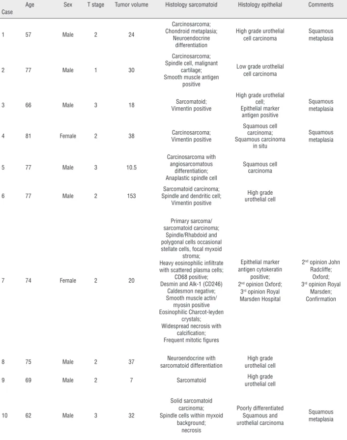
Table 1 - Patient characteristics and histopathological information.
Case
Age Sex T stage Tumor volume Histology sarcomatoid Histology epithelial Comments
1 57 Male 2 24
Carcinosarcoma; Chondroid metaplasia;
Neuroendocrine differentiation
High grade urothelial cell carcinoma
Squamous metaplasia
2 77 Male 1 30
Carcinosarcoma; Spindle cell, malignant
cartilage; Smooth muscle antigen
positive
Low grade urothelial cell carcinoma
3 66 Male 3 18 Sarcomatoid;
Vimentin positive
High grade urothelial cell; Epithelial marker
antigen positive
Squamous metaplasia
4 81 Female 2 38 Carcinosarcoma;
Vimentin positive
Squamous cell carcinoma; Squamous carcinoma
in situ
Squamous metaplasia
5 77 Male 3 10.5
Carcinosarcoma with angiosarcomatous
differentiation; Anaplastic spindle cell
Squamous cell carcinoma
6 77 Male 2 153
Sarcomatoid carcinoma; Spindle and dendritic cell;
Vimentin positive
High grade urothelial cell
7 74 Female 2 20
Primary sarcoma/ sarcomatoid carcinoma;
Spindle/Rhabdoid and polygonal cells occasional stellate cells, focal myxoid
stroma; Heavy eosinophilic infiltrate with scattered plasma cells;
CD68 positive; Desmin and Alk-1 (CD246)
Caldesmon negative; Smooth muscle actin/ myosin positive Eosinophilic Charcot-leyden
crystals; Widespread necrosis with
calcification; Frequent mitotic figures
Epithelial marker antigen cytokeratin
positive; 2nd opinion Oxford;
3rd opinion Royal
Marsden Hospital
2nd opinion John
Radcliffe; Oxford; 3rd opinion Royal
Marsden; Confirmation
8 75 Male 2 37 Neuroendocrine with
sarcomatoid differentiation
High grade urothelial cell
9 69 Male 2 7 Sarcomatoid High grade
urothelial cell
10 62 Male 3 32
Solid sarcomatoid carcinoma; Spindle cells within myxoid
background; necrosis
Poorly differentiated Squamous and urothelial carcinoma
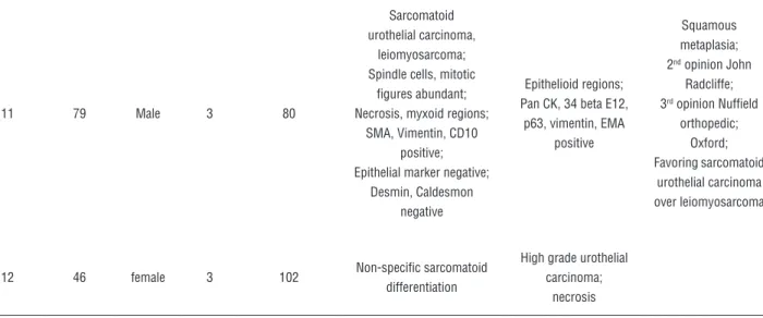
11 79 Male 3 80
Sarcomatoid urothelial carcinoma,
leiomyosarcoma; Spindle cells, mitotic
figures abundant; Necrosis, myxoid regions;
SMA, Vimentin, CD10 positive; Epithelial marker negative;
Desmin, Caldesmon negative
Epithelioid regions; Pan CK, 34 beta E12,
p63, vimentin, EMA positive
Squamous metaplasia; 2nd opinion John
Radcliffe; 3rd opinion Nuffield
orthopedic; Oxford; Favoring sarcomatoid
urothelial carcinoma over leiomyosarcoma
12 46 female 3 102 Non-specific sarcomatoid
differentiation
High grade urothelial carcinoma;
necrosis
Table 2 - Demographic and clinicopathological characteristics of urothelial and sarcomatoid bladder tumurs.
Sarcomatoid Urothelial cell P value
Male/female 9/3 185/45 0.726
Age years 65.3 67.67 0.37
Grade high 10 213 0.269
Intermediate 2 17
Tumour volume cc 44 14 0.0015
T stage localized 7 140 0.771
Locally advanced 5 90
Carcinoma in-situ 2/12 105/230 0.0428
Nodal involvement 1/12 51/230 0.3096
Nodal density 12/22 = 0.545 128/2299 = 0.055 0.0001
Extracapsular extension 1/12 = 0.083 22/128 = 0.44 0.69
Prostate cancer 5/7 = 0.71 83/185 = 0.44 0.14
complications 2/12= 0.17 59/230 = 0.25 0.044
Additional treatment (chemo or radiotherapy)
3/12 = 0.25 44/230 = 0.19 0.29
Table 3 - Survival rates among urothelial and sarcomatoid bladder tumors.
Urothelial Sarcomatoid Log rank significance test
All causes of mortality
(mean survival in months) 89 (CI 79-100) 102 (CI 57.756 to 146.385) P=0.52
Disease specific survival
(mean survival in months) 110 (CI 99-120) 104 (CI 61-149) P=0.61
Progression free survival
(mean survival in months) 106 (CI 95-117) 74 (CI 40.8 – 106.8) P=0.61
Figure 1 - A) Kaplan-Meier curve demonstrating all cause mortality. 1B) Kaplan-Meier curve demonstrating disease specific mortality. 1C) Kaplan-Meier curve demonstrating time to progression.
All Cause Mortality
0 50 100 150 200
Time
Sur
vival probality (%)
12 6 4 1 0 Histology
Sarcoma
Number at risk Group: Sarcoma
Group: TCC
TCC
230 90 44 1 0
A B
12 6 4 1 0
Number at risk Group: Sarcoma
Group: TCC
230 90 44 1 0
Disease_Specific_Mortality
Time
100
95
90
85
80
75
70
65
60
55
0 50 100 150 200
Sur
vival probality (%)
Histology Sarcoma TCC
0 50 100 150 200
Sur
vival probality (%)
12 6 3 0 0
Number at risk Group: Sarcoma
Group: TCC
230 88 44 11 0
progression
100
90
80
70
60
50
40
Histology Sarcoma TCC
led to a complete response and markedly impro-ved survival (4). Four of our surgical patients had multimodal treatment. Two had adjuvant gemci-tabine and cisplatin and they are still alive at 118 and 8 months respectively. One patient had neo--adjuvant radiotherapy (20 Gray) but died after 45 months. Another had neo-adjuvant MVAC and died 9 months post surgery.
Despite the initial appearance of worse survival outcomes for carcinosarcoma patients, we have not found any significant difference be-tween the two cohorts Figure 1a: all cause mor-tality, Figure 1b for disease specific mortality and Figure 1c for progression free survival.
Sarcomatous tumors were three times the volume of urothelial cell tumors in this report (mean 43cc v 14cc), which may contribute to its reputation as an aggressive tumour. However, the-re was less CIS associated, which may contribu-te to the betcontribu-ter outcome for the patients reporcontribu-ted here. On cox regression analysis, histology does not, regardless of type, bestow a worse prognosis. The most important prognostic factors are stage related. Sarcomatoid elements should not darken the attitude of physicians, or patients, and allow them to better assess the risks and potential bene-fits of treatment. Sarcomatoid tumours, like high grade TCC is an aggressive tumour, but no more so than its urothelial counterpart. We hope that our study adds to the very small pool of studies done sporadically over the last 3 decades and stimulates further debate on this subject. Further advances in the molecular biology of this disease may lead to development of targeted treatment strategies for this very rare but dangerous disease.
LIMITATIONS
Limitations of our study include the retros-pective nature of its design and the small number of patients, which is unavoidable due to the rarity of the disease. We do plan to compare all TURBT specimens showing sarcomatoid components to illustrate the range of treatments and outcomes compared to those receiving cystectomy. This, ho-wever, is not too dissimilar to the study sizes of previously published case series (Lahoti et al. n=5, Wang et al. n=14) (7, 15).
CONCLUSIONS
From this study, it does not appear that sarcomatoid tumors of the bladder bestow a worse prognosis.
ACKNOWLEDGEMENTS
Dr. Shaila Suvarna, Dr. Mabel Thyveetel and Dr. Mufeed Ali for their kind assistance in providing the histological slides
CONFLICT OF INTEREST
None declared.
REFERENCES
1. Moch H, Humphrey PA, Ulbright TM, Reuter VE. WHO Classification of tumours of the urinary system and male genital organs, WHO/IARC Classification of Tumours, 4th Edition, Volume 8.
2. Völker HU, Zettl A, Schön G, Heller V, Heinrich E, Rosenwald A, et al. Molecular genetic findings in two cases of sarcomatoid carcinoma of the ureter: evidence for evolution from a common pluripotent progenitor cell? Virchows Arch. 2008;452:457-63.
3. Gorstein F, Anderson TL. Malignant mixed mesodermal tumors: carcinoma, sar-coma, or both? Hum Pathol. 1991;22:207-9.
4. Halachmi S, DeMarzo AM, Chow NH, Halachmi N, Smith AE, Linn JF, et al. Genetic alterations in urinary bladder carcinosarcoma: evidence of a common clonal origin. Eur Urol. 2000;37:350-7.
5. Armstrong AB, Wang M, Eble JN, MacLennan GT, Montironi R, Tan PH, et al. TP53 mutational analysis supports monoclonal origin of biphasic sarcomatoid urothelial carcinoma (carcinosarcoma) of the urinary bladder. Mod Pathol. 2009;22:113-8.
6. Thompson L, Chang B, Barsky SH. Monoclonal origins of malignant mixed tumors (carcinosarcomas). Evidence for a divergent histogenesis. Am J Surg Pathol. 1996;20:277-85. 7. Lahoti C, Schinella R, Rangwala AF, Lee M, Mizrachi H.
8. Lopez-Beltran A, Pacelli A, Rothenberg HJ, Wollan PC, Zincke H, Blute ML, et al. Carcinosarcoma and sarcomatoid carcinoma of the bladder: clinicopathological study of 41 cases. J Urol. 1998;159:1497-503.
9. Perret L, Chaubert P, Hessler D, Guillou L. Primary heterologous carcinosarcoma (metaplastic carcinoma) of the urinary bladder: a clinicopathologic, immunohisto-chemical, and ultrastructural analysis of eight cases and a review of the literature. Cancer. 1998;82:1535-49.
10. Graphpad Software. Available at. <http://www.graphpad. com> (last accessed 10.09.2016).
11. MedCalc. easy-to-use statistical software. Available at <http://www.medcalc.org> (last access 10.09.2016). 12. Helpap B. Nonepithelial neoplasms of the urinary bladder.
Virchows Arch. 2001;439:497-503. Erratum in: Virchows Arch 2002;440:342.
13. Ikegami H, Iwasaki H, Ohjimi Y, Takeuchi T, Ariyoshi A, Kikuchi M. Sarcomatoid carcinoma of the urinary bladder: a clinicopathologic and immunohistochemical analysis of 14 patients. Hum Pathol. 2000;31:332-40.
14. Wang J, Wang FW, Lagrange CA, Hemstreet Iii GP, Kessinger A. Clinical features of sarcomatoid carcinoma (carcinosarcoma) of the urinary bladder: analysis of 221 cases. Sarcoma. 2010;2010.
15. Wang J, Gilespie C, Kunadharaju R, Taimon GA, Enke C. Sarcomatoidurothelial carcinoma: a single center experience. World Journal of Oncology. 2011;2:175-1180.
_______________________ Correspondence address: