The insulin re ce pto r substrate 1
asso ciate s with pho spho tyro sine
pho sphatase SHPTP2 in live r and
m uscle o f rats
Departamentos de 1Clínica Médica, Faculdade de Ciências Médicas, and 2Fisiologia e Biofísica, Instituto de Biologia, Universidade Estadual de Campinas,
Campinas, SP, Brasil M.H.M. Lima2,
J.E. Zambelli1,
C.R.O . Carvalho1
and M.J.A. Saad1
Abstract
Insulin stimulates the tyrosine kinase activity of its receptor resulting in the phosphorylation of its cytosolic substrate, insulin receptor substrate-1 (IRS-1) which, in turn, associates withproteins containing SH2 domains. It has been shown that IRS-1 associates with the tyrosine phosphatase SHPTP2 in cell cultures. While the effect of the IRS-1/SHPTP2 association on insulin signal transduction is not com-pletely known, this association may dephosphorylate IRS-1 and may play a critical role in the mitogenic actions of insulin. However, there is no physiological demonstration of this pathway of insulin action in animal tissues. In the present study we investigated the ability of insulin to induce association between IRS-1 and SHPTP2 in liver and muscle of intact rats, by co-immunoprecipitation with anti-IRS-1 antibody and anti-SHPTP2 antibody. In both tissues there was an increase in IRS-1 association with SHPTP2 after insulin stimulation. This association occurred when IRS-1 had the highest level of tyrosine phosphorylation and the decrease in this association was more rapid than the decrease in IRS-1 phosphorylation levels. The data provide evidence against the participation of SHPTP2 in IRS-1 dephosphory-lation in rat tissues, and suggest that the insulin signal transduction pathway in rat tissues is related mainly to the mitogenic effects of the hormone.
Co rre spo nde nce M.J.A. Saad
Departamento de Clínica Médica FCM, UNICAMP
13081-970 Campinas, SP Brasil
Fax: + 55-19-239-3114
Presented at the XIII Annual Meeting of the Federação de Sociedades de Biologia Experimental, Caxambu, MG, Brasil, August 26-29, 1998.
Research supported by FAPESP and CNPq (PRO NEX).
Received April 14, 1998 Accepted August 13, 1998
Ke y wo rds
•Insulin action
•Insulin receptor substrate
•Phosphotyrosine phosphatase
•SHPTP2
The insulin receptor is the principal me-diator of insulin action on cellular and meta-bolic processes. The insulin receptor ß-sub-unit, which contains an intrinsic tyrosine kinase, undergoes tyrosyl autophosphoryla-tion and is activated in response to insulin binding to the extracellular α-subunit (1). Moreover the discovery of the tyrosine ki-nase activity of the insulin receptor
1 (IRS-1). Evidence from different sources has shown that the phosphotyrosine-con-taining form of IRS-1 binds to the enzyme phosphatidylinositol 3-kinase (PI 3-kinase) through the Src homology 2 (SH2) domains of the latter (4,5) and that this association activates the enzyme.
In addition to PI 3-kinase, other proteins containing SH2 domains such as SHPTP2, Nck, Grb2 have been shown to bind to IRS-1 (IRS-1). SHPTP2 is an SH2 domain-containing tyrosine phosphatase that associates with the COOH-terminal tyrosine phosphorylation sites of IRS-1 in cell cultures (6), and this association increases the phosphatase activ-ity of SHPTP2 in vitro (7). However, there is no physiological demonstration of this path-way of insulin action in animal tissues. In the present study we investigated the ability of insulin to induce association of IRS-1 and SHPTP2 in liver and muscle of intact rats, two of the main target tissues for insulin action.
The rats were anesthetized with sodium amobarbital (15 mg/kg body weight, intra-peritoneally) and used in the experiments 10-15 min later, as soon as anesthesia was assured by the loss of foot and corneal re-flexes. The abdominal cavity was opened, the vena cava exposed and 0.5 ml saline (0.9% NaCl), containing or not 6 µg of insu-lin, was injected. After the indicated time, the liver or muscle was removed, minced coarsely and immediately homogenized in extraction buffer (1% Triton X-100, 100 mM Tris, pH 7.4, containing 100 mM so-dium pyrophosphate, 100 mM soso-dium fluo-ride, 10 mM EDTA, 10 mM sodium vana-date, 2 mM PMSF and 0.1 mg aprotinin/ml) at 4oC with a Polytron PTA 20S generator
(Brinkmann Instruments model PT 10/35; Westbury, NY) operated at maximum speed for 30 s. Both extracts were centrifuged at 15,000 rpm and 4oC in a Beckman 70.1 Ti
rotor (Palo Alto, CA) for 45 min to remove insoluble material, and the supernatant of these tissues was used for
immunoprecipita-tion with anti-IRS-1 antibody and protein A Sepharose 6 MB. The samples were treated with Laemmli sample buffer (8) containing 100 mM DTT, heated in a boiling water bath for 4 min and submitted to SDS-PAGE (6.5% Tris/acrylamide). Electrotransfer of proteins from the gel to nitrocellulose was performed for 2 h at 100 V (constant) in the Bio-Rad miniature transfer apparatus (Mini-protean), as described by Towbin et al. (9) but with 0.02% SDS added to the transfer buffer to enhance the elution of high-molecular mass protein. Nonspecific protein binding to ni-trocellulose was reduced by pre-incubating the filter overnight at 4oC in blocking buffer
(3% BSA, 10 mM Tris, 150 mM NaCl and 0.02% Tween 20). The pre-stained molecu-lar mass standards used were myosin (205 kDa), galactosidase (116 kDa), BSA (80 kDa) and ovalbumin (49.5 kDa). The nitro-cellulose blot was incubated with anti-phos-photyrosine (1 µ/ml) or anti-SHPTP2 (1:100) antibodies for 4 h at 22oC. Monoclonal
anti-phosphotyrosine antibody and anti-IRS-1 and anti-SHPTP2 antibodies were from Santa Cruz Biotechnology, Santa Cruz, CA. The blots were subsequently incubated with 2 µCi [125I]-protein A (30 µCi/µg) in 10 ml of
blocking buffer for 1 h at 22oC and washed
again. [125I]-protein A bound to the
antibod-ies was detected by autoradiography using preflashed Kodak XAR film with Cronex Lightning Plus intensifying screens at -70oC
for 12-48 h. Band intensities were quantified by optical densitometry (Molecular Dynam-ics) of the developed autoradiogram (10-12).
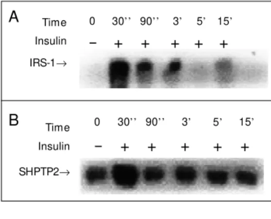
thereafter and had almost vanished by 15 min. In order to investigate the association of SHPTP2 with IRS-1 the same membrane was stripped and re-blotted with anti-SHPTP2 antibody. Figure 1B shows that there is a basal association between IRS-1 and SHPTP2 which increases 30 s after insulin infusion and returns to basal levels thereafter. It is interesting to note that at 90 s and 3 min after insulin infusion there still was IRS-1 ty-rosine phosphorylation, but the association with SHPTP2 was just basal.
The results obtained for muscle were similar to those obtained for liver but high tyrosine phosphorylation levels of IRS-1 were observed at 30 and 90 s after insulin infusion (Figure 2A). When these samples previously immunoprecipitated with anti-IRS-1 antibody were blotted with anti-SHPTP2 antibody, an important association was observed 30 s
after insulin infusion, which was dissociated from IRS-1 tyrosine phosphorylation, whose levels continued to be high up to 90 s.
The results presented here show that SHPTP2 associates with phosphorylated IRS-1 in liver and muscle of rats. In cell culture, SHPTP2 is activated during association with IRS-1. While the effect of IRS-1/SHPTP2 association on signal transmission is not com-pletely known, there is a possibility that this interaction autoregulates IRS-1 phosphory-lation. The tyrosine phosphatase SHPTP2 may dephosphorylate signaling intermedi-ates located either in the IRS-1 signaling complex or at distant sites, thus downregu-lating signaling. In this regard, there is a study showing that SHPTP2 is able to de-phosphorylate IRS-1 in vitro (13), but this phenomenon was not observed in vivo (14-16). Our results showing that IRS-1/SHPTP2
Figure 1 - Time course of insulin-stimulated IRS-1 ty-rosine phosphorylation (panel A) and association w ith SHPTP2 in rat liver (panel B). Rats w ere anesthetized and the abdominal w all w as incised to expose the viscera. Saline or insulin w as infused at the indicated time. The tissues w ere excised and homogenized in extraction buffer at 4oC as described in the text. After
centrifugation, aliquots containing equal amounts of protein w ere immunoprecipitated w ith IRS-1 anti-body and protein A Sepharose 6 M B and then resolved on 6.5% SDS-polyacrylamide gels. The protein bands w ere subsequently transferred to a nitrocellulose mem-brane and detected w ith anti-phosphotyrosine antibody (A) or anti SHPTP2 (B) and [125I]-protein A, after w hich
the membrane w as subjected to autoradiography. The data are representative of five experiments.
Time Insulin
IRS-1→
Time Insulin
SHPTP2→
- + + + + +
- + + + + +
0 30’’ 90’’ 3’ 5’
0 30’’ 90’’ 3’ 5’ 15’ A
B
Time Insulin IRS-1→
Time Insulin SHPTP2→
A
B
- + + + + +
0 30’’ 90’’ 3’ 5’ 15’
- + + + + +
0 30’’ 90’’ 3’ 5’ 15’
Figre 2 - Time course of insulin-stimulated IRS-1 ty-rosine phosphorylation (panel A) and association w ith SHPTP2 in rat muscle (panel B). Rats w ere anesthe-tized and the abdominal w all w as incised to expose the viscera. Saline or insulin w as infused at the indicated time. The tissues w ere excised and homogenized in extraction buffer at 4oC as described in the text. After
centrifugation, aliquots containing equal amounts of protein w ere immunoprecipitated w ith IRS-1 anti-body and immunoblotted as described in the legend to Figure 1. The data are representative of four experi-ments.
association occurs when IRS-1 has the high-est level of tyrosine phosphorylation are ev-idence against the effect of SHPTP2 dephos-phorylating IRS-1, and suggest that this as-sociation depends on an important increase in IRS-1 tyrosine phosphorylation.
The role of SHPTP2 in insulin signaling has been examined by several approaches. Microinjection of the SH2 domains of SHPTP2 or antibodies against SHPTP2 blocks insulin-stimulated DNA synthesis (14,17). Similarly, overexpression of a cata-lytically inactive mutant SHPTP2 molecule inhibits mitogenesis and p21ras and MAP
kinase activation (14-17). It is not clear how SHPTP2 transmits signals to p21ras and MAP
kinase or mediates mitogenesis during insu-lin signainsu-ling, but there is no doubt that the activity of SHPTP2 plays a critical role in the mitogenic actions of insulin.
It is known that insulin activates hexose transport via at least two mechanisms: a p21ras-dependent pathway leading to an
in-crease in the amount of cell surface GLUT 1,
and a metabolic, p21ras-independent
path-way leading to translocation of the insulin-responsive transporter GLUT 4 to the cell surface (1,7). Hausdorff et al. (17), using microinjection of a glutathione S-transferase fusion protein encoding the N- and C-termi-nal SH2 domains of SHPTP2 or anti-SHPTP2 antibodies into 3T3-L1 adipocytes, demon-strated that SHPTP2 is important for the expression of GLUT 1, but is not required for activation of GLUT 4 translocation. These data suggest that SHPTP2 plays a role in the insulin-induced transcription of immediate early genes such as GLUT 1, but is not required for the metabolic increase in trans-port mediated by GLUT 4 translocation.
In summary, the present results demon-strate that after insulin stimulation of liver and muscle of intact rats there is an increase in the association of IRS-1 with SHPTP2, a pathway that has been shown to play an important role in insulin-induced DNA syn-thesis in cell culture.
Re fe re nce s
1. White M F & Kahn CR (1994). The insulin signaling system. Journal of Biological Chemistry, 269: 1-5.
2. White M F, M aron R & Kahn CR (1985). Insulin rapidly stimulates tyrosine phos-phorylation of an M r 185 000 protein in intact cells. Nature, 318: 183-186. 3. Keller SR, Aebersold RH, Garner CW &
Lienhard GE (1993). The insulin-elicited 160 kDa phosphot yrosine prot ein in mouse adipocytes is an insulin receptor substrate 1: identification by cloning. Bio-chimica et Biophysica Acta, 1172: 323-326.
4. Sun XJ, Rothenberg PL, Kahn CR, Backer JM , Araki E, W ilden P, Cahill DA, Goldstein BJ & White M F (1991). Struc-ture of the insulin receptor substrate IRS-1 defines a unique signal transduction pro-tein. Nature, 352: 73-77.
5. Carvalho CRO, Brenelli SL, Silva AC, Nunes ALB, Velloso LA & Saad M JA (1996). Effect of aging on insulin receptor, insulin receptor substrate-1 and phospha-tidylinositol 3-kinase in liver and muscle
of rats. Endocrinology, 137: 151-159. 6. Kuhné M R, Paw son T, Lienhard GE &
Feng GS (1993). The insulin receptor sub-strate 1 associates w ith the SH2-contain-ing phosphotyrosine phosphatase syp.
Journal of Biological Chem istry, 268: 11479-11481.
7. M yers M G & White FM (1996). Insulin signal transduction and the IRS-1 proteins.
Annual Review of Pharmacology and Toxi-cology, 36: 615-658.
8. Laemmli UK (1970). Cleavage of struc-tural proteins during the assembly of the head of bacteriophage T4. Nature, 227: 680-685.
9. Tow bin H, Staehlin J & Gordon J (1979). Electrophoretic transfer of proteins from polyacrylam ide gels t o nit rocellulose sheets. Procedure and some applications.
Proceedings of the National Academy of Sciences, USA, 76: 4350-4354.
10. Saad M JA, Carvalho CRO, Thirone ACP & Velloso LC (1996). Insulin induces tyrosine phosphorylation of Jak2 in insulin-sensi-tive tissues of the intact rat. Journal of
Biological Chemistry, 271: 22100-22104. 11. Thirone ACP, Carvalho CRO, Brenelli SL,
Velloso LA & Saad M JA (1997). Effect of chronic grow th hormone treatment on signal transduction in rat tissues. M olecu-lar and Celluolecu-lar Endocrinology, 130: 33-42.
12. Saad M JA, M aeda L, Brenelli SL, Carvalho CRO, Paiva RS & Velloso LA (1997). De-fects in insulin signal transduction in liver and muscle of pregnant rats. Diabetolo-gia,40: 179-186.
13. Sugimoto S, Wandless TJ, Shoelson SE, Nell BG & Walsh CT (1994). Activation of the SH2-containing protein tyrosine phos-phatase, SHPTP2, by phosphotyrosine-containing peptides derived from insulin receptor substrate-1. Journal of Biological Chemistry, 269: 13614-13622.
15. Yamauchi K, Ribon V, Saltiel AR & Pessin JE (1995). Identification of the major SHPTP2-binding protein that is tyrosine-phosphorylated in response to insulin.
Proceedings of the National Academy of Sciences, USA, 270: 17716-17722.
16. Yamauchi K, M ilarski KL, Saltiel AR & Pessin JE (1995). Protein-tyrosine-phos-phatase SHPTP2 is a required positive ef-fector for insulin dow nstream signaling.
Proceedings of the National Academy of Sciences, 92: 664-668.
17. Hausdorff FS, Bennett AM , Neel BG & Biirnbaum M J (1995). Different signaling roles of SHPTP2 in insulin-induced GLUT1 expression and GLUT4 t ranslocat ion.
The David Rockefeller Center for Latin American Studies, Harvard University, invites candidates for its “Visiting Schol-ars and Fellows Program”. The “Visiting ScholSchol-ars and Fel-lows Program” is a program for academic and non-academic professionals interested in developing research for a deter-mined period of time (one or two academic semesters) while residing at Harvard University. The selection of profession-als is made by an examination.
With the support of Mr. Jorge Paulo Lemann, the Center recently created the “Lemann Visiting Fellowship”. This is a fellowship for a professional for the above mentioned pro-gram whose research project has Brazil as the study objec-tive. This fellowship is awarded once each academic year (September-June) and covers the administrative expenses of the Center and airline tickets. The professional will also receive financial assistance of US$15,000 (fifteen thousand American dollars, subject to income tax), regardless of the number of semesters for which he is accepted.
Registration Process: Candidates must be academic or non-academic professionals who have completed their doctorate degree or equivalent academic preparation, fluent in English and have a substantial number of publications. Registration can be made for one academic semester or for the entire academic year (1 September 1998 to 30 June 1999). The deadline for registration for the 2nd semester of 1998 and for the above cited academic year is 1 April 1998. The deadline
The Alexander Von Humboldt Foundation, which exists more than 130 years and whose actual structure was established a few years after WWII (1953), is offering fellowships for post-doctorate study and for specialized research projects in all areas of knowledge. There are 500 fellowships annually, distributed without limitation of nationality, and 150 fellow-ships (Feodor Lynen Research Fellowfellow-ships), only for Ger-man researchers and professors, to work as invited research-ers of ex-fellows of all nationalities and their Univresearch-ersities. The Alexander Von Humboldt Foundation Fellowships are open to all professors who meet the above-mentioned requi-sites and who are less than 40 years of age. There is also one specialized fellowship for candidates between 40 and 45 years of age. These fellowships are for 6 to 24 months, receiving 3400 to 4200 German marks monthly.
for registration for the 1st semester of 1999 is 31 October 1998. All documentation will be analyzed by an interdiscipli-nary panel of professors of the Harvard University, affiliated with the David Rockefeller Center of Latin American Stud-ies.
Candidates must include, in English:
- Curriculum vitae;
- One page explaining why the candidate wishes to study at Harvard;
- Three or four pages describing the proposed research project;
- Two letters of reference from persons who can prove the qualifications of the candidate and the importance of the proposed project.
For additional information, contact:
Visiting Scholars and Fellows Program Coordinator David Rockefeller Center for Latin American Studies Harvard University
61 Kirkland Street
Cambridge, MA 02138 USA
Tel: (617) 495-3366 Fax: (617) 496-2802 e-mail: drclas@fas.harvard.edu
Web site: http://www.fas.harvard.edu/~drclas
Interested candidates should contact:
Alexander Von Humboldt-Stiftung Jean Paul Strasse 12
D-53173 BONN (Germany) Telefax: 0049-228-833-199 Internet: http://www.avh.de
Ex-fellows of the Foundation are represented in Brazil by:
“Clube Humboldt do Brasil”
Av. Brig. Faria Lima, 1572 - 7o andar, sala 705 01463-900 São Paulo, SP
President Prof. Dr. Bruno Konig Junior
Department of Anatomy, ICB, USP, São Paulo, Brasil Tel: (011)818-7258