Tissue-specific regulation of IRS-1 in
unilaterally nephrectomized rats
Departamento de Clínica Médica, Faculdade de Ciências Médicas, Universidade Estadual de Campinas, Campinas, SP, Brasil A.D. Sasse, E. Chen,
C.R.O. Carvalho, J.A.R. Gontijo, S.L. Brenelli and M.J.A. Saad
Abstract
Insulin stimulates the tyrosine kinase activity of its receptor, resulting in the phosphorylation of its cytosolic substrate, insulin receptor substrate 1 (IRS-1). IRS-1 is also a substrate for different peptides and growth factors, and a transgenic mouse “knockout” for this protein does not have normal growth. However, the role of IRS-1 in kidney hypertrophy and/or hyperplasia was not investigated. In the present study we investigated IRS-1 protein and tyrosine phosphorylation levels in the remnant kidney after unilateral nephrectomy (UNX) in 6-week-old male Wistar rats. After insulin stimulation the levels of insulin receptor and IRS-1 tyrosine phosphorylation were reduced to 79 ± 5% (P<0.005) and 58 ± 6% (P<0.0001), respectively, of the control (C) levels, in the remnant kidney. It is possible that a circulat-ing factor and/or a local (paracrine) factor playcirculat-ing a role in kidney growth can influence the early steps of insulin action in parallel. To investigate the hypothesis of a circulating factor, we studied the early steps of insulin action in liver and muscle of unilateral nephrecto-mized rats. There was no change in pp185 tyrosine phosphorylation levels in liver (C 100 ± 12% vs UNX 89 ± 9%, NS) and muscle (C 100 ± 22% vs UNX 91 ± 17%, NS), and also there was no change in IRS-1 phosphorylation levels in both tissues. These data demonstrate that after unilateral nephrectomy there is a decrease in insulin-induced insulin receptor and IRS-1 tyrosine phosphorylation levels in kidney but not in liver and muscle. It will be of interest to investigate which factors, probably paracrine ones, regulate these early steps of insulin action in the contralateral kidney of unilaterally nephrectomized rats. Correspondence
M.J.A. Saad
Departamento de Clínica Médica Faculdade de Ciências Médicas Universidade Estadual de Campinas 13083-970 Campinas, SP Brasil
Fax: 55 (019) 239-3114
Research supported by FAPESP and CNPq.
Received April 10, 1997 Accepted July 29, 1997
Key words
•Insulin receptor
•Insulin receptor substrate
•Unilateral nephrectomy
•Insulin action
One of the earliest cellular responses to stimulation by insulin is the activation of insulin receptor kinase and tyrosine phos-phorylation of insulin receptor ß subunit and pp185, a cytoplasmic phosphoprotein found in most cells and tissues (1). A component of the pp185 band was purified and cloned from several sources (2,3). The cloned
including IGF-1 and GH, and a transgenic mouse “knockout” for this protein does not have normal growth. IGF-1 and its receptor have been involved in kidney compensatory hypertrophy after unilateral nephrectomy (UNX) or diabetes. However, the role of IRS-1 in kidney hypertrophy and/or hyper-plasia was not investigated. The first aim of this study was to investigate IRS-1 protein and tyrosine phosphorylation levels in the remnant kidney after UNX.
Male Wistar rats (6 weeks old) under-went unilateral left nephrectomy (the kid-neys were trimmed of fat and capsule and the adrenal glands were left intact), as described previously (6), or sham operation, where a flank incision was made and the left kidney was manipulated, but not removed. The ani-mals recovered and received food and water
ad libitum. After seven days, the rats were anesthetized with sodium amobarbital (15 mg/kg body weight, intraperitoneally) and used in experiments 10-15 min later, as soon as anesthesia was confirmed by the loss of pedal and corneal reflexes. The abdominal cavity was opened, the vena cava exposed and 0.5 ml of normal saline (0.9% NaCl) containing or not 10 µM insulin was in-jected. After 30 or 90 s, fragments of liver and muscle, respectively, were removed, minced coarsely and immediately homog-enized in approximately 6 volumes of solu-bilization buffer A using a Polytron PTA 20S generator (Brinkmann Instruments, mo-del PT 10/35) operated at maximum speed for 30 s in a water bath maintained at 100oC
as previously described (7-9). To investigate IRS-1 phosphorylation in kidney, saline with or without insulin was infused into the vena cava and the remnant kidney was extracted 90 s later in the same way as described for liver and muscle. Solubilization buffer A contained 1% SDS, 50 mM HEPES, pH 7.4, 100 mM sodium pyrophosphate, 100 mM sodium fluoride, 10 mM EDTA, and 10 mM sodium vanadate. The homogenates were
then boiled for 10 min and cooled in an ice bath for 40 min. In some experiments, kid-ney, liver and muscle were excised and ho-mogenized with a Polytron apparatus in 6 volumes of homogenization buffer B cooled in an ice bath. The composition of buffer B was the same as buffer A except that 1% Triton X-100 replaced 1% SDS and 2 mM PMSF and 0.1 mg/ml aprotinin were added. Both extracts were centrifuged at 100,000 g
(55,000 rpm) at 4oC in a Beckman 70.1 Ti
rotor for 30 min to remove insoluble mate-rial, and the resulting supernatant was used for the experiments. The kidney, liver and muscle homogenized in buffer B were used for immunoprecipitation with anti-IRS-1 antibody and Protein A-Sepharose 6 MB.
The samples were treated with Laemmli sample buffer (10) containing 100 mM DTT and heated in a boiling water bath for 4 min. For total extracts, similar size samples (150 µg of protein) were submitted do SDS/PAGE (6.5% Tris/acrylamide) in a Bio-Rad minia-ture slab gel apparatus (11). Electrotransfer of proteins from the gel to nitrocellulose was performed for 2 h at 100 V (constant) in the Bio-Rad miniature transfer apparatus (Mini-protean), as described by Towbin et al. (12) but with 0.02% SDS added to the transfer buffer to enhance the elution of high molec-ular mass protein. Nonspecific protein bind-ing to the nitrocellulose was reduced by pre-incubating the filter overnight at 4oC in
block-ing buffer (3% BSA, 10 mM Tris, 150 mM NaCl, and 0.02% Tween 20). The prestained molecular mass standards used were myosin (205 kDa), β-galactosidase (116 kDa), BSA (80 kDa) and ovalbumin (49.5 kDa). The nitrocellulose blot was incubated with anti-phosphotyrosine antibody (1 µg/ml) for 4 h at 22oC. The blots were subsequently
incu-bated with 2 µCi of 125I-Protein A (30 µCi/
µg) in 10 ml of blocking buffer for 1 h at 22oC and washed for 2 h. 125I-Protein A
XAR film with Cronex Lightning Plus inten-sifying screens at -70oC for 12-48 h. Band
intensities were quantified by densitometry (Molecular Dynamics) of the developed au-toradiogram.
In the present study we evaluated the effect of UNX on insulin receptor and IRS-1 phosphorylation in the remnant kidney. There was no change in insulin receptor protein levels as determined by immunoblotting with an antibody to the C-terminus of the insulin receptor (data not shown). Following insulin infusion into the vena cava, a phosphotyro-sine band of 95 kDa, previously identified as the insulin receptor ß subunit (1,13,14), ap-peared and became prominently phospho-rylated. The level of phosphorylation of this band was reduced to 79 ± 5% (P<0.005) in the remnant kidney. In addition to the 95-kDa band seen after insulin injection, a broad band migrating between 165 and 185 kDa was also detectable (Figure 1A). This band is known as pp185 and IRS-1 is one compo-nent of this band (15). The phosphorylation of pp185 was reduced to 58 ± 6% (P<0.0001) in the remnant kidney. In order to character-ize IRS-1 phosphorylation after insulin stim-ulation, we immunoprecipitated kidney ex-tracts with anti-IRS-1 antibody and immu-noblotted these with IRS-1 and anti-phosphotyrosine antibody (Figure 1B). There was no change in IRS-1 protein levels in the remnant kidney. However, after insulin stim-ulation IRS-1 tyrosine phosphorylation was reduced to 41 ± 5% (P<0.0001) in the kidney of UNX rats compared to the kidney of control rats. The mechanism(s) responsible for the reduction in insulin receptor and IRS-1 tyrosine phosphorylation in the remnant kidney after 7 days of UNX have not been elucidated. The regulation of insulin recep-tor and IRS-1 is under the control of differ-ent situations such as fasting, obesity, hor-mones and diabetes. We have previously demonstrated that in other tissues the excess of cortisol (16), glucagon (7), epinephrine
(9), and GH (17) and also hyperinsulinemia (13) can modulate the early steps of insulin action by inducing a reduction in insulin receptor and IRS-1 phosphorylation. How-ever, none of these hormones is in excess in our animal model of kidney growth. It is possible that a circulating factor and/or a local (paracrine) factor that plays a role in kidney growth can influence the early steps of insulin action in parallel. If there is a
Figure 1 - A, Insulin receptor (IR) and pp185 tyrosine phosphorylation in remnant kidney tissue after saline (-) or saline plus 10 µM insulin (+) in unilaterally nephrectomized (UNX) rats, compared with controls. The tissue was extracted as described in Methods and immunoblotting (Blot) of total tissue extracts was performed with anti-phosphotyrosine antibody (PY) and 125I-Protein A. B, Effect of unilateral nephrectomy on insulin receptor substrate 1 (IRS-1) in the rat remnant kidney. Kidney extracts from controls and unilaterally nephrectomized rats were immunoprecipitated (IP) with anti-IRS-1 antibody (α IRS-1) and then immunoblotted with anti-phosphotyrosine antibody.
A - Blot: PY
UNX Control
- + - +
Insulin
pp185
IR
205
116
80
B - IP: α IRS-1 Blot: PY
UNX Control
- + - +
Insulin
kDa
→
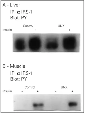
Figure 2 - Effect of unilat-eral nephrectomy (UNX) on insulin receptor substrate 1 (IRS-1) in the rat liver and muscle. The liver extracts from controls and unilater-ally nephrectomized rats were immunoprecipitated (IP) with anti-IRS-1 antibody (α IRS-1) and then immuno-blotted (Blot) with anti-phosphotyrosine antibody (PY) (A). The same proce-dure was applied to muscle (B).
unilateral nephrectomized rats. The experi-ments were performed as described above, except that we infused insulin into the portal vein, and extracted liver and muscle as de-scribed above. The results of total tissue extracts showed that in liver there was no change in pp185 phosphorylation levels (C 100 ± 12% vs UNX 89 ± 9%, NS). In accor-dance, after immunoprecipitation with anti-IRS-1 antibody and blotting with anti-phos-photyrosine antibody there was also no change in IRS-1 tyrosine phosphorylation in the liver of UNX rats (Figure 2A). In muscle the results were very similar with no change in insulin-induced pp185 phosphorylation (C 100 ± 22% vs UNX 91 ± 17%, NS) and also in IRS-1 tyrosine phosphorylation (Fig-ure 2B) in UNX rats.
In summary, the data demonstrated that after UNX there was a decrease in insulin-induced insulin receptor and IRS-1 tyrosine phosphorylation levels in the remnant kid-ney but not in liver and muscle. It will be of interest to determine which factors, prob-ably paracrine ones, regulate these early steps of insulin action in the contralateral kidney of UNX rats.
References
1. White MF & Kahn CR (1994). The insulin signalling system. Journal of Biological Chemistry, 269: 1-5.
2. Sun XJ, Rothenberg PL, Kahn CR, Baker JM, Araki E, Wilden P, Cahill DA, Goldstein BJ & White MF (1991). Struc-ture of the insulin receptor substrate-1 (IRS-1) defines a unique signal transduc-tion protein. Nature, 352: 73-77. 3. Araki E, Sun XJ, Haag III BL, Chuang LM,
Zhang Y, Yang-Feng TL, White MF & Kahn CR (1993). Human skeletal muscle insulin receptor substrate-1: characterization of the cDNA, gene and chromosomal local-ization. Diabetes, 42: 1041-1054. 4. Saad MJA, Velloso LA & Carvalho CRO
(1995). Angiotensin II induces tyrosine phosphorylation of insulin receptor sub-strate 1 and its association with phospha-tidylinositol 3-kinase in rat heart. Bio-chemical Journal, 310: 741-744.
5. Argetsinger LS, Hsu GW, Myers MG, Billestrup N, White MF & Carter-Su C (1995). Growth hormone, interferon-γ and leukemia inhibitory factor promoted tyrosyl phosphorylation of insulin recep-tor substrate-1. Journal of Biological Chemistry, 270: 14685-14692.
6. Flyvbjerg A, Ussing OT, Naera R, Ingerslev J & Orskov H (1988). Kidney tissue so-matomedin C and initial renal growth in diabetic and uninephrectomised rats. Dia-betologia, 31: 310-314.
7. Saad MJA, Hartmann LGC, Carvalho DS, Galoro CAO, Brenelli SL & Carvalho CRO (1995). Effect of glucagon on insulin sub-strate-1 (IRS-1) phosphorylation and asso-ciation with phosphatidylinositol-3 kinase (PI-3 kinase). FEBS Letters, 370: 131-134.
8. Carvalho CRO, Brenelli SL, Silva AC, Nunes ALB, Velloso LA & Saad MJA (1996). Effect of aging on insulin receptor, insulin receptor substrate-1, and phospha-tidylinositol-3 kinase in liver and muscle of rats. Endocrinology, 137: 151-159. 9. Saad MJA, Hartmann DSC, Galoro CAO,
Brenelli SL & Carvalho CRO (1995). Modu-lation of the early steps in insulin action in liver and muscle of epinephrine treated rats. Endocrine, 3: 755-759.
10. Laemmli UK (1970). Cleavage of struc-tural proteins during the assembly of the head of bacteriophage T4. Nature, 227: 680-685.
11. Bradford MM (1976). A rapid and sensi-tive method for quantitation of microgram quantities utilizing the principle of protein dye binding. Analytical Biochemistry, 227: 248-254.
circulating factor that can regulate the early steps of insulin action in kidney, it may also modulate insulin receptor and IRS-1 in other tissues such as liver and muscle. To investi-gate this possibility we studied the early steps in insulin action in liver and muscle of
A - Liver
Control
- +
Insulin - +
UNX
IP: α IRS-1 Blot: PY
B - Muscle
Control
- +
Insulin - +
UNX
12. Towbin H, Staehlin J & Gordon J (1979). Electrophoretic transfer of proteins from polyacrylamide gels to nitrocellulose sheets. Procedure and some applications.
Proceedings of the National Academy of Sciences, USA, 76: 4350-4354.
13. Saad MJA, Araki E, Miralpeix M, Rothenberg PL, White MF & Kahn CR (1992). Regulations of insulin receptor substrate-1 in liver and muscle of animal models of insulin resistance. Journal of Clinical Investigation, 90: 1839-1849.
14. Kasuga M, Karlson FA & Kahn CR (1982). Insulin stimulates the phosphorylation of the 95,000 dalton subunit of its own re-ceptor. Science, 215: 185-187.
15. Sun XJ, Wang LM, Zhang Y, Yenush LP, Myers MG, Glasheen EM, Lane WS, Pierce JH & White MF (1995). Role of IRS-2 in insulin and cytokine signalling.
Nature, 377: 173-177.
16. Saad MJA, Folli F, Kahn JA & Kahn CR (1993). Modulation of insulin receptor sub-strate-1 (IRS-1) and phosphatidylinositol 3-kinase in liver and muscle of dexameth-asone treated rats. Journal of Clinical In-vestigation,92: 2065-2072.