Thrombomodulin and interleukin 6 as potential
biomarkers of endothelial dysfunction and
inflammation after renal transplant
Trombomodulina e interleucina 6 como potenciais biomarcadores da disfunção
endotelial e da inflamação pós-transplante renal
Ana Paula L. Mota; Suellen R. Martins; Lorraine V. Alves; Carolina N. Cardoso; Patrícia N. Alpoim; Ieda de Fátima O. Silva; Fernando das Mercês-de-Lucas-Júnior; Cristiano X. Lima; Karina B. Gomes; Luci Maria S. Dusse
Universidade Federal de Minas Gerais (UFMG), Minas Gerais, Brazil.
First submission on 08/22/18; last submission on 10/08/18; accepted for publication on 10/23/18; published on 12/20/18
ABSTRACT
Introduction: Endothelial dysfunction may contribute to hypercoagulable and inflammation states presents in renal transplant, chronic kidney disease (CKD) and its causes. These disorders can be evaluated by markers, such as thrombomodulin (TM), von Willebrand factor (vWF) and interleukin 6 (IL-6). Objectives: The aim of this study was to assess TM, vWF and IL-6 in renal transplant recipients (RTR) and associate their plasma levels with primary cause of end-stage renal disease (ESRD) and allograft function. Methods: 160 RTR were grouped according to the primary cause of CKD (G1: glomerulopathy; G2: hypertensive nephrosclerosis; G3: diabetic nephropathy; and G4: other causes/unknown etiology); creatinine plasma levels (C1 < 1.4 and C2 ≥ 1.4 mg/dl); and the estimated glomerular filtration rate (eGFR) (R1< 60 and R2 ≥ 60 ml/min/1.73 m2). TM and vWF were determined by the enzyme-linked immunosorbent assay (ELISA)
and IL-6 by flow cytometry. The results were presented as median, minimum and maximum; p-value < 0.05 was considered statistically
significant. Results: TM levels were significantly higher in the G1 group compared to the others (G1: 8.38; G2: 5.51; G3: 5.88; G4: 6.33 ng/ml, p < 0.0001), and in the R1 group compared to R2 (R1: 6.65; R2: 6.19 ng/ml, p = 0.02). The concentration of IL-6, measured by the mean fluorescence intensity, was higher in C2 group when compared to C1 (C1: 7.9; C2: 13.35, p = 0.03). There was no difference in vWF levels among groups. TM correlated positively with IL-6 and creatinine, and negatively with eGFR. IL-6 also correlated positively with vWF. Conclusion: TM and IL-6 can be identified as potential markers for evaluating renal graft function. TM was more related to the primary cause of CKD compared to vWF and IL-6.
Key words: kidney transplant; endothelium; thrombomodulin; interleukin 6; von Willebrand factor.
INTRODUCTION
Hypertension, diabetes mellitus (DM) and primary
glomerulopathy are the main causes of chronic kidney disease (CKD) in Brazilian subjects(1). These diseases can also occur after
renal transplantation and are related to endothelial dysfunction, vascular fragility and tissue hypoxia(2). All these conditions
strongly contributes to increase the cardiovascular mortality risk
among renal transplant recipients (RTR)(3, 4).
In patients with hypertension and diabetes, an increased production of reactive oxygen species (ROS) and others
inflammatory markers are found, proving that endothelial dysfunction is a relevant mechanism of these diseases(5, 6). It
is important to report that immunosuppressive therapy and
impairedvascular graft can be associated to hypertension and its
complications in transplant patients(7-9).
The primary glomerulopathy as a cause of CKD is also characterized by endothelial dysfunction, which is related to inflammatory response. The inflammatory glomerular lesions could be caused by circulating inflammatory cells, proliferating glomerular cells or binding of antibodies to the glomerular
endothelial cells induced by complement activation(10).
In order to better evaluate these primary causes of CKD in RTR, some biomarkers have been investigated, such as thrombomodulin (TM), von Willebrand factor (vWF) and interleukin 6 (IL-6). TM is a membrane glycoprotein, present on the surface of endothelial cells which participates in the physiological mechanism of natural
anticoagulation mediated by proteins C and S(11, 12). The damaged
endothelial cell releases soluble TM that can be identified in plasma
samples(13). The vWF is a multimeric glycoprotein synthesized by
endothelial cells and megakaryocytic and is essential to the platelet
adhesion and aggregation(14, 15). High levels of vWF are associated
with thrombotic and atherosclerotic processes and endothelial function(16, 17). IL-6 is a pro-inflammatory cytokine that play an
important role in allograft function. Inflammatory lesions and extracellular matrix proteins deposition could be influenced by IL-6 production(18). The precise role of these biomarkers in renal
transplantation and endothelial dysfunction has not been fully
investigated yet.
In this context, we aimed to evaluate TM, vWF and IL-6 in Brazilian RTR and correlate them to the primary cause of end-stage renal disease (ESRD), creatinine plasma levels and estimated glomerular filtration rate (eGFR). The evaluation of these unusual biomarkers can contribute to the understanding of endothelial dysfunction after renal transplantation. This research could provide relevant biomarkers as new laboratorial tools for prognostic and
clinical monitoring.
METHODS
Ethical aspects
This study was approved by the Research Ethics Committee at the Universidade Federal de Minas Gerais (UFMG), Brazil (Protocol no. ETIC 387/09). All subjects signed the informed consent form. The research protocol did not interfere with any
medical recommendation or prescription.
Study population
This cross-sectional pilot study included 160 RTR, selected in two Brazilian Renal Transplant Centers (Clinical Hospital of UFMG and Felício Rocho Hospital, Belo Horizonte, Minas Gerais, Brazil) from 2010 to 2011. The study population consisted of 103 men and 57 women aged from 19 to 73 years, the period after transplant ranged from 1 to 229 months and plasma creatinine
level ranged between 0.59 and 2.98 mg/dl. Patients were allocated
into four groups, according to the primary cause of ESRD, as follows: G1: glomerulopathy (n = 27), G2: hypertensive
nephrosclerosis (n = 38), G3: diabetic nephropathy (n = 19), and
G4: other causes (urinary tract infection, vesicoureteral reflux and polycystic kidney) or unknown etiology (n = 76). The diagnosis of G1 and G2 groups were performed based on urine analysis,
creatinine and plasma ureia concentration, proteinuria and renal
biopsy. G3 and G4 groups were identified by the same tests and other complementary examinations (glucoses levels, urine culture and ultrasonography). These data were obtained from medical records of all patients. For a secondary analysis, all patients were allocated into two groups according to creatinine plasma levels, as
follows: C1: creatinine < 1.4 mg/dl (n = 82) and C2: creatinine ≥ 1.4 mg/dl (n = 78)(19). Finally, all transplant patients were
distributed into two groups according to eGFR, as follows: R1 < 60 ml/min/1.73 m2 (n = 48) and R2 ≥ 60 ml/min/1.73 m2
(n = 112)(20). Main clinical and demographic characteristics of
the study population are presented in Table 1.
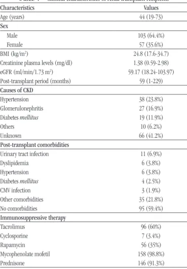
TABLE 1 − Clinical characteristics of renal transplant recipients
Characteristics Values
Age (years) 44 (19-73)
Sex
Male 103 (64.4%)
Female 57 (35.6%)
BMI (kg/m2) 24.8 (17.6-34.7)
Creatinine plasma levels (mg/dl) 1.38 (0.59-2.98) eGFR (ml/min/1.73 m2) 59.17 (18.24-103.97)
Post-transplant period (months) 59 (1-229) Causes of CKD
Hypertension 38 (23.8%)
Glomerulonephritis 27 (16.9%)
Diabetes mellitus 19 (11.9%)
Others 10 (6.2%)
Unknown 66 (41.2%)
Post-transplant comorbidities
Urinary tract infection 11 (6.9%)
Dyslipidemia 6 (3.8%)
Hypertension 6 (3.8%)
Diabetes mellitus 4 (2.5%)
CMV infection 3 (1.9%)
Other comorbidities 35 (21.8%)
No comorbidities 95 (59.4%)
Immunosuppressive therapy
Tacrolimus 96 (60%)
Cyclosporine 7 (3.4%)
Rapamycin 56 (35%)
Mycophenolate mofetil 158 (98.8%)
Prednisone 146 (91.3%)
Inclusion criteria
We have defined as inclusion criteria: patients clinically stable, according to medical evaluation at the time of blood collection; adults older than 18 years of age at time of recruitment; patients who had received kidney from living donors and were submitted to the same protocol of immunossupression, which initially consisted of combined corticosteroid, calcineurin inhibitor (tacrolimus or cyclosporine) and mycophenolic acid, according to general
guidelines on renal transplantation(21, 22).
Exclusion criteria
Exclusion criteria were: acute or chronic allograft rejection defined by histological diagnosis, recent bonefracture and surgery (less than three months before blood collection), coagulopathy, thrombotic diseases, acute infection, recipients under hemodialysis treatment and acute allograft dysfunction. Acute allograft dysfunction was defined by an absolute enhance of 0.3 mg/dl or percentage increase (≥ 50%) over baseline creatinine values, or oliguria (urine output of less than 0.5 ml/Kg/h for more than 6 h)(20, 23-25).
Samples
Whole blood samples were drawn into sodium citrate 0.109 mol/l (VacuetteÒ) and EDTA-K2 1.8 mg/ml (VacuetteÒ) from all patients. The samples were processed within 1 hour after collection and centrifuged at 2.000 g for 20 min at 4oC to obtain plasma samples [platelet poor plasma (PPP)]. Plasma aliquots were separated immediately after centrifugation and stored at -80oC until the enzyme-linked immunosorbent assay (ELISA) and flow cytometry.
TM and vWF measurements
Plasma TM and vWF levels were measured by ELISA,
IMUNOBIND® Thrombomodulin ELISA and IMUNOBIND® vWF ELISA, respectively, following the manufacturer’s instructions (American Diagnostica® Inc., USA). The TM reference range
(provided by the manufacturer) was 4 to 5.35 ng/ml and vWF reference range was 683 to 1012 mU/ml (not provided by the
manufacturer). We used the vWF reference range determined by our research group of a previous study(26).
IL-6 measurement
The determination of plasma IL-6 levels was performed by flow cytometry method/cytometric bead array (CBA) (BD™ FACScalibur cytometer, California, USA) using the diagnosis
Human Inflammation kit (BD® Biosciences Pharmingen, USA). The CBA immunoassay was standardized by our workforce as
described in a previous study(27) with minor modifications of the
manufacturer’s instructions. The results were expressed in mean fluorescence intensity (MFI). The expression of IL-6 results in MFI shows a better sensitivity to differentiate patients whose plasma cytokine levels are outside the linear range, especially when IL-6 are very low(28). Standard curves were performed daily to IL-6 as a
quality control.
Creatinine plasma levels and eGFR
Plasma creatinine levels were measured by a specific enzymatic method (VITROS® 5.1 FS) according to the manufacturer’s instructions and eGFR was calculated using the modification of diet in renal disease (MDRD) equation: 186 × plasma creatinine
level−1.154 × age−0.203 × 0.742 (if female) × 1.212 (if black)(29).
Statistical analysis
For statistical analysis, we used the GraphPad PRISM® software (version 5.0). The normal distribution was tested by Shapiro-Wilk. The comparison among groups was performed using Kruskal-Wallis followed by Dunn’s test. The Spearman test was used for the correlation analysis (TM, vWF, IL-6, creatinine and eGFR). Results were expressed as median, minimum and maximum values. Statistically significance was defined as p < 0.05.
RESULTS
In this study, we compared different groups of RTR according to the primary cause of ESRD, plasma creatinine levels and eGFR. The demographic characteristics of the study population are showed in Table 1.
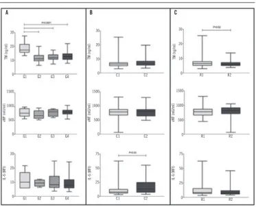
Median plasma TM concentration were significantly higher in G1 group (8.38 ng/ml; p < 0.0001) compared to the others
(G2: 5.51 ng/ml; G3: 5.88 ng/ml; G4: 6.33 ng/ml). There were no differences between groups for plasma vWF (p = 0.3) and
IL-6 (p = 0.74) concentrations regarding primary cause of ESRD
(Figure 1A).
Considering the patients distribution by plasma creatinine
level, plasma IL-6 level were significantly higher in the C2 subgroup (MFI = 13.35; p = 0.03) compared to C1 (MFI = 7.9)
(Figure 1B). On the other hand, no differences were found for TM (p = 0.21) and vWF (p = 0.16) values, between C1 and C2
In the final analysis, the plasma eGFR, TM concentration were significantly higher in R1 (6.65 ng/ml; p = 0.02) when compared
to R2 (6.19 ng/ml). Similarly to TM, IL-6 levels were higher in patients with low eGFR (R1), however not significantly (p = 0.06). No differences were observed for vWF (p = 0.11) levels comparing
R1 and R2 (Figure 1C).
In correlation analysis, plasma TM concentration were positively correlated with concentrations of IL-6 (r = 0.23; p =
0.01) and creatinine (r = 0.2; p = 0.01) in plasma, and there was a negative correlation between TM and eGFR (r = -0.17; p = 0.03).
However, all correlations were weak. Plasma vWF concentration were weakly correlated with IL-6 (r = 0.23; p = 0.01). The other correlations were not significant (Table 2).
DISCUSSION
Endothelial dysfunction is associated with systemic inflammation, insulin resistance and hypertension in patients with CKD. These patients have evidence of endothelial damage,
measured by circulating markers, such as TM and IL-6(30). TM is a
marker of endothelial function and it is present in cells along the
vascular territory(31). This marker is also an indicator of vascular
damage(32).
In the present study, TM levels were significantly increased in G1 group, which presented chronic glomerulopathy as the primary cause of CKD, when compared to other groups (G2, G3 and G4). It is important to mention that there were no differences between groups related to age and gender in this analysis. It should also be noted that the medians for the four groups were above the reference range for TM (4 to 5.35 ng/ml), showing rising levelof this marker in plasma of RTR.
The evaluation of endothelial dysfunction in patients with CKD or RTR is complex due to its multiple nature. In glomerulopathy, many mechanisms as proteinuria, hypoalbuminemia, dyslipidemia
and insulin resistance may result in endothelial damage and promote a procoagulant state in these patients(33-35). In our study,
patients with hypertension or DM did not show increased levels of TM compared to patients with chronic glomerulopathy. It seems that TM levels are more influenced by chronic glomerulopathy than by other comorbidities associated with CKD. An increase in plasma TM concentrations in RTR has been previously reported in other
studies(4, 31, 33, 34, 36).
In this study, high levels of TM have also been found in patients with reduced eGFR (< 60 ml/min/1.73 m2) compared to
patients with eGFR ≥ 60. Liu et al. (2014)(33),and we also noted
high plasma TM levels in patients with low eGFR. Other previous studies have reported that the endothelial damage is more marked
in transplant patients with low eGFR(37-39). Levels of eGFR should
be evaluated along with creatinine plasma levels since creatinine may be influenced by muscle mass, tubular secretion, diet and patient’s hydration status(40, 41).
Endothelial damage is one of the most important factors involved in the development of cardiovascular and thrombotic FIGURE 1 − Plasma concentration of TM, vWF and IL-6 from renal transplant recipients
(RTR)
Plasma levels of TM, vWF and IL-6 according to primary cause of end-stage renal disease (ESRD) (A), creatinine plasma concentrations (B) and eGFR (C) in groups of RTR. All results are presented as median, minimum and maximum values. Data are expressed in ng/ml (TM), mU/ml (vWF) and MFI (IL-6); G1: glomerulopathy; G2: hypertensive nephrosclerosis; G3: diabetic nephropathy; G4: other causes or unknown etiology; C1: creatinine lower than 1.4 mg/dl; C2: creatinine lower or equal to 1.4 mg/dl; R1: eGFR lower than 60 ml/min/1.73 m2; R2: eGFR lower or equal 60 ml/min/1.73 m2. Significant
differences (p < 0.05) are highlighted by connecting lines.
TM: thrombomodulin; vWF: von Willebrand factor; IL-6: interleukin 6; CKD: chronic kidney disease; eGFR: estimated glomerular filtration rate; RTR: renal transplant recipients; MFI: mean fluorescence intensity.
TABLE 2 − Correlation analysis between TM, vWF,
IL-6 and plasma creatinine levels
Correlations r coefficient p-value
TM × IL-6 0.23 0.01
TM × creatinine 0.2 0.01
TM × eGFR -0.17 0.03
TM × vWF -0.09 0.2
vWF × IL-6 0.23 0.01
vWF × creatinine -0.1 0.23
vWF × eGFR 0.1 0.23
TM: thrombomodulin; vWF: von Willebrand factor; IL-6: interleukin 6; eGFR: estimated glomerular filtration rate; p < 0.05 was significant.
A B C
C1 C2
C1 C2
C1 C2
R1 R2
R1 R2
R1 R2
G1 G2 G3 G4
G1 G2 G3 G4
G1 G2 G3 G4
TM (ng/ml)
vWF (mU/ml) vWF (mU/ml)
vWF (mU/ml)
IL-6 (MFI) IL-6 (MFI) IL-6 (MFI)
complications. Additionally, patients with high thrombotic risk also have increased levels of inflammatory markers(39). There
are many endothelial and inflammatory biomarkers, but in our study we chose to evaluate TM, IL-6 and vWF due to the previous experience of our research group(26, 42).
On this background, we found a correlation between the concentrations of TM and IL-6 in plasma. Our findings showed a possible link between renal disease, inflammation and endothelial dysfunction in RTR. Indeed, pro-inflammatory cytokines such as IL-6 and tumor necrosis factor alpha (TNF-α) influence not only the mechanisms that lead to the development of CKD, but also the rejection process after renal transplantation. On the other hand, modulating cytokines, such as IL-4 and IL-5, are involved in mechanisms of immunological tolerance(26, 37, 43-45). Correlations
were also found between concentrations of TM and creatinine in plasma (positive correlation) and there was a negative correlation between TM and eGFR. Similarly, a previous study reported a correlation between concentrations of TM and creatinine in
plasma(46).
Renal transplantation is a clinical intervention that causes
an inflammatory response, regardless the primary cause of CKD. This may result in increase of cytokine synthesis in RTR after transplantation. In our data, an increase in plasma IL-6 concentrations in patients with creatinine ≥ 1.4 mg/dl was observed. We also found higher levels of IL-6 in the group of patients with lower eGFR (R2), which corroborates the results obtained for creatinine, although this finding was not significant. IL-6 was previously demonstrated as a sensitive inflammatory marker to monitor the post-renal transplant and weather the creatinine level rises, IL-6 levels increases in the same proportion. The decrease in filtration rate may also affect the clearance of pro-inflammatory substances directly due to high production or indirectly by renal retention of these markers(4, 41).
Correlations were found between IL-6 and vWF levels (positive
correlation). Bolton et al.(2001)(37) also reported a positive
correlation between IL-6 and vWF levels. These results may be explained by the ability of cytokines to regulate vWF production in endothelial cells. Cytokines have effects on the endothelium and can reduce the clearance of endothelial products.
In our study, no significant differences were found in vWF levels according to primary cause of CKD, creatinine plasma levels and eGFR. It is known that endothelial damage causes an increase in vWF secretion, which considerably increases the circulating levels of this factor. Damage of microvascular endothelial cells is a condition present in CKD and acute
vascular rejection(47). In agreement, other studies showed high
levels of vWF in patients with CKD(37) and RTR(4, 36). However,
another study demonstrated that treatment with corticosteroids
or mycophenolate might reduce vWF levels(48). In our research,
91.3% of patients have used corticosteroids and 98.8% have used mycophenolate. In fact, the immunosuppressive therapy can be a negative influence on immune response and inflammation, since
it prevents cellular immune defense avoiding the activation of T-cells, which could also reduce endothelial dysfunction(26, 49, 50).
However, these immunosuppressive drugs could be associated with adverse effects related to endothelial cells damage and
toxicity(50). Once the effects of immunosuppressive regimen
appear to be unpredictable, the absence of significance for
plasma vWF levels could be explained in part by the type
immunosuppressive therapy used. Thus, we have as perspective
of study to evaluate the influence of immunosuppressive drugs in TM, vWF and IL-6 levels.
We observed that the endothelial damage markers, such as TM and inflammatory markers, as IL-6, could play an important role in the evaluation of endothelial function in RTR. The results suggest that TM and IL-6 are associated with eGFR and creatinine
plasma levels in these patients.
It is important to mention that the relatively low number of subjects and heterogeneity of the cohort, with time post-transplant ranging from 1 month to more than 20 years, limited this study. Despite the influence of the period post-transplant on TM, vWF and IL-6 levels has not been considered as a variable of interest in this study, this analysis was performed and showed no difference. The cross-sectional design of the study may also have interfered in the establishment of the influence of other accumulated comorbidities by patients on the primary cause of CKD.
CONCLUSION
These results indicate that TM and IL-6 could be used as biomarkers of allograft function in RTR. TM was also correlated to primary cause of CKD, vWF was not associated with any study variables. Further analysis is required to elucidate the role of these
biomarkers in renal transplantation.
ACKNOWLEDGEMENT
REFERENCES
1. Sesso RC, Lopes AA, Thomé FS, Lugon JR, Santos DR. Relatório do censo brasileiro de diálise de 2010. J Bras Nefrol. 2011; 33(4): 442-7.
2. Puddu P, Puddu GM, Zaca F, Muscari A. Endothelial dysfunction in hypertension. Acta Cardiol. 2000; 55(4): 221-32.
3. Harada KM, Mandia-Sampaio EL, de Sandes-Freitas TV, et al. Risk
factors associated with graft loss and patient survival after kidney
transplantation. Transplant Proc. 2009; 41(9): 3667-70.
4. Zbroch E, Małyszko J, Małyszko J, Koc-Żórawska E, Myśliwiec M.
Renalase, kidney function, and markers of endothelial dysfunction in
renal transplant recipients. Pol Arch Med Wewn. 2012; 122(1-2): 40-4. 5. Dharmashankar K, Widlansky ME. Vascular endothelial function and hypertension: insights and directions. Curr Hypertens Rep. 2010; 12(6): 448-55.
6. Nesto RW. Correlation between cardiovascular disease and diabetes mellitus: current concepts. Am J Med. 2004; 116(2004): 11S-22S. 7. Opelz G, Döhler B; Collaborative Transplant Study. Improved long-term outcomes after renal transplantation associated with blood pressure
control. Am J Transplant. 2005; 5(11): 2725-31.
by the Conselho Nacional de Desenvolvimento Científico e Tecnológico (CNPq) and Fundação de Amparo à Pesquisa do Estado de Minas Gerais (FAPEMIG). Karina Braga Gomes is grateful for CNPq grant.
CONFLICT OF INTERESTS
The authors declare that there is no conflict of interests.
RESUMO
Introdução: A disfunção endotelial pode contribuir para estados de hipercoagulabilidade e inflamação presentes no transplante renal e na doença renal crônica (DRC) e suas causas, podendo ser avaliada por marcadores como trombomodulina (TM), fator de von Willebrand (FvW) e interleucina 6 (IL-6). Objetivos: Avaliar TM, FvW e IL-6 em receptores do transplante renal (RTR) e associar seus níveis com a causa primária de DRC pré-transplante e função do enxerto. Métodos: Foram alocados 160 RTR em grupos de acordo com a causa primária da DRC (G1: glomerulopatias; G2: nefroesclerose hipertensiva; G3: nefropatia diabética; e G4: outras causas/etiologia desconhecida), os níveis plasmáticos de creatinina (C1 < 1.4 e C2 ≥ 1.4 mg/dl) e o ritmo de filtração glomerular estimado (eRFG) (R1< 60 e R2 ≥ 60 ml/min/1.73 m2). A TM e o FvW foram determinados pelo ensaio de
imunoabsorção enzimática (ELISA) e a IL-6, por citometria de fluxo. Os resultados foram apresentados como mediana, mínimo e máximo; p < 0,05 foi considerado significativo. Resultados: Níveis de TM foram significativamente maiores no grupo G1 em comparação com os demais (G1: 8,38; G2: 5,51; G3: 5,88; G4: 6,33 ng/ml, p < 0,0001), e no grupo R1 comparado com o R2 (R1: 6,65; R2: 6,19 ng/ml, p = 0,02). A concentração de IL-6, avaliada pela intensidade média de fluorescência, foi maior no grupo C2 quando comparada com o C1 (C1: 7,9; C2: 13,35, p = 0,03). Não houve diferença entre os grupos para o FvW. TM correlacionou-se positivamente com IL-6 e creatinina e negativamente com eRFG. A IL-6 foi positivamente correlacionada com o FvW. Conclusão: TM e IL-6 podem ser apontadas como potenciais marcadores para avaliar a função do enxerto renal. A TM relacionou-se mais com a causa primária da DRC, se comparada com FvW e IL-6.
Unitermos: transplante de rim; endotélio; trombomodulina; interleucina 6; fator de von Willebrand.
8. Zhang R, Leslie B, Boudreaux JP, Frey D, Reisin E. Hypertension after kidney transplantation: impact, pathogenesis and
therapy. Am J Med Sci. 2003; 325(4): 202-8.
9. Curtis JJ, Luke RG, Jones P, Diethelm AG, Whelchel JD. Hypertension after successful renal transplantation. Am J Med. 1985; 79(2): 193-200.
10. Couser WG. Pathogenesis of glomerular damage in glomerulonephritis. Nephrol Dial Transplant. 1998; 13(1): 10-5.
11. Kavanagh D, Goodship T. Genetics and complement in atypical HUS. Pediatr Nephrol. 2010; 25(12): 2431-42.
12. Malyszko J, Koc-Zorawska E, Malyszko J. YKL-40, a marker of cardiovascular disease and endothelial dysfunction, in kidney transplant
recipients. Transplant Proc. 2014; 46(8): 2651-3.
13. Martin FA, Murphy RP, Cummins PM. Thrombomodulin and the vascular endothelium: insights into functional, regulatory, and therapeutic aspects. Am J Physiol Heart Circ Physiol. 2013; 304(12): H1585-97.
14. Perutelli P, Molinari AC. von Willebrand factor, von Willebrand factor-cleaving protease, and shear stress. Cardiovasc Hematol Agents Med
15. Tonaco L, Rios DRA, Vieira LM, Carvalho MG, Dusse LMS. Púrpura trombocitopênica trombótica: o papel do fator von Willebrand e da ADAMTS13. Rev Bras Hematol Hemoter. 2010; 32(2): 155-61.
16. Dayananda KM, Singh I, Mondal N, Neelamegham S. von Willebrand factor self-association on platelet GpIbalpha under hydrodynamic shear: effect on shear-induced platelet activation. Blood. 2010; 116: 3990-8. 17. Crawley JT, de Groot R, Xiang Y, et al. Unraveling the scissile bond: how ADAMTS13 recognizes and cleaves von Willebrand factor. Blood. 2011; 118: 3212-21.
18. Sonkar GK, Singh S, Sonkar SK, Singh U, Singh RG. Evaluation of serum interleukin 6 and tumor necrosis factor alpha levels, and their association with various non-immunological parameters in renal transplant recipients. Singapore Med J. 2013; 54(9): 511-5.
19. Hosgood SA, Barlow AD, Johari Y, Bankart MJ, Nicholson ML. Early graft function defined by area under the curve serum creatinine 7 days post-transplant in a series of live donor kidney transplantation. J Surg Res. 2011; 171(2): 838-43.
20. Jung YJ, Lee HR, Kwon OJ. Comparison of serum cystatin C and creatinine as a marker for early detection of decreasing glomerular filtration rate in renal transplants. J Korean Surg Soc. 2012; 83(2): 69-74. 21. Abto.org. Protocolo clínico e diretrizes terapêuticas – imunossupressão no transplante renal [Internet]. Brasil: Ministério da Saúde. Portaria no. 666 de 17 de julho de 2012 [cited 2018, Feb 8] Available at: http://www. abto.org.br/abtov03/Upload/file/Noticias/anexo1.pdf.
22. Womer KL, Kaplan B. Recent developments in kidney transplantation - a critical assessment. Am J Transplant. 2009; 9(6): 1265-71.
23. Humar A, Johnson EM, Payne WD, et al. Effect of initial slow graft function on renal allograft rejection and survival. Clin Transplant. 1997; 11(6): 623-7.
24. Humar A, Ramcharan T, Kandaswamy R, Gillingham K, Payne WD, Matas AJ. Risk factors for slow graft function after kidney transplants: a
multivariate analysis. Clin Transplant. 2002; 16(6): 425-9.
25. Bellomo R, Ronco C, Kellum JA, Mehta RL, Palevsky P; the ADQI workgroup. Acute renal failure – definition, outcome measures, animal models, fluid therapy and information technology needs: the Second International Consensus Conference of the Acute Dialysis Quality Initiative (ADQI) Group. Crit Care. 2004; 8(4): R204-12.
26. Mota AP, Alpoim PN, de Figueiredo RC, Simões e Silva AC, Gomes KB, Dusse LM. Hemostatic parameters according to renal function and time after transplantation in Brazilian renal transplanted patients. Dis Markers. 2015; 2015: 472750.
27. Mota APL, Vilaça SS, Mercês-Júnior FL, et al. Cytokines signatures in short and long-term stable renal transplanted patients. Cytokine. 2013; 62(2): 302-9.
28. Konijnenberg A, Van Der Post JA, Mol BW, et al. Can flow cytometric detection of platelet activation early in pregnancy predict the occurrence of preeclampsia? A prospective study. Am J Obstet Gynecol. 1997; 177(2): 434-42.
29. Levey AS, Bosch JP, Lewis JB, Greene T, Rogers N, Roth D. A more accurate method to estimate glomerular filtration rate from serum creatinine: a new prediction equation. Modification of Diet in Renal Disease Study Group. Ann Intern Med. 1999; 130(6): 461-70.
30. Adams MJ, Irish AB, Watts GF, Oostryck R, Dogra GK. Hypercoagulability in chronic kidney disease is associated with coagulation activation but not endothelial function. Thromb Res. 2008; 123(2): 374-80.
31. Walker FJ, Fay PJ. Regulation of blood coagulation by the protein C
system. FASEB J. 1992; 6(8): 2561-7.
32. Sharain K, Hoppensteadt D, Bansal V, Singh A, Fareed J. Progressive increase of inflammatory biomarkers in chronic kidney disease and end-stage renal disease. Clin Appl Thromb Hemost. 2013; 19(3): 303-8. 33. Liu KL, Lee KT, Chang CH, Chen YC, Lin SM, Chu PH. Elevated plasma
thrombomodulin and angiopoietin-2 predict the development of acute kidney injury in patients with acute myocardial infarction. Crit Care. 2014; 18(3): R100.
34. Małyszko J, Małyszko JS, Brzosko S, Wołczynski S, Myśliwiec M. Markers
of endothelial cell activation/injury: CD146 and thrombomodulin are related to adiponectin in kidney allograft recipients. Am J Nephrol. 2005; 25(3): 203-10.
35. El-Minshawy O, El-Bassuoni E. Validity of current equations to estimate glomerular filtration rate in kidney transplant recipients. Transplant Proc. 2013; 45(6): 2165-70.
36. Sun Q, Zhang M, Xie K, et al. Endothelial injury in transplant glomerulopathy is correlated with transcription factor T-bet
expression. Kidney Int. 2012; 82(3): 321-9.
37. Bolton CH, Downs LG, Victory JG, et al. Endothelial dysfunction in chronic renal failure: roles of lipoprotein oxidation and pro-inflammatory
cytokines. Nephrol Dial Transplant. 2001; 16(6): 1189-97.
38. Malyszko JS, Malyszko J, Hryszko T, Koźminski P, Pawlak K, Mysliwiec M. Markers of endothelial damage in patients on hemodialysis and hemodiafiltration. J Nephrol. 2006; 19(2): 150-4.
39. Malyszko J, Malyszko JS, Pawlak K, Mysliwiec M. Endothelial function and novel adhesion molecule CD44 in kidney allograft recipients. Transplant Proc. 2008; 40(10): 3470-3.
40. Burtis CA, Ashwood ER, Bruns DE. Tietz – fundamentos de química clínica. 6 ed. Rio de Janeiro (RJ): Elsevier; 2008.
41. Dahle DO, Mjøen G, Oqvist B, et al. Inflammation-associated graft loss in renal transplant recipients. Nephrol Dial Transplant. 2011; 26(11): 3756-61.
42. Domingueti CP, Fóscolo RB, Reis JS, et al. Association of haemostatic and inflammatory biomarkers with nephropathy in type 1 diabetes mellitus. J Diabetes Res. 2016; 2016: 2315260.
43. Manzano AMC, Buenrostro LEM, Espinoza LG, et al. Markers of inflammation before and after renal transplantation. Transplantation. 2005; 80(1): 47-51.
44. Karczewski M, Karczewski J, Poniedzialek B, Wiktorowicz K, Glyda M. Cytometric analysis of TH1/TH2 cytokines in the urine of patients
undergoing kidney transplantation. Ann Transplant. 2009; 14(3): 25-8. 45. Pereira AB, Rezende NA, Teixeira Junior AL, Teixeira MM, Simões e Silva AC. Citocinas e quimiocinas no transplante renal. J Bras Nefrol. 2009; 31(4): 286-96.
47. Woywodt A, Schroeder M, Gwinner W, et al. Elevated numbers of circulating endothelial cells in renal transplant recipients. Transplantation. 2003; 76(1): 1-4.
48. Lu GY, Shen L, Wang ZY, et al. Significance of plasma von Willebrand factor level and von Willebrand factor-cleaving protease activity in patients with chronic renal diseases. Chin Med J (Engl). 2008; 121(2): 133-6.
49. Duncan MD, Wilkes DS. Transplant-related immunosuppression: a review of immunosuppression and pulmonary infections. Proc Am Thorac Soc. 2005; 2(5): 449-55.
50. Thoms S, Ali AI, Jonczyk R, Scheper T, Cornelia B. Tacrolimus inhibits angiogenesis and induces disaggregation of endothelial cells in spheroids – toxicity testing in a 3D cell culture approach. Toxicol In Vitro. 2018; 53: 10-9.
CORRESPONDING AUTHOR
Ana Paula Lucas Mota
Avenida Presidente Antônio Carlos, 6.627; Campus Pampulha; CEP: 31270-901; Belo Horizonte-MG, Brasil; e-mail: [email protected].