On the Effects of Mechanical
Stress of Biological Membranes in
Modeling of Swelling Dynamics of
Biological Systems
Igor Khmelinskii
1& Vladimir I. Makarov
2✉
We highlight mechanical stretching and bending of membranes and the importance of membrane deformations in the analysis of swelling dynamics of biological systems, including cells and subcellular organelles. Membrane deformation upon swelling generates tensile stress and internal pressure, contributing to volume changes in biological systems. Therefore, in addition to physical (internal/ external) and chemical factors, mechanical properties of the membranes should be considered in modeling analysis of cellular swelling. Here we describe an approach that considers mechanical properties of the membranes in the analysis of swelling dynamics of biological systems. This approach includes membrane bending and stretching deformations into the model, producing a more realistic description of swelling. We also discuss the effects of membrane stretching on swelling dynamics. We report that additional pressure generated by membrane bending is negligible, compared to pressures generated by membrane stretching, when both membrane surface area and volume are variable parameters. Note that bending deformations are reversible, while stretching deformation may be irreversible, leading to membrane disruption when they exceed a certain threshold level. Therefore, bending deformations need only be considered in reversible physiological swelling, whereas stretching deformations should also be considered in pathological irreversible swelling. Thus, the currently proposed approach may be used to develop a detailed biophysical model describing the transition from physiological to pathological swelling mode.
Electrodiffusion and swelling operate in a wide variety of biological systems1–6. These processes are important
in mammalian cells and subcellular organelles such as mitochondria, nuclei and liposomes, where ion trans-port takes place through specialized membranes. Biophysical models of electrodiffusion with or without swelling have been presented and extensively analyzed7–13. However, the role of membrane stretching has been largely
ignored in previous studies. Recently developed models14–20 included membrane elasticity and the mechanisms
of ion diffusion/electrodiffusion to the membrane surface keeping the electrolyte balance between cell cyto-plasm and intercellular fluid. The theoretical models developed earlier14–20 were based on the Euler-Lagrange
equation describing mechanical tension of the bent membrane, i.e. the only mechanical property addressed was bending tension developed of the deformed membrane. Thus, considering a bilayer phospholipid membrane, its elasticity was characterized by a set of bending tensile parameters. The previous theoretical approaches14–20
considered dynamics of the S/V evolution, where S is the membrane surface area and V is the volume enclosed by the membrane, and S was constant during swelling. However, the effects of stretching deformations (with S being a variable parameter) have not been considered before. Thus, this theory should be upgraded to take into consideration the membrane stretching stress created by swelling. However, presently it is not possible to use the classical membrane elasticity theory together with the model of biophysical processes going on in the system, due to mathematical complexities inherent to the resulting description with a set of partial differential equations21,22.
Therefore, the proposed theoretical approach has only a limited application in the more complex models capable of simulating swelling dynamics of real systems. Note that stretching stress may induce structural changes in the membrane. As it was shown earlier23, transition pores are created in a vesicular membrane by stretching. These
1Faculty of Science and Technology, Department of Chemistry and Pharmacy, and Center of Electronics,
Optoelectronics, and Telecommunications, University of Algarve, Faro, Portugal. 2Department of Physics, University
of Puerto Rico, Rio Piedras Campus, San Juan, USA. ✉e-mail: [email protected]
open
transition pores are an important factor controlling swelling dynamics, and permeability transition pore (PTP) dynamics was studied in mitochondria24,28. However, the mechanism of PTP opening in mitochondria is poorly
understood on molecular level.
Several computational models describing changes in the mitochondrial volume have been developed, repro-ducing swelling dynamics of these organelles26–28. Recently, we have proposed a simplified mathematical approach
to the analysis of mitochondrial swelling dynamics, which includes stretching mechanical properties of the inner mitochondrial membrane24,25. This approach allowed to describe transition from the reversible (physiological) to
the irreversible (pathological) mitochondrial swelling mode.
Presently we report a simple approach incorporating membrane mechanical properties into the description of swelling dynamics of biological systems in both reversible and irreversible modes. Our current focus was on the tools describing the membrane mechanical properties, and on the role of these properties in swelling dynamics. To facilitate the analysis of this problem, we simulated swelling dynamics of a closed membrane (modeling e.g. erythrocytes), with an emphasis on the initial Na+ and Cl− ionic concentrations inside/outside the cell, and water
transport across the membrane. The cell was initially shaped as an oblate axisymmetric ellipsoid. Currently we present the resulting biophysical model, as an example of using the membrane-property tools for a simplified biological system. These tools should be useful in the evaluation of biophysical models of different levels of com-plexity and in the study of volume changes of biological structures. We also evaluated the contribution of these tools to the models of equilibrium (quasistationary) conditions, comparing their results with those obtained by the usage of the classical biomembrane theory17–20, and discussing the relations between the key parameters of
the two theoretical approaches.
Model description
We tested our theoretical approach on a model of closed membrane that describes an erythrocyte. An erythrocyte is approximately a disk 6–8 μm in diameter and 2–3 μm thick29. For simplicity, we modeled an erythrocyte by an
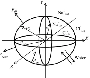
oblate axisymmetric ellipsoid, shown in Fig. 1.
In equilibrium initial conditions, the ellipsoid geometrical parameters were a = b = a0 = 8 μm and c = 2 μm,
with the ellipsoid axes coinciding with the respective x, y and z axes of the Cartesian referential. The rigidity parameters describing the mechanical properties of the membrane will be introduced below. Note that classical membrane elasticity theory developed earlier17–20 is mainly applied to the analysis of biomembrane shape (S/V is
variable with S = constant), when stretching stress may be neglected in a direct problem. However, presently we are considering an inverse problem, where the membrane shape is given, stretching stress is significant, and for the given shape, bending rigidity parameters and pressure induced by the bending tension should be deduced.
Classical elastic membrane model.
To simplify the analysis of membrane bending tension, we used the classical theory of elastic membranes developed earlier30,31 for phospholipid bilayer membranes, with the freeenergy of the membrane given by:
F f dA pdV f k H c k K ( ) 2( ) (1) S c V c c 02 d
∫
λ∫
= + + Δ = + +here, H and K are the respective mean and Gaussian curvatures, which were determined and calculated using the procedure developed earlier32,33, dA and dV are the area and volume elements, of the membrane surface,
and of the space enclosed by the membrane, respectively. Also, kc and kd are the bending modulus values, c0 is
the spontaneous curvature of the membrane, which was neglected in our analysis due to planar spontaneous membrane structure, λ is the Lagrangian multiplier and Δp is the additional pressure inside the membrane,
Z
Y
X
a
0a
0c
Na
+inCl
-inNa
+ outCl
-outWater
P
osP
bendP
strcaused by bending tension. Note that phospholipid bilayer model for the membrane was only used to calculate the bending-induced pressure. The Euler-Lagrange equation corresponding to this energy functional for a spherical vesicle is given by34:
p 2λH k Hc(2 c0)(2H2 c H0 2 )K 2 (kc 2H) 0 (2)
Δ − + + − − + ∇ =
The equations for the energy functional of an oblate axisymmetric erythrocyte are quite complex and may only be analyzed numerically. These equations have four unknown parameters: kc, kd, Δp, and λ, where Δp and λ
depend on the membrane shape. Note that presently we limit ourselves to the analysis of a simple biomembrane elasticity theory, which neglects all of the effects of the respective cytoskeleton30,31. Our objective was thus to
cal-culate the values of the Δp, kc and kd parameters at different levels of cell swelling.
Stretching stress in a biomembrane.
As we already noted, our model neglects the transport of neutral or ionic species through the membrane and the resulting concentration difference of Na+ and Cl− across themem-brane is therefore controlled by water transport only. Thus, here we focus on the analysis of tools that describe membrane stretching.
Osmotic pressure may be described by the following relationship:35
∑
ΔP =k TN (C −C ) (3) os B i i in outinwhere kB is the Boltzmann constant, N is the Avogadro number, T is the absolute temperature, Ciin and Ciout are
the concentrations of the i-th species at the two sides of the membrane. In a biological system, osmotic pres-sure induces cell swelling through the influx of water. The membrane properties defining its mechanical stress due to swelling may be described by a second-rank rigidity tensor, in a biological object with an arbitrary 3D configuration24,25.
For simplicity, we shall model erythrocytes by oblate ellipsoids (see Fig. 1). Figure 1 also shows a referential that is rigidly connected to the biological system, with its Cartesian x-, y-, and z-axes directed respectively along with the a, b, and c axes of the oblate ellipsoid. The rigidity tensor becomes diagonal in this referential24,25, with
only the diagonal components g’ii (i = x, y, z) different from zero. Note that these tensor components are directly
dependent on the membrane properties including its biochemical composition and structure, its permeability with respect to transport of different ionic and neutral species, its mechanical stability, and its physicochemical activity. Besides, there are external parameters (concentration gradient of ions/neutral species, protein concen-trations, temperature, metabolic activity, etc.) that are strongly coupled to the internal membrane parameters. For an axisymmetric oblate ellipsoid (a = b), we obtain g’xx = g’yy24,25. Using the material resistance theory36, diagonal
tensor components may be presented as:
β β β β ′ = ′ = = − +ΔΔ = − + ΔΔ g g g g r r g g z z 1 1 1 1 (4) xx yy n n zz zz z n z n 0 00 0 0 ,0 1 1 1 1
These expressions of the rigidity tensor components include membrane disruption at large mechanical defor-mations (irreversible pathological swelling), where βi (i = 0 and z) and n1 are the respective phenomenological
parameters, introduced to describe the reduction in stress at large deformations24,25. The ellipsoid deformations
are Δr along the x and y axes, and Δz along the z axis, respectively. Here g00 and gzz,0 are the membrane rigidity
parameters describing the membrane properties in the x, y and z directions, respectively, at infinitely small defor-mations. According to Eq. (4), the membrane rigidity approaches zero asymptotically at large mechanical defor-mations, to describe irreversible pathological swelling and cell membrane mechanical disruption. The parameter values were determined by fitting the available experimental data with biophysical models of different complexity levels, where swelling dynamics was described taking into account the membrane mechanical properties23,25. Note
that we used phenomenological rigidity tensor parameters, dependent on membrane composition and structure. These rigidity parameters may be affected e.g. by viscous elements present in the membrane. Note that the rigidity tensor formalism was already used to describe swelling dynamics of biological objects37,38. However, presently
we produced an improved version of this formalism. As we already noted, membrane rigidity should depend on membrane composition and structure, and will be explored in a follow-up study.
The internal pressure induced by membrane stretching stress may be represented as follows:24,25
ΔP t = g t r tΔ +g t z tΔ
S t
( ) 2 ( ) ( ) ( ) ( )
( ) (5)
IMM 0 zz
where S(t) is the time-dependent surface area of the membrane, g0(t) and gzz(t) are the rigidity tensor components
represented by Eq. (4), which are time-dependent due to the time dependence of the Δr(t) and Δz(t) deforma-tions. The surface area and volume of an axisymmetric oblate ellipsoid were given by Tikhonov and Samarskii39,
and we have discussed the respective calculations earlier24,25.
= Δ − Δ
dV
dt S t p t( ) ( )(w Pos PIMM) (6)
Here, pw(t) is the time-dependent membrane permeability for water. The changes in the internal
concentra-tions of the i-th ionic or neutral species due to swelling were described by: = − dC dt C V t dV t dt ( ) ( ) (7) iin iin
Equations (6, 7) provide a correct description of swelling by taking into account the mechanical properties of the biological membrane, primarily as regards stretching stress. Similar equations combined with a compre-hensive description of the respective biophysical processes may be used to analyze the swelling of cells or cellular organelles such as mitochondria24,25. Other membrane-specific biophysical processes may include the generation
of concentration gradients of ionic and neutral species and transport of these species through the membrane. The usage of such combined models to predict and model the experimental data could generate quantitative estimates for the rigidity tensor components, as has already been done for the inner mitochondrial membrane24,25.
Numerical simulations.
Numerical simulations of a closed membrane (erythrocyte) surface area and volume dynamics using the theory of subsection 2.2. Since the parameter values describing the rigidity tensorcompo-nents, Eq.(4), are unknown, we used the values of the respective parameters estimated earlier24,25 for the inner
mitochondrial membrane isolated from murine heart. The values of these parameters are listed in Table 1. Note that this selection of the parameter values is quite reasonable, because the protein-to-lipid ratio equals 80:20 in the inner mitochondrial membrane (IMM), and 50:50 in the outer membrane40. The IMM rigidity is determined
by the amount of cholesterol in the IMM, which depends on the type of cells used as the mitochondrial source41.
All of the simulations were carried out in a system, where the cell volume was much smaller than that of the surrounding medium, equivalent to assuming constant external concentration of Na+ and Cl−, equal to 1 mM in
all of the numerical experiments. However, the initial internal concentrations of the same ions varied from 10 mM to 50 mM, and the dynamics of the cell surface area and volume were calculated for each of the sets of the initial concentrations. The results of calculations are presented as dimensionless parameters:
ζ( )t =a t a0( )/ (0)0 −1, and ( )ξt =c t c( )/ (0)−1, (8) describing relative changes in the lengths of the two axes of the ellipsoid. Numerical analysis was carried out using homemade FORTRAN code, and the results are shown in Fig. 2.
We observe saturation effects for the inner concentrations of the Na+ and Cl− ions in all cases. The saturation
effects originate in the equality of the water fluxes going in and out of the cell. The incoming water flux is gener-ated by the osmotic pressure, causing a dilution of ionic concentrations due to the growing cellular volume, while the outgoing water flux is generated by the internal cell pressure caused by the membrane stretching. Presently we did not analyze the transition conditions from the reversible to the irreversible swelling, as our model system is insufficiently comprehensive to appropriately describe a real erythrocyte. As we noted above, our main objective was to draw attention to the importance of the mechanical stretching of the membrane in its swelling behavior. Therefore, here we considered a simple model simulating the shape of an erythrocyte. In future, a more detailed biophysical model including exchange of additional ionic and neutral solutes in an erythrocyte and mechanical properties of its membrane will be considered in swelling modeling.
We have to note that curves in Fig. 2a have very similar rise time. This result follows directly from Eq. (6), which in an approximate form may be presented as follows:
α α Δ = Δ − Δ Δ ≈ Δ d V dt Sp P V P V ( ) (9) w os IMM
where S and pw are the average surface area and water permeability values and α is the coefficient coupling the pressure induced by mechanical stretching and volume changes. The solution of Eq.(9) may be presented as follows:
Parameter Value Units
n1 4 – gzz,0 0.008 ± 0.001 dyn/nm g00 0.0101 ± 0.0011 dyn/nm βz (1.6 ± 0.2)×104 μm–n1 β0 (1.8 ± 0.2)×104 μm–n1 pw (3.1 ± 0.7)×102 μl·min–1Pa–1μm–2
Table 1. The membrane rigidity tensor components24,25. The values of the tensor components were exchanged
as compared to our previous publications23,25, since a prolate axisymmetric ellipsoid was considered earlier,
α
ΔV t( )= ΔP (1−e−α )
(10) os Sp tw
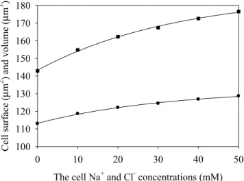
Equation (10) shows that the risetime is αSpw, and it is independent on the osmotic pressure, while the satura-tion value is dependent on the osmotic pressure. The same is valid for the data of Fig. 2b. Using the ellipsoid parameter dynamics shown in Fig. 2, we calculated the saturation values of the cell surface area and volume, keeping ellipsoidal shape at all values of the internal ionic concentrations during swelling. The calculated satura-tion values of the surface area and volume are shown in Fig. 3.
Note the nonlinearity of the plots, which should be due to dilution of the solutes and the linearly growing membrane stretching, thus, no significant deviations of the mechanical stress from linearity were detected in these numerical experiments. Next, we shall reanalyze the results of Fig. 3 using the classical membrane elasticity theory (Section 2.1).
Classical elastic membrane theory used to analyze membrane stretching.
The equilibrium shape of our model system was described by an oblate axisymmetric ellipsoid, with the parameter values listed in Table 2at different solute concentrations.
Taking into account the information in Table 1 and the relationship coupling the stretching rigidity parame-ters with the respective bending rigidity constants for a homogeneous sheet42, we carried out numerical
optimi-zation of the kc and kd parameter values (see Table 3). Taking into account the estimated values of the bending
rigidity constants and Eq. (2), we additionally calculated the pressure induced by the membrane bending stress. The numerical analysis was carried using home-made FORTRAN code, and the respective results are listed in Table 3.
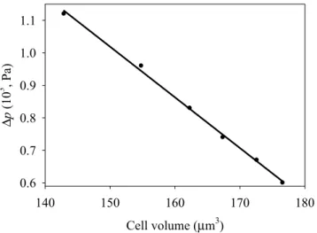
We see that bending rigidity is independent on the swelled cell shape within the range of the parameter values tested. The pressure Δp induced by the membrane bending stress decreases with increasing volume, as shown in Fig. 4.
(a) Time (s) 0 200 400 600 800 1000 1200
ζ
0.00 0.01 0.02 0.03 0.04 0.05 0.06 0.07 (b) Time (s) 0 200 400 600 800 1000 1200ξ
0.00 0.02 0.04 0.06 0.08 0.10Figure 2. Dynamics of the (a) ζ and (b) ξ parameters induced by the cell swelling, bottom to top: 10 mM NaCl
– black line; 20 mM NaCl – red line; 30 mM NaCl – blue line; 40 mM NaCl – green line and 50 mM NaCl – yellow line.
The cell Na+ and Cl- concentrations (mM)
0 10 20 30 40 50 Cell surface (µ m 2 ) and volume (µ m 3 ) 100 110 120 130 140 150 160 170 180
Figure 3. The saturated values of the cell surface area (circles) and volume (squares) versus the initial
Note that the Δp vs. V dependence may be fitted by a linear function, with the intercept and slope equal to 3.359 Pa and –0.0156 Pa/μm3, respectively. These results are in qualitative agreement with those reported
earlier19, where it was shown that bending-induced pressure varies as r–3 for a spherical membrane. Note that
bending-induced pressure is directed outside of the cell and it is perpendicular to the membrane as shown in Fig. 1, while the pressure induced by membrane stretching is directed inside the cell. In other words, bending-generated pressure attempts to restore the flat shape of the membrane, and remove the bending strain, while the stretching-induced pressure attempts to restore the equilibrium conditions. However, bending-induced pressure (Δpbend = 6 Pa at 50 mM NaCl) is typically much lower than stretching-induced pressure (Δpstr = 124 Pa
at 50 mM NaCl) during swelling, whereupon both S and V parameters are changed. Therefore, we may neglect bending-induced pressure in our swelling models, where reversible to irreversible swelling transition is analyzed. In the next section, we will discuss the currently developed approach to the analysis of cell swelling dynamics.
Discussion
Most biophysical models of cell swelling reported to date have ignored the internal pressure generated in the system by membrane stretching (as we already noted, bending-induced pressure is negligible during swelling, when both S and V parameters are changing). Swelling dynamics in these models was described by the equation:27
∑
η = − dV t dt S t k TN C t C t ( ) ( ) [ ( ) ( )] (11) w B i i in outin [Na+] = [Cl−], mM a0, μm C, μm 0 8.00 2.00 10 8.20 2.06 20 8.32 2.10 30 8.39 2.13 40 8.47 2.15 50 8.53 2.17Table 2. Equilibrium shape of an oblate axisymmetric ellipsoid modeling an erythrocyte. [Na+], [Cl−], mM k c, 10−5 erg kd, 10−5 erg Δp, Pa 0 1.38 0.71 11.2 10 1.40 0.73 9.6 20 1.37 0.70 8.3 30 1.36 0.76 7.4 40 1.38 0.71 6.7 50 1.34 0.72 6.0
Table 3. Optimized values of the membrane bending rigidity constants kc and kd, and the bending contribution
to pressure Δp. Cell volume (µm3) 140 150 160 170 180 ∆p (10 3 , Pa ) 0.6 0.7 0.8 0.9 1.0 1.1
Figure 4. Bending-induced pressure inside the cell in function of its volume for an oblate axisymmetric
where ηw is the membrane permeability coefficient for water, and other parameters of Eq. (11) were defined above. The term:
∑
η = J S t( ) k TN C t( ) (12) win w B i i in describes the inflow of water, while the term:∑
η = J S t( ) k TN C ( )t (13) wout w B i i outdescribes the outflow of water from the cell. Assuming that the equilibrium was achieved, we have: =
Jwin Jwout (14)
whereupon no more swelling will occur, corresponding to saturated swelling. As we already noted, this approach does overlook the water flux induced by the internal pressure due to membrane stretching. Therefore, Eq. (9) may produce an incorrect description of swelling. As we also noted above, dynamic effects of the internal pressure induced by bending stress were extensively studied earlier14–20. However, the developed model does not
include the mechanisms for solute transport through the biomembrane, typically considering diffusion-limited solute transport to and from the membrane. In all of those models, as we already noted, the system volume is only changed during swelling, while the membrane surface area is kept constant, thus positive swelling dynamics cor-responds to a decrease in the S/V ratio, while negative dynamics corcor-responds to growing S/V ratio at a constant value of S. Bending tension and pressure will be created by bending deformation of the membrane. These effects may be understood considering an elastic spherical shell filled by a liquid, which provides the minimum S/V ratio. If we will pump some liquid out of the shell, its shape will change, generating additional bending tension and negative internal pressure in our deformed system. Thus, the S/V will increase, while keeping the surface area constant. Note that bending pressure is directed outside of the shell (Fig. 1), and for a water-permeable shell, such negative pressure induces an influx of water. This effect is of opposite sign, compared to the effects generated by stretching stress of our shell.
Although the errors introduced by Eq. (9) could be negligible when the cells are incubated in physiologi-cal solution ([NaCl]0 ≈ 145 mM), they could become significant in cells of the renal epithelium bathed in dilute
luminal fluid ([NaCl]0 < 50 mM)33. Also, Eq. (9) does not describe correctly the irreversible swelling of biological
systems, which is important in pathological states of cells or subcellular organelles such as mitochondria. This problem is addressed in the present study through the analysis of the membrane stretching properties. Indeed, membrane disruption at large stretching deformations may be described using the rigidity tensor components of Eq. (4). We should consider the reversibility of membrane deformations together with its internal parameters that affect its rigidity, directly or indirectly. These include, among others, biochemical composition of the membrane, its shape, its permeability for ionic and neutral species, temperature, and biophysical/biochemical activity. The rigidity tensor parameters describing membrane stretching deformations could be evaluated experimentally by analyzing swelling dynamics based on the presently developed mathematical models. Indeed, membrane disrup-tion should be included into the equadisrup-tions at large membrane strain values, when we model and analyze irre-versible (pathological) swelling. The rigidity data thus obtained could be critical to understanding the membrane internal properties.
We showed above that the tools developed for the analysis of stretching deformations of biomembranes allow to describe the swelled membrane shape in equilibrium conditions, and to calculate bending rigidity parameters and bending-induced pressure in the system by applying the classical membrane elasticity theory to the equilib-rium configuration. Presently we demonstrated the utility of this approach for a phospholipid bilayer membrane. However, considering a real membrane, cytoskeletal properties should be taken into account for a more correct analysis of the membrane bending tension30 in equilibrium shape. The tools for such an analysis were already
developed earlier14–20, and should be combined with those discussed presently to obtain a complete description
of the cellular membrane properties. We shall provide such analysis in a follow-up publication, including addi-tionally the analysis of the irreversible cell swelling. As we already noted, the knowledge of the rigidity parameter values may help to understand the structure of biological membranes. We additionally note that both bending and stretching rigidity parameters contribute to modeling the membrane structure and properties. We also noted that bending tension effects are only important, when the S/V ratio is changed at constant S. On the other hand, stretching deformation of membrane dominates when S grows, and here bending stress effects may be neglected compared to those induced by stretching stress.
Thus, membrane mechanical properties should be included for the correct analysis of swelling dynamics in biological systems. Swelling caused by osmotic pressure deforms the membrane, generating a stress-dependent membrane contribution to the internal pressure in the system. This additional pressure could partially compen-sate for the existence of an inward osmotic gradient, leading to dissipation of water fluxes and maintenance of cell volume. The effect of membrane stress promoting the outflow of water would be independent of the acti-vation of mechano-sensitive ion-channels that promote the exchange of osmolytes and prevent further volume increase43–45. Nevertheless, both mechanisms could operate to reduce the membrane tension and preserve cell
function during osmotic stress. Additional studies are needed to evaluate whether the proposed model adequately simulates the experimental data on the osmotic behavior of cells. This information is critical for the contribution of the membrane mechanical properties to the osmotic behavior of cells, in particular, during alterations of extra-cellular tonicity (hypotonic-isotonic-hypertonic), as could be experienced by the kidney tubular cells, or during cell activation in inflammation, cell culture, and cryopreservation.
Conclusion
In the current study, we discussed tools that describe reversible and irreversible swelling dynamics of a biomem-brane induced by osmotic water influx, where membiomem-brane stretching plays a key role. It was shown that bending effects (Δpbend = 6 Pa at 50 mM NaCl) in swelling dynamics are much less important in comparison to stretching
effects (Δpstr = 124 Pa at 50 mM NaCl)... shown that bending effects (Δpbend = 6 Pa at 50 mM NaCl) in swelling
dynamics are much less important in comparison to the stretching effects (Δpstr = 124 Pa at 50 mM NaCl), when
both volume and surface area are changed. However, bending effects should be taken into consideration, when the cell volume is a variable parameter, while keeping its surface area constant. Note that bending deforma-tions are reversible, while stretching deformadeforma-tions may be irreversible, leading to membrane disruption when they exceed a certain threshold level. Therefore, bending deformations need only be considered for reversible physiological swelling, whereas stretching deformations should also be considered for pathological irreversible swelling. Thus, realistic biophysical models should include both bending and stretching deformations of biomem-branes. It should be made clear that different membrane deformations are important at different levels of swelling: bending is the most important when S/V ≥ (S/V)min at S constant; while stretching is the most important when
S/V < (S/V)min at both S and V variable, where (S/V)min is the minimum ratio value at S constant.
Limitations.
The currently discussed approach includes numerous approximations, lacking dynamics of sol-ute transport, membrane potential, intrasystem metabolism and other processes that should be all taken into con-sideration in real biological systems. However, presently we are developing tools that take into account membrane mechanical properties, and explore their impact on detailed biophysical models.Received: 16 December 2019; Accepted: 20 April 2020; Published: xx xx xxxx
References
1. Jakobsson, E. Interactions of cell volume, membrane potential, and membrane transport parameters. Am. J. Physiol. 238, C196–206, https://doi.org/10.1152/ajpcell.1980.238.5.C196 (1980).
2. Kay, A. R. How cells can control their size by pumping ions. Front. Cell & Develop. Biol. 5, 41, https://doi.org/10.3389/fcell.2017.00041 (2017).
3. Knocikova, J. A., Bouret, Y., Argentina, A. & Counillon, L. Mathematical modeling of cell volume alterations under different osmotic conditions. Biophysics and Medical Physics Computing 8, 1164–1168 (2015).
4. Raspaud, E., da Conceicao, M. & Livolant, F. Do free DNA counterions control the osmotic pressure? Phys. Rev. Lett. 84, 2533–2536, https://doi.org/10.1103/PhysRevLett.84.2533 (2000).
5. Somjen, G. G. Ions in the brain: normal function, seizures, and stroke. ISBN: 9780195151718 (Oxford University Press, 2004). 6. Vereninov, I. A., Yurinskaya, V. E., Model, M. A., Lang, F. & Vereninov, A. A. Computation of pump-leak flux balance in animal cells.
Cell Physiol. Biochem. 34, 1812–1823, https://doi.org/10.1159/000366382 (2014).
7. Hill, A. E. Fluid transport: a guide for the perplexed. J Membr. Biol. 223, 1–11, https://doi.org/10.1007/s00232-007-9085-1 (2008). 8. Hoppensteadt, F. C. & Peskin, C. S. Modeling and simulation in medicine and the life sciences. ISBN 978-0-387-21571-6 (Springer
Science & Business Media, 2002).
9. Keener, J. P. & Sneyd J., Mathematical physiology. ISBN 978-0-387-09419-9 (Springer, 1998).
10. Mathias, R. T. Steady-state voltages, ion fluxes, and volume regulation in syncytial tissues. Biophys. J. 48, 435–448, https://doi. org/10.1016/S0006-3495(85)83799-1 (1985).
11. Shapiro, B. E. Osmotic forces and gap junctions in spreading depression: a computational model. J. Comput. Neurosci 10, 99–120, https://doi.org/10.1023/a:1008924227961 (2001).
12. Weinstein, A. M. Mathematical models of tubular transport. Annu. Rev. Physiol. 56, 691–709 (1994).
13. Yi, C. S., Fogelson, A. L., Keener, J. P. & Peskin, C. S. A mathematical study of volume shifts and ionic concentration changes during ischemia and hypoxia. J. Theor. Biol. 220, 83–106, https://doi.org/10.1006/jtbi.2003.3154 (2003).
14. Zaal, M. Cell swelling by osmosis: A variational approach. Interfaces and Free Boundaries 14, 487–520, https://doi.org/10.4171/ IFB/289 (2012).
15. Lehrenfeld, C. & Rave, S. Mass conservative reduced order modeling of free boundary cell swelling problem. Adv. Comp. Math. 45, 2215–2239, https://doi.org/10.1007/s10444-019-09691-z (2019).
16. Mori, Y. Mathematical properties of pump-leak models of cell volume control and electrolyte balance. J. Math. Biol. 65, 875–918, https://doi.org/10.1007/s00285-011-0483-8 (2012).
17. Safran, S. Statistical thermodynamics of surfaces, interfaces, and membranes (Frontiers in physics) ISBN: 978-0813340791 (Westview Press, 2003).
18. Alimohamadi, H. & Rangamani, P. Modeling membrane curvature generation due to membrane–protein interactions. Biomolec 8, 120, https://doi.org/10.3390/biom8040120 (2018).
19. Tu, Z. C. Elastic theory of membranes. AAPPS Bull 16, 30–33 (2006).
20. Campelo, F., Arnarez, C., Marrink, S. J. & Kozlov, M. M. Helfrich model of membrane bending: from Gibbs theory of liquid interfaces to membranes as thick anisotropic elastic layers. Adv. Colloid & Interface Sci. 208, 25–33, https://doi.org/10.1016/j. cis.2014.01.018 (2014).
21. Agueh, M. Existence of solutions to degenerate parabolic equations via the Monge-Kantorovich theory. Adv. Differ. Eqs 10, 309–360; https://projecteuclid.org/euclid.ade/1355867881 (2005).
22. Almgren, F., Tayler, J. E. & Wang, L. Curvature-drive flows: a variational approach. SIAM J. Control & Optimiz. 31, 387–438, https:// doi.org/10.1137/0331020 (1993).
23. Sanore, O., Moreaux, L. & Brochard-Wyart, E. Dynamics of transient pores in stretched vesicles. PNAS 96, 10591–10596, https://doi. org/10.1073/pnas.96.19.10591 (1999).
24. Makarov, V. I., Khmelinskii, I. V. & Javadov, S. Computational modeling of in vitro swelling of mitochondria: a biophysical approach.
Molecules 23, 783–803, https://doi.org/10.3390/molecules23040783 (2018).
25. Makarov, V. I., Khmelinskii, I. V., Khuchua, Z. & Javadov, S. In silico simulation of reversible and irreversible swelling of mitochondria: the role of membrane rigidity. Mitochondrion 50, 71–81, https://doi.org/10.1016/j.mito.2019.09.006 (2020). 26. Bazil, J. N., Buzzard, G. T. & Rundell, A. E. Modeling mitochondrial bioenergetics with integrated volume dynamics. PLoS Comput.
Biol. 6, e1000632, https://doi.org/10.1371/journal.pcbi.1000632 (2010).
27. Pokhilko, A. V., Ataullakhanov, F. I. & Holmuhamedov, E. L. Mathematical model of mitochondrial ionic homeostasis: three modes of Ca2+ transport. J. Theor. Biol. 243, 152–169, https://doi.org/10.1016/j.jtbi.2006.05.025 (2006).
28. Selivanov, V. A. et al. A model of mitochondrial Ca2+-induced Ca2+ release simulating the Ca2+ oscillations and spikes generated by mitochondria. Biophys. Chem. 72, 111–121 (1998).
29. Stockmann, E. Biological membranes architecture and function in Handbook of biological physics (eds. Lipowsky, R. and Sackmann, E.) vol. 1; ISBN: 9780444819758 (Elsevier, 1993).
30. Helfrich, W. Elastic properties of lipid bilayers - theory and possible experiments. Z. Naturforsch. C 28, 693–703 (1973).
31. Asgari, M. & Biria, A. Free energy of the edge of an open lipid bilayer based on the interactions of its constituent molecules. Int. J.
Non-linear Mech. 76, 135–143, https://doi.org/10.1016/j.ijnonlinmec.2015.06.001 (2015).
32. Jia, Y-B, Gaussian and Mean Curvatures from Elementary differential geometry (Pressley, A.), http://web.cs.iastate.edu/~cs577/ handouts/gaussian-curvature.pdf, (Springer-Verlag, 2019).
33. DoCarmo, M. P., Differential Geometry of Curves and Surfaces; ISBN: 978-0132125895 (Prentice-Hall, 1976).
34. Zhong-Can, Ou-Yang & Helfrich, W. Instability and deformation of aspherical vesicle by pressure. Phys. Rev. Lett. 59, 2486–2488, https://doi.org/10.1103/physrevlett.59.2486 (1987).
35. Atkins, P. & De Paula, J. Physical chemistry for the life sciences; ISBN: 9780199564286 (Oxford University Press, 2011).
36. Beer, F. P. & Russell E., Johnston, Jr., Mazurek D.F. & DeWolf, J. T. Mechanics of materials, 7e SI. ISBN: 9789814595247 (McGraw-Hill, 2014).
37. Simmons, N. L. & Naftalin, R. J. Membrane and intracellular modes of sugar-dependent increments in red cell stability. BBA -
Biomembr. 419, 493–511, https://doi.org/10.1016/0005-2736(76)90261-3 (1976).
38. Naftalin, R. J., Seeman, P., Simmons, N. & Symons, M. A sugar-dependent increase in red cell stability. Biochim. Biophys. Acta 352, 146–171, https://doi.org/10.1016/0005-2736(74)90187-4 (1974).
39. Tikhonov, A.N. & Samarskii, A.A. Equations of mathematical physics. ISBN: 978-0486664224 (Dover books on physics, 2011). 40. Krauss, S. Mitochondria: structure and role in respiration, in eLS; https://doi.org/10.1038/npg.els.0001380 (2001).
41. Mitchell, P. Chemiosmotic coupling in energy transduction: a logical development of biochemical knowledge. J. Bioenerg. Biomembr.
3, 5–24, https://doi.org/10.1007/BF01515993 (1972).
42. Atanackovic, T. M. & Guran A. Theory of elasticity for scientists and engineers; ISBN 978-1-4612-7097-3 (Springer, 2000). 43. Alpern, R. J. & Hebert S. C., eds., Seldin and Giebisch’s The kidney, 4th edition, physiology & pathophysiology 1-2, 253-261; ISBN:
9780120884889 (Elsevier, 2007).
44. Casula, E. et al. Osmotic behaviour of human mesenchymal stem cells: implications for cryopreservation. PLoS One 12, e0184180, https://doi.org/10.1371/journal.pone.0184180 (2017).
45. Casula, E. G. et al. Modelling the osmotic behaviour of human mesenchymal stem cells. Biochem. Eng. J. 151, 107296, https://doi. org/10.1016/j.bej.2019.107296 (2019).
Acknowledgements
This project was partially supported by PR NASA EPSCoR (NASA Cooperative Agreement 80NSSC19M0049), and PR Space Grant (NASA Training Grant Number NNX15AI11H); The authors are grateful to Prof. S. Javadov and Prof. N. Escobales for useful discussions.
Author contributions
Conceptualization: I.K., V.M.; methodology, validation, formal analysis, investigation, writing original draft I.K. and V.M., supervision V.M.
Competing interests
The authors declare no competing interests.
Additional information
Correspondence and requests for materials should be addressed to V.I.M. Reprints and permissions information is available at www.nature.com/reprints.
Publisher’s note Springer Nature remains neutral with regard to jurisdictional claims in published maps and
institutional affiliations.
Open Access This article is licensed under a Creative Commons Attribution 4.0 International
License, which permits use, sharing, adaptation, distribution and reproduction in any medium or format, as long as you give appropriate credit to the original author(s) and the source, provide a link to the Cre-ative Commons license, and indicate if changes were made. The images or other third party material in this article are included in the article’s Creative Commons license, unless indicated otherwise in a credit line to the material. If material is not included in the article’s Creative Commons license and your intended use is not per-mitted by statutory regulation or exceeds the perper-mitted use, you will need to obtain permission directly from the copyright holder. To view a copy of this license, visit http://creativecommons.org/licenses/by/4.0/.